1. Introduction
The Leguminosae (Fabaceae) is the third-largest plant family among angiosperms that comprises 20,000 species and 750 genera1)(2)(3)(4. Legumes establish a symbiotic association with Gram-negative soil bacteria known as rhizobia1)(5) that results in the development of nodules in roots and stems, where atmospheric nitrogen is reduced (fixed) to ammonia, that is assimilated by plants into organic compounds. The family of legumes is divided into three subfamilies that include mostly plants adapted to tropical regions, Cesalpinoidea, Papilinoidea and Mimosoidea; within the former one, only 11 genera form nitrogen-fixing nodules1)(6. Rhizobia are clustered within the Alpha and Betaproteobacteria7, intermingled with non-symbiotic, photosynthetic and plant pathogenic genera and distributed in at least 14 genera7. Rhizobia vary in specificity as well as legumes, while some of them nodulate several hosts, others nodulate only one or two species. Furthermore, several hosts are nodulated by several rhizobia, including both Alpha and Betaproteobacteria. In other cases, the rhizobial strain-host genotype interaction is highly specific, to the point that two closely related species of plants are nodulated by different species of rhizobia7.
Legume nodules induced by rhizobia also might be co-inhabited by a set of various Gram-positive and Gram-negative bacteria, some of them unable to induce nodule development1)(6)(7)(8. These endophytic bacteria can promote plant growth through many different mechanisms and they can do so in symbiosis or as free-living cells. Plant Growth-Promoting Bacteria (PGPB) may act directly by increasing the availability of nutrients such as P solubilization or N2 fixation, by influencing plant hormone levels, or indirectly by attenuating the effects of pathogens9)(10, increasing in this way host fitness. In line with this, rhizobia also may act as PGPB in non-legume cash-crops such as rice or wheat, which turn out to be some of the best beneficial interaction between endophytic rhizobia and plants11.
Both tree legumes and actinorricic plants contribute to increasing N in soils, which is the main reason to consider such systems as a sustainable strategy to improve the productive potential of many different agroecosystems12, to manage silvicultural systems or to prevent soil degradation and N depletion as well. Many legume trees contribute N to tropical wetlands and rainforests, and because of this, it is necessary to have a better understanding of such systems to improve the use and conservation of natural resources13. Additionally, such systems might be exploited in reforestation as well as in land restoration14. A good example of such systems is the Acacia sensu lato species, highly tolerant to drought, due to their large root systems that not only lead plants to explore more soil but also to access deeper layers of water. Vachellia sp. are fast-growing plant species pioneers in extreme environments due to their nitrogen-fixing ability, and also to other symbioses they establish with fungi (e.g. endo and/or ectomycorrhiza), which are critical for their role in poor and eroded soils14. Many legumes might be relevant tools to increase soil organic matter in such systems. Species of Poecilanthe grow in northeast Brazil, in arid areas where water is the main limiting factor. In such ecosystems, a subsistence agriculture mainly based in extensive livestock prevail, where pastures of native legumes are among the most important sources of cow food.
Considering the potential of tree legumes, several studies have been conducted in South America12)(13)(14)(15)(16)(17)(18)(19)(20)(21)(22)(23)(24)(25)(26)(27)(28)(29)(30. They consisted in the isolation of rhizobia and the study of nodule morphology, biochemical and genetic characteristics, systematics and phylogenetic relationships of bacterial symbionts, as well as their ability to nodulate and fix N. However, there are not very many reports regarding species like P. parviflora (Lapachillo), an endemic tree to South America, Vachellia caven (Espinillo) and E. contorsiliquum (Timbó). Regarding the genus Poecilanthe, there are only a few reports of bacteria isolated from nodulated plants growing in Brazil13)(30. Thus, it is of great interest to study the rhizobia as well as other bacteria (nodule microbiome) associated with Poecilanthe sp., Vachellia sp., and Enterolobium sp. in South-American ecosystems, considering their role in nature, particularly regarding legumes growth and development31.
The aim of this work was to isolate and characterize, based on microbiological as well as molecular markers, bacteria within nodules of three native trees species from South America: Poecilanthe parviflora Benth, Vachellia caven (Molina) Seigler & Ebinger, and Enterolobium contortisiliquum (Vell.) Morong. Such information might lead to make a better use of the potential of these native legumes in complex environments.
2. Materials and methods
2.1 Source of bacterial endophytes and biological material
Composite samples of soil were collected from places located under the canopy of P. parviflora plants. Nodules were collected from roots of plants growing within the marginal rain forest of Martín García Island, a protected area located at 34°11′19″S, 58°15′00″W; 14 m.a.s.l (meters above sea level), Buenos Aires, Argentina, belonging to the Delta and Islas del Paraná ecoregion32. Nodules were maintained in hermetic containers until they were processed, which occurred within 48 h. Collected plants were transferred and maintained in 3 L pots filled with soil from Martín García Island and vermiculite at a 1:1 ratio. Pots were watered every other day with distilled water.
Nodules collected from plant roots were surface sterilized by immersing them 2 min. in ethanol 50% followed by 2 min. in sodium hypochlorite 4 g L-1. Then, they were rinsed at least 10 times with sterile distilled water. Sterilization of nodule surfaces was checked by rolling them on plates filled with: a) Yeast Extract Mannitol Agar (YMA)33 K2HPO4 0,5 g L-1, MgSO4. 7 H2O 0,2 g L-1, NaCl 0,1 g L-1, CO3Ca 1 g L-1, yeast extract 1 g L-1, mannitol 10 g L-1, congo red (1/400) 10 ml, Agar 15 g L-1, Ph 6.8; b) Cetrimide agar34 (gelatin peptone 20 g L-1, magnesium chloride 1.4 g L-1, potassium sulfate 10 g L-1, cetyltrimethylammonium bromide 0.3 g L-1, Agar 15 g L-1), and c) Nutrient agar35 (peptone 5 g L-1, meat extract 3 g L-1, sodium chloride 8 g L-1, Agar 15 g L-1).
Isolations were made according to Vincent33 using the described YMA and Nutrient agar media and inoculated plates were incubated at 28 °C. After 7-day-incubation period colonies were subcultured until pure cultures of the isolates were obtained. Then, isolated bacteria were grown in YEM broth33, until saturation and aliquots were mixed with glycerol to make a final concentration of 10% in stocks that were maintained at -80 °C.
V. caven and E. contorsiliquum nodule bacteria were obtained as described before, from nodules developed by inoculated plants. Seeds of these species were surface scarified and sterilized with sulfuric acid for 15 min., rinsed with sterile water and germinated in 10% water-agar in Petri dishes in the dark. Seedlings were transplanted into pots with sterile vermiculite and inoculated with 1 ml of soils decimal dilution in sterile water. Soils were collected at 24°48’52.3’’S 65°29’59.6’’W, 1.300 m.a.s.l. for V. caven. and at 24°35’18.7’’S 65°02’50’’W, 750 m.a.s.l. for E. contorsiliquum, from the dry Chaco ecoregion32, Salta, Argentina. Stock cultures of isolated bacteria were made and kept as described before. Also, reference strains B. japonicum E109, SEMIA 5079, B. elkanii SEMIA 5019 and SEMIA 587, as well as the type strain Mesorhizobium Chacoense Pr5 (=LMG19008=CECT5336) isolated from Prosopis alba36 at the dry Chaco ecoregion32, Chancaní Reserva, Córdoba, Argentina, were included in this work.
2.2 Microbiological characterization
All isolates were observed with the light field optical microscope (1000X) after Gram staining37. Alkalinization or acidification of the culture medium was determined in non-buffered YEM broth supplemented with bromothymol blue, after 10 days of growth, at 28 °C in a rotary shaker at 250 rev. min-1. Isolates were classified as developing a strong, moderately or slightly acid reaction based on the yellow colour developed, while neutral and basic reactions were associated with green and blue colours, respectively.
Bacterial isolates were grown for 10 days at 28 °C on NBRI-P (National Botanical Research Institute's phosphate)38) medium (glucose 10g L-1, Ca3PO4 5 g L-1, MgCl2.6H2O 5 g L-1, KCl 0,2 g L-1, (NH4)2SO4 0.1g L-1, SO4Mg.7 H2O, Agar 20g L-1) to evaluate their ability to solubilize inorganic phosphorous that was determined by the development of a transparent halo around bacterial colonies.
The antimicrobial activity of isolates was assayed by co-culturing them with two model plant pathogens, Stemphylium lycopersici CIDEFI 216, the causal agent of grey leaf spot39, and Fusarium graminearum SP1, the etiological agent of head blight40. These two pathogens have a wide host range, representatives of them have been reported to provoke diseases on legumes and are used as model systems to study plant-microbe interactions in our laboratory. Both fungi were grown on PDA (Potato Dextrose Agar, potato 250 g L-1, dextrose 20 g L-1, agar 20 g L-1)41) medium, for 3 (F. graminearum) or 10 (S. lycopersici) days at 25 °C in the darkness. Antimicrobial activity was evaluated by placing bacteria on PDA medium in two equidistant parallel streaks. After an incubation period of approximately 7 days at 25 °C, a disc of the fungus was placed at the center of each Petri dish. Control treatment consisted of plates inoculated with fungi in the absence of bacteria. All plates were sealed with parafilm and incubated at 25 °C. The experiment finished within 3 (F. graminearum) or 10 (S. lycopersici) days.
The inhibitory activity of bacteria was estimated by measuring mycelial growth on the surface of the Petri dishes using the software Image tool 3.0 (Image Tool Software Copyright)42. The percentage of inhibition (InC) was calculated with the formula: % InC = [(control mycelial growth area - treatment mycelial growth area)/control mycelial growth area]*10043. Bacterial isolates ability to solubilize inorganic phosphorous and their antagonistic effect against S. lycopersici and F. graminearum were evaluated in three independent experiments. The collected data were subjected to a one-way analysis of variance (ANOVA), means were compared by the Tukey test. All statistical analyses were performed using Infostat version 201544.
2.3 Molecular analysis
2.3.1 DNA extraction and genetic diversity analysis
Isolates DNA was extracted using the Wizard® Genomic DNA Purification Kit (Promega)45. Briefly, isolated bacteria were cultured in liquid media until a cell concentration of 1×10⁹cells mL−1 was obtained. Aliquots of 5 mL were used to extract DNA, whose quality and quantity was checked by electrophoresis in 0.7% agarose gels stained with ethidium bromide that included a molecular marker46.
Endophytic bacterial isolates were fingerprinted using a BOX-PCR with the universal BOXA1R primer (5′-CTACGGCAAGGCGACGCTGACG-3′). PCR contained 1x amplification buffer (Inbio Highway), 2.5mM MgCl2 (Inbio Highway), 50 pmol BOXA1R primer, 2 mM each dNTP (Inbio Highway), 50-100 ng of genomic DNA, and 0.6 U Taq DNA polymerase (Inbio Highway) in a 15 ul volume. Reactions were performed in a PTC-1152 Mini Cycler (MJ Research) programmed as follows: an initial step at 94 (C for 7 min, followed by 35 cycles at 94 (C for 1 min, 53 (C for 1 min, and a final step at 65 (C for 8 min, and a final cycle at 65 (C for 16 min47. The PCR amplicons were separated on 1.5% agarose gels, stained with ethidium bromide.
The sizes of the fragments were normalized according to the MW of the DNA markers (λ HindIII marker, Invitrogen). Fingerprints were analyzed in a binary data matrix, scoring 0 for the absence and 1 for the presence of bands. A multivariate analysis was carried out using Past3 software48.The Dice similarity index was used to create a similarity matrix that was used to generate a dendrogram using the Unweighted Pair Group Method with Arithmetic Mean (UPGMA) algorithm39. All those bacterial cultures that had a unique fingerprint were selected for 16S rDNA analysis.
2.3.2 16S rDNA amplification and sequencing
Bacterial isolates were identified using a partial sequence of the 1500 bp 16SrDNA. Such fragments were amplified by PCR in a thermocycler (MinicyclerTM, MJ Research Inc., Waltham, MA, USA), using primers 27F (5´-AGAGTTTGATCMTGGCTCAG-3´) and 1492R (5´-ACGGTTACCTTGTTACGACTT-3´) as described by Reysenbach and others49. PCR products were purified as described by Sambrook and others50, precipitated and sequenced at MACROGEN Inc. (Seoul, South Korea). Sequences were analysed and trimmed with Geneious R9 software version R9.0.5, Biomatters51. Sequences were analysed by Basic Local Alignment Search Tool (BLAST) against 16S rDNA sequences of type strains.
The phylogenetic analysis was conducted using Geneious R9 software51. Sequences were aligned using the default parameters of the ClustalW algorithm (gap opening penalty 15, gap extension penalty 6.66)52. Phylogenetic analysis was performed using the genetic distance model as described by Tamura- Nei using the neighbor-joining method53.
3. Results and discussion
3.1 Isolation of endophytic bacteria from nodules of tree legumes from Argentina
The isolates collected from nodules of native legume trees included in this work are presented in Table 1. All P. parviflora isolates were obtained from root nodules belonging to marginal rain forest plants, which was the only environment in the Martín García Island, among the three sampled sites (riparian forest, marginal rain forest and the coronillo forest). We found P. parviflora plants with functional indeterminate nodules whose interior were red, suggesting they contain leghemoglobin and are fixing nitrogen. In the riparian and the coronillo forests, samples of P. parviflora plants had no nodules or non-functional ones. This may be due to the different soil texture classes, frequent flooding of these soils and/or variations in legume species composing the flora at the different vegetative units, associated to the environmental gradient of the island54)(55, which might influence and/or determine the pools of resident bacteria in the soil.
Among isolated bacteria, 15 were obtained from P. parviflora, 6 from V. caven and 1 from E. contortisiliquum. Interestingly, the isolates from P. parviflora were recovered, some of them on one media and the other ones on other media, suggesting that nodules probably harbour a diverse array of bacteria.
3.2 Microbiological characteristics of the isolates
Four bacteria among those collected from P. parviflora and the E. contorsiliquum isolate alkalinize the culture medium, while 1 from M. chacoense, 7 from P. parviflora and all 6 isolated from nodules of V. caven acidified the media. However, while some of them generated a slight acid reaction, others generated a strong one on the same media. The other 4 bacteria isolated from P. parviflora did not alter the pH of the medium. The main microbiological characteristics of the isolates are presented in supplementary material (Table S1). It is known that while Bradyrhizobium sp. alkalinize the YEM broth, Ensifer (Sinhorhizobium) acidified it and Mesorhizobium species activity on the culture broth is dependent on the carbon source of the media36.
Table 1: Bacterial isolates obtained from nodulated plants and control strains used in our experiments. Host species and the media used to isolate bacteria as well as the site of material source (soil sample or nodule plant collected in situ) used for the bacterial isolation are presented. YMA: Yeast Extract Mannitol Agar supplemented with congo red; NA: Nutrient Agar. The isolates were named according to the host of origin E: Enterolobium, AC: Acacia (Vachellia) caven and LA: Lapachillo. Numbers before and after the letters indicate the number of the isolate, except for the denominations as an "S" followed by number (e.g. S43), which indicate different soil sites.
| Isolates | Host | Isolation media | Site of material source |
|---|---|---|---|
| 4ES6-1 | E. contorsiliquum | YMA | Salta |
| ACS43 | V. caven | YMA | Salta |
| 9ACS43 | V. caven | YMA | Salta |
| 11AC3 | V. caven | YMA | Salta |
| ACS41 | V. caven | YMA | Salta |
| AC4S41 | V. caven | YMA | Salta |
| 5ACS4-2 | V. caven | YMA | Salta |
| LA33A2 | P. parviflora | YMA | Buenos Aires |
| LA11 | P. parviflora | YMA | Buenos Aires |
| LA13 | P. parviflora | YMA | Buenos Aires |
| LA2 | P. parviflora | YMA | Buenos Aires |
| LA32B1 | P. parviflora | YMA | Buenos Aires |
| LA32A2 | P. parviflora | YMA | Buenos Aires |
| LA32A1 | P. parviflora | YMA | Buenos Aires |
| LA35B | P. parviflora | YMA | Buenos Aires |
| LA33A1 | P. parviflora | YMA | Buenos Aires |
| LA32B2 | P. parviflora | YMA | Buenos Aires |
| LA26 | P. parviflora | YMA | Buenos Aires |
| LA21 | P. parviflora | NA | Buenos Aires |
| LA4 | P. parviflora | NA | Buenos Aires |
| LA10 | P. parviflora | NA | Buenos Aires |
| LA8 | P. parviflora | YMA | Buenos Aires |
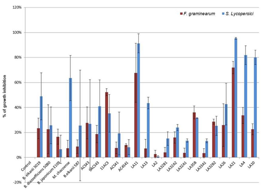
We first screened the isolates to identify their capacity to inhibit S. lycopersici and F. graminearum growth (data no show). B. japonicum SEMIA5079 and isolates 4ES6-1, 5ACS4-2, LA33A2 and LA8 inhibited fungal growth at values <10%. Based on these results we run additional experiments with selected isolates and reference strains that inhibited mycelial growth ≥10% of any of the pathogenic fungi assayed. Among isolates of P. parviflora, V. caven and E. contorsiliquum analyzed (Figure 1), 11 of them inhibited the growth of F. graminearum and/or S. lycopersici within a range of 19% to 72% and 41% to 95%, respectively. Both fungal pathogens F graminearun and S. lycopersici were inhibited by the following 5 isolates of P. parviflora: LA4, LA10, LA 11, LA21 and LA26. The former pathogen was inhibited in a 34%, 23%, 68%, 72% and 26%, and the latter one in an 82%, 80%, 91%, 95% and 43%, respectively. Also, isolate 9ACS43 obtained from V. caven inhibited F. graminearum and S. lycopersici in a 19% and 41%, respectively. The commercial strain B. elkanii 5019 behaved like the isolates described, inhibiting F. graminearum and S. lycopersici by a 23 and 49%, respectively (Figure 1).
Two isolates collected from V. caven (ACS43 and 11AC3) and three from P. parviflora (LA13, LA32B2, LA35B) as well as M. chacoense only inhibited the growth of S. lycopersici or F. graminearum (Figure1) with values on S. lycopersici of 44% (LA13) and 64% (M. chacoense), and 28% (ACS43), 29% (LA32B2), 36% (LA35B) and 52% (11AC3) for F. graminearum. Among the isolates that proved to have a considerable capacity to inhibit fungal growth, only isolate 11AC3 that was collected from nodules of V. caven was able to solubilize phosphorus, suggesting that it might be a promising plant growth-promoting bacteria that not only control plant pathogens but also might help plants to access more P, that most of the time is in unavailable forms in the soil.
3.3 Genetic studies: diversity and identification of endophytic bacteria.
Molecular biology provided researchers with powerful methods for the typing and genetic identification of organisms. Such information is crucial and is used to perform phylogenetic analysis, since universal conserved gene sequences such as the 16S rDNA are used. DNA-based methods have become increasingly important to identify and characterize rhizobia, in particular, phylogenetic analyses of sequences of the 16S ribosomal rDNA (rRNA) gene, a range of “housekeeping” genes and genes involved in symbiosis have been used and proposed as a "standard approach" for the final identification of organisms. Concomitantly, the 16S rDNA gene sequence on its own turn out to be a reliable tool mainly for the preliminary identification of microorganisms56. However, several gene sequences like those of the nif and nod genes involved in N2 fixation and legume host specificity are often carried on plasmids or symbiotic islands in many nitrogen-fixing bacteria, and therefore, might be horizontally transferred between different species within a genus and less frequently across genera57)(58; because of this their relevance or role in identification has to be cautiously considered.
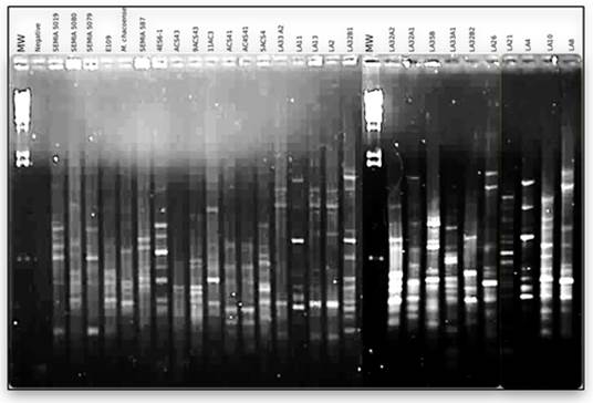
Based on the reasons described we compared the genome fingerprints generated using the BOXAR1 primer of isolated bacteria with those of B. Diazoeficiens (SEMIA 5080), B. japonicum (E109 and SEMIA 5079), B. elkanii (SEMIA 5019 and SEMIA 587), and M. Chacoense type strain using the BOXAR1 primer PCR (Figure 2).

Figure 1: Growth inhibition in vitro of fungal pathogens F. graminearum and S. lycopersici by 18 bacterial isolates. B elkanii 5019 and 587, B. diazoefficiens SEMIA5080, B japonicum E109 and M chacoense were included as controls

Figure 2: BOXPCR fingerprints of bacteria isolated from nodules of from P. parviflora, V. caven and E. contorsiliquum
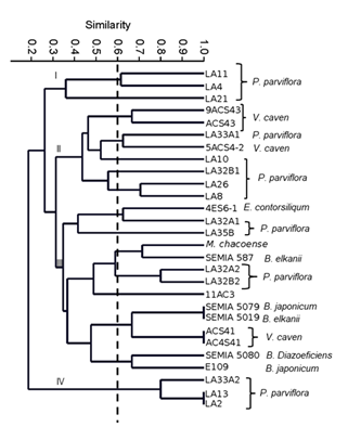
Isolates and type strains were grouped into 4 major clusters (Figure 3) at a ~0.35 similarity level, two of them only containing isolates of P. parviflora. Cluster I was formed by isolates LA11, LA4 and LA21, and Cluster IV included isolates LA33A2, LA13 and LA2. Clearly, these two groups of bacteria were quite different. The other 16 isolates were grouped within cluster II and III. Cluster II included isolates of P. parviflora LA33A1, LA10 and LA32B1 that were distributed into 3 different subgroups, LA26 and LA8 were together and in a different subgroup than isolate 5ACS4-2 of V. caven, which also was different from V. caven 9ACS43 and ACS43, that were grouped at a ~0.7 similarity level. Cluster III included isolates of V. caven, P. parviflora, E. contorsiliqum, M. Chacoense, B. elkanii, B. japonicum and B. diazoeficiens, so most probably, as shown below, these bacteria were representatives of nitrogen-fixing ones. At a 0.6 similarity level, the isolate of P. parviflora LA32A1 was grouped with bacteria isolated from E. contorsoliquum in a different subgroup, apart from isolates LA35B, LA32A2 and LA32B. The isolate 11AC3 of V. caven differed from ACS41 or AC4S41, although additional genetic studies should be performed to accurately establish similarity between the isolates. Cluster IV included two isolates that had an identical fingerprint and therefore might be siblings of the same strain, while the other isolate proved to be quite diverse. In any case, organisms within this cluster are the more diverse compared to the other bacteria. It is evident that even though isolates were collected from the same hosts, they were genetically quite diverse, and even nitrogen fixing bacteria might belong to different rhizobial species as well.
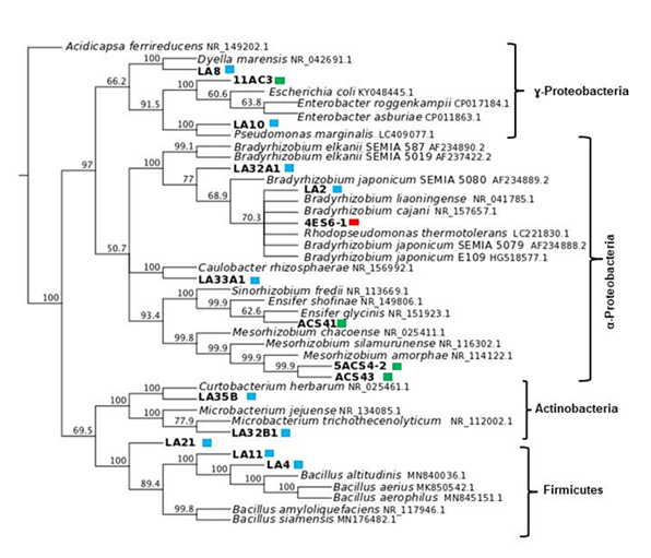
Based on the BOX dendrogram we proceeded to identify the most diverse bacteria by amplifying and sequencing their 16S rDNA gene. The BLAST analysis of these sequences suggested that root nodule endophytic bacteria belonged to 10 genera that were either within the Alphaproteobacteria, Gammaproteobacteria, Firmicutes or Actinobacteria (Figure 4).
Within nodules we isolated bacteria that based on the 16S rDNA sequence were clustered in 4 main groups (Figure 4). Since they were isolated from nodules, they might either be nitrogen-fixing bacteria, plant growth-promoting bacteria that live within nodules, or opportunistic saprophytic bacteria that alone or by interacting with the plant or other microbes, provide plants with a biological advantage for the ecological niche. One group of bacteria consisted in Gammaproteobacteria and included isolates LA8 and LA10 that were isolated from nodules of P. parviflora. These bacteria belonged to the families Rhodanobacteraceae (Dyella sp.) and Pseudomonadaceae (Pseudomonas sp.), with 99.86% and 100% of identity, respectively. This same cluster also included isolate 11AC3, that was collected from nodules of A. caven and belonged to Enterobacteraceae, being an Enterobacter sp. However, the sequence identity was too low, therefore, more sequences or a larger 16SrDNA sequence should be analyzed to accurately identify the isolate.

Figure 3: Dendrogram generated by UPGMA cluster analysis using the Dice similarity coefficient based on the BOXAR1 fingerprint of studied isolates

Figure 4: Phylogenetic relationship of the isolates based on aligned sequences of the 16S rDNA gene. Sequences accession number representing the genera is on the tree. Isolates from the same legume species are indicated as coloured squares: blue for P. parviflora, green for V. caven, and red for E. contorsiliquum. Acidicapsa ferrireducens was the outgroup
Among isolated bacteria, the blast analysis showed that at least 6 were nitrogen-fixing bacteria that belonged to Alphaproteobacteria. These were isolates LA2 from P. parviflora; 4-ES6-1 from E. contorsiliquum; isolates ACS41, 5ACS4-2, ACS43 that were collected from nodules of A. caven; and isolate LA32A1, whose 16SrDNA sequence, unfortunately, had a too low similarity level, indicating that we need to go back to more precisely identify the isolate. These bacteria, except for the latter one, were clustered separately with a high bootstrap value of 100% and 93.4%. Furthermore, they are most probably representatives of Bradyrhizobium, Mesorhizobium and Ensifer since their sequences shared 99.66 % and 100% identity with type strains of these genera. It has already been reported that nodules induced by rhizobia might contain other bacterial taxa such as representatives of the Gammaproteobacteria. Pantoea agglomerans, Enterobacter kobei, Enterobacter cloacae, Leclercia adecarboxylata, Escherichia vulneris, Pseudomonas sp., Bacillus sp., Pseudomonas sp., Curtobacterium herbarum and Microbacterium sp., among others, which were frequent bacteria within nodules of Hedysarum mediterranean species59)(60. This might be related to the nutrient level and/or particular environment of nodules that evidently harbour a wide variety of non-symbiotic bacteria, which could be either necessary partners or opportunistic competitors.
Other isolates were clustered within the Actinobacteria group, like LA32B1 and LA35B organisms, that were isolated from P. parviflora and identified one as Microbacterium and the other as Curtobacterium. Representatives of the Firmicutes LA4, LA11 and LA21 were identified as Bacillus species, since their 16SrDNA sequence was 100% identical to that of Bacillus species. Bacteria recovered from native legume trees live endophytically within nitrogen-fixing nodules induced by rhizobia, but might be either cooperative microorganisms, plant growth promoting bacteria or nitrogen fixing ones. It has already been described that Pseudomonas (Gammaproteobacteria) developed N2 fixing nodules on Robinia pseudoacacia61 and Acacia confusa62. Geobacillus (Firmicutes), Paenibacillus (Firmicutes) and Rhodococcus (Actinobacteria) also were described as rhizobial symbionts of Lotus corniculatus63. These bacterial species had similar nodA gene sequences to Mesorhizobium representatives isolated from the same plants; most probably these bacterial species received these genes by lateral gene transfer from Mesorhizobium. Although such process might not be so frequent, the analysis should be done to clarify this issue. As reported by Saïdi and others64, Pseudomonas, Enterobacter, Bacillus, Staphylococcus, Serratia, Stenotrophomonas and Xanthomonas have already been found within Vicia faba root nodules, though these bacteria failed to nodulate it and did not have nifH or nodC genes. However, they also cited reports of Alphaproteobacteria, Betaproteobacteria and Gammaproteobacteria that were isolated from root nodules as nodulating bacteria or nodule associated bacteria in a wide array of legumes. All this together suggests that legume nodulation may not be exclusively triggered by Alphaproteobacteria8. Also, the other organisms living inside nodules provide plants with additional advantages that might be related to nutrition or plant health. This is not surprising considering that rhizobia free legumes like alfalfa can develop nodules even in axenic conditions65, suggesting that legumes have preprogram sets of events that allow them to develop nodules. Probably, there are many more organisms than rhizobia alone that can trigger nodule formation in legumes66.
Rhizobia isolates were clustered in three groups belonging to the families Bradyrhizobiaceae, Rhizobiaceae and Phyllobacteriaceae (Figure 4), among them, Mesorhizobium is closer to Sinorhizobium than to Bradyrhizobium, which is in agreement with previous reports11. Isolates LA2, LA32A1 and 4ES61 that were isolated from P. parviflora and E. contorsiliquum, respectively, belonged to a cluster that included B. japonicum strains, suggesting that they most probably are Bradyrhizobium sp. Interestingly, B. Elkanii SEMIA 6403 (=BR 8205) has been recommended in Brazil to inoculate P. parviflora67. Nodules of E. contorsiliquum contained a Bradhyrhizobium sp. that was characterized by an extremely slow growth rate and very small cells, such that they were hardly visible in the light microscope. This and the 16S rDNA sequence homology with the 16SrDNA sequence of B. liaoningense type strain (data not shown) suggested that it is most probably a B. liaoningense representative. In line with this, cultures of such isolate alkalinize the culture media. Isolates from P. parviflora belonged to Bradyrhizobium genus and, even though they were different, additional sequences should be examined to accurately establish their identity, particularly because there are other native legumes within the area suggesting that they might be promiscuous bacteria.
V. Caven was nodulated by Mesorhizobium strains since isolates 5ACS4-2 and ACS43 clustered with Mesorhizobium representatives with a high bootstrap value of 99.88 %. The 16S rDNA sequence of the isolated strains were 99.79 % and 100 % identical to that of Mesorhizobium strains. Unlike other genera of rhizobia, species within Mesorhizobium show relatively low sequence divergence at core loci, this is such that all Mesorhizobium type strains presented a high level of sequence homology11)(68. Laranjo and others11) grouped 30 Mesorhizobium species in four clusters, which as in this work showed differences between them. Furthermore, MLSA phylogeny analysis divided isolates in three main groups that contained M. plurifarium and M. chacoense, suggesting, in a way, the low resolution of the 16SrDNA gene sequences11. They stated that lateral gene transfer, as well as duplication, might bring additional problems for phylogenetic studies within the genus Mesorhizobium. Because of this, the phylogenetic analysis of Mesorhizobium might disagree with broader phylogenies based on multiple core genes68)(69. However, Laranjo and others11 found that there is no gene transfer of core loci between some specific groups of Mesorhizobium.
Vachellia is a genus that includes promiscuous species that might be nodulated by several species of rhizobia70. Our results confirmed this since isolate ACS41 was clustered with Sinorhizobim sp. (Ensifer sp.) at 100% bootstrap value and a 99.85% of sequence identity, suggesting that it belongs to the genus Ensifer. Interestingly, Ensifer representatives have been found to have the widest host range so far studied71, and Vachellia species were nodulated by diverse and promiscuous Ensifer species72)(73.
The rhizobia isolated for the native trees included in our study are in line with previous findings that stated that 59 Mesorhizobium species nodulate a wide array of different legumes74: M. amorphae from Amorpha fruticosa75, M. silamurunense from Astragalus membranaceus76, and Mesorhizobium acaciae from Acacia melanoxylon R. Br.(77, among others. Beukes and others70 studied Vachellia karroo root nodules that contained the genera Bradyrhizobium, Ensifer (32 isolates), Mesorhizobium (27 isolates), and Rhizobium, as well as Betaproteobacteria; and more recently, Pereira-Gómez and others22 reported 8 V. caven isolates, affiliated to the genus Mesorhizobium. Thus, this diversity may contribute to the ecological success of Vachellia sp. as pioneer species, as previously noted, for example, for V. Karoo70.
Both genera Ensifer and Mesorhizobium include several species that nodulate Acacia sensu lato species; this is in agreement with previous data reported for other African Acacias (i.e., Vachellia sp. and Senegalia sp.), which also are predominantly nodulated by isolates of Mesorhizobium, Rhizobium and Ensifer70. Also, Cordero and others14 found that V. macracantha, a legume tree distributed across neotropical regions, spreading from the Southern United States to Northern Argentina, was nodulated by Ensifer numidicus, Ensifer kummerowiae, Ensifer fredii, Ensifer americanus and Ensifer saheli. A range of genera in the Proteobacteria (most commonly Bradyrhizobium, Ensifer (Sinorhizobium), Mesorhizobium and Rhizobium) can form functional (N2 fixing) nodules on specific legumes, and for the Mimoseae tribe genera Acacia, Senegalia, Prosopis and Vachellia, and a complete list of legume -rhizobia symbiosis- is available in Andrews and Andrews56.
4. Conclusions
South American native tree legumes V. caven and P. parviflora harbour a great diversity of endophytic bacteria and alpha-rhizobia in their root nodules.
E. contorsiliquum may establish a nitrogen-fixing symbiosis with Bradyrhizobium lianonginese, and P. parviflora may develop nodules with Bradyrhizobium spp strains that need to be studied further to determine the species. V. caven within the environments sampled may be nodulated by both Ensifer spp and Mesorhizobium strains, suggesting that it is a promiscuous legume species that interacts mainly with broad host range rhizobia.
It is known that nodules are rich in nutrients providing a habitat for bacteria, not only for nitrogen-fixing rhizobia, but for other microorganisms (associated or endophytic) as well, and interestingly, the native legumes studied contained Gammaproteobacteria (Pseudomonas, Dyella, Caulobacter), Actinobacteria (Curtobacterium, Micobacterium) and Firmicutes (Bacillus), that in in-vitro studies showed they can control fungal pathogens or solubilize P. These non-rhizobia endophytes might play a role in nutrition, promoting plant growth and health. In this regard, more work is needed to understand the role of the endophytic bacteria in root nodules of these legumes in combination with rhizobia, as well as to establish their condition as micro-symbiont or endophytic bacteria.
These biological roles of symbiotic and endophytic microorganisms' interactions as well as the knowledge of nodule microbiomes might contribute to have a better understanding of the system, and a better management of natural resources.


