Serviços Personalizados
Journal
Artigo
Links relacionados
Compartilhar
Odontoestomatología
versão impressa ISSN 0797-0374versão On-line ISSN 1688-9339
Odontoestomatología vol.25 no.42 Montevideo 2023 Epub 01-Dez-2023
https://doi.org/10.22592/ode2023n42e417
Case report
Cone beam computed tomography as an aid in the treatment of a maxillary incisor with unusual anatomy: a case report.
1 Facultad de Odontología UJED. Durango, México
The failure in root canal treatment can lead to the loss of the dental organ. Various factors have been reported as causes of failure in endodontic therapy. Among these factors, omitted canals have a greater influence, as they allow colonization and multiplication of bacteria within the root canal. Abnormal anatomical variations can increase the chances of failure due to the inability to diagnose accurately. In these cases, cone-beam computed tomography has proven to be of great assistance in their interpretation.
Keywords: Root canal; CBCT; Complex anatomy
El fracaso en el tratamiento de conductos puede conducir a la pérdida del órgano dental, distintos factores han sido reportados como causantes del fracaso en la terapia endodóntica, dentro de estos factores los conductos omitidos tienen un influencia mayor debido a que permiten una colonización y multiplicación de las bacterias dentro del conducto radicular. Variaciones anatómicas anormales pueden aumentar las posibilidades de fracaso debido a la imposibilidad de diagnosticar de manera correcta, es en estos casos que la tomografía computarizada de haz cónico ha demostrado ser de gran ayuda para interpretarlas.
Palabras clave: Conducto radicular; TCHC; anatomía compleja
O fracasso no tratamento de canais radiculares pode levar à perda do órgão dental. Vários fatores têm sido relatados como causas de fracasso na terapia endodôntica. Dentre esses fatores, os canais omitidos têm uma influência maior, pois permitem a colonização e multiplicação de bactérias dentro do canal radicular. Variações anatômicas anormais podem aumentar as chances de fracasso devido à dificuldade de diagnóstico preciso. Em tais casos, a tomografia computadorizada de feixe cônico demonstrou ser de grande auxílio para a interpretação.
Palavras-chave: Tratamento de canal radicular; TCFC; Anatomia complexa
Introduction and background
Endodontic lesions develop from pulp tissue exposure to oral bacteria and their toxins, primarily from untreated caries processes. Once the infection sets in the root canal and the toxins released by the microorganisms (e.g., Lipopolysaccharides, lipoteichoic acid) reach the periapex, many innate and adaptive immune cells produce large amounts of different inflammatory mediators such as neuropeptides 1,2,3 cytokines, chemokines, tumor necrosis factor (TNF-α), interleukin 6 (IL-6), interleukin 17 (IL-17), interferon gamma (IFN-γ) and interleukin 1 α and β 4,5,6,7. Thus, periapical inflammation and bone resorption, for the most part, is a consequence of the interaction between the microbial infection and the host response (8,9,10.
Root canal treatment focuses on solving two of the main endodontic objectives: preventing and eliminating apical periodontitis (11. This concept is based on the elimination of irritants from infected root canals both mechanically and chemically and the following obturation of the root canal system to eliminate or reduce the entry of microorganisms (10, providing an environment that favors the healing of periapical tissues12,13.
There are several factors involved to obtain a good prognosis, the main ones being: knowledge of the internal morphology, adequate interpretation of the X-rays at different angles, performing an adequate access, a detailed exploration of the root canals (14, as well as reducing the microorganisms load in the infected root canals (15.
It has been reported that omitted canals can be the main cause of post-treatment apical periodontitis (15,16,17,18) due to non-removed intraradicular infection during cleaning and shaping. Also, if the canal was not infected prior to endodontic therapy, it can become an important reinfection niche (18,19,20. Accurate diagnostic images are an essential part of the evaluation and endodontic treatment; the use of periapical X-ray has been the main weapon of the endodontist to make diagnosis, evaluate root anatomy and plan treatment, but since it is a 2D image of 3D structures, it is known to have limitations that hinder a proper diagnosis (21.
Cone beam computed tomography (CBCT) has overcome the distortion problem by allowing the endodontist to handle the image in several spatial planes for the reconstruction of three-dimensional (3D) and non-invasive images (23. The limited volumes allow high-resolution scans with minimal distortion, an important aspect to evaluate the most accurate details needed during treatment. Its use has expanded in endodontics as it helps in the diagnosis of odontogenic or non-odontogenic diseases, internal and external root resorption, and in the evaluation of the extension of the periapical lesion, diagnosis of trauma, surgical treatment planning, and guided endodontics (24,25,26. In addition, root morphology can be analyzed in three dimensions, as well as the number of root canals and their trajectory inside the root (27,28. This work aims to demonstrate the great contribution of CBCT in the diagnosis of target dental organs, since correct diagnosis is a cornerstone for the success of endodontic therapy.
Clinical case
A 36-year-old female patient attended the diagnostic clinic of the Faculty of Dentistry of Juarez University in Durango, reporting chewing pain in the upper central incisor area, as well as soft tissue inflammation in that area. The patient reported having received root canal treatment 2 years ago, but the symptomatology did not subside, and due to the COVID-19 pandemic she could not be treated again.
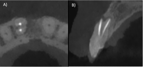
Upon clinical examination, we observed swelling of the tissues in the anterosuperior area and the presence of sinus tract, a misadjusted restoration corresponding to a metal-free crown, invasion of the biological width, gingival inflammation, chewing pain, horizontal and vertical percussion (Fig. 1B). In the periapical X-ray of the dental organ #11 (ortho-radial and mesio-radial), we observed a radiolucent lesion in the root at the apical and mesial level, loss of the interproximal bone crest, and non-visible lamina dura. In the apical portion, an underfilling was observed (Fig. A,C). A CBCT was performed, and the CT scan confirmed that there were two roots in said dental organ, and that only the vestibular root was endodontically treated (Fig. 2 A-B). Diagnosis: Dental organ previously treated, with chronic apical abscess (29. Anesthesia was administered supraperiosteally using 4% articaine with epinephrine 1: 100,000 in cul-de-sac anesthetizing the anterior alveolar nerve. The coronary restoration was removed using a diamond bur and high-speed handpiece, and once removed, absolute isolation was achieved with a rubber dam. Then, the gutta-percha was removed with Wave One Gold files (Dentsply Sirona, Tulsa Dental), the palatal canal was explored with type K 6 and 8 files until it was permeabilized, the working length of both roots was obtained with the help of the Apex ID apical locator (SybronEndo, Orange, CA) (Fig.3 A). It was performed with Wave One Gold files (Dentsply Sirona, Switzerland) Large (45/05) for the vestibular canal, and medium (35/06) for the palatal canal, irrigation was performed with hypochlorite at 5.25% throughout the treatment to finish with an irrigation protocol of three cycles of 20 seconds, sodium hypochlorite 5.25%, distilled water and EDTA 17% (Smear clear Sybron Endo, CA), activated with ultrasound (Ultra X, Eighteeth Medical). Calcium hydroxide was placed as intra-ductal medication for 15 days.
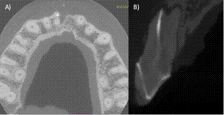
In the second appointment, the irrigation protocol was performed again, the canals were dried with sterile paper tips and obturated with continuous wave technique (Fast fill and Fast Pack, Eighteeth Medical) and AH-Plus cement (Dentsply Maillefer, Switzerland) (Fig. 3 B-C). Finally, flowable resin was used for sealing the coronal access. A second CBCT was requested to evaluate the correct localization and obturation of the dental organ (Fig. 4 A-B).

Figure 1: A) Preoperative ortho-radial X-ray. B) Preoperative clinical photograph, volume increase and discoloration changes are observed. C) Preoperative mesio-radial X-ray.

Figure 2: A) Axial section of the CT scan, two roots belonging to dental organ #11 are observed, the vestibular root shows obturation material, but the palatal root was not treated, B) Sagittal section where a permeable palatal canal can be seen.

Figure 3: A) Intraoperative X-ray showing that the canal is completely open, and the determination of the working length. B) Primary tip testing C) Immediate postoperative X-ray.
Discussion
Odontogenesis is a complex biological process involving diverse epithelial-mesenchymal interactions. Alterations in these interactions can alter normal development causing anomalies in the number of roots, their direction, number or shape of canals (30). Although the etiology of these anomalies is unknown, an abnormal growth of the epithelial Hertwig's root sheath (31 has been stated as a possible cause. Dental trauma (32 such as avulsion33 and intrusive luxation34,35) has been linked to root duplication in upper incisors, but, in our case, the patient did not mention a history of trauma during her childhood, as in other reported studies36,37, so trauma could be only one of the factors involved. Therefore, clinical history is an important factor to consider during the consultation. However, not identifying complex anatomical variations has been reported as a possible cause of failure. Mustafa, Almuhaiza16 assessed the causes of root canal treatment failure in Saudi Arabian patients and concluded that 14.4% of failures were due to omitted canals. Baruwa, Martins 17 analyzed the prevalence of omitted canals in previously treated dental organs, finding that 12.0% were associated with periapical pathology in 82.6% of cases, thus having a significant impact on the prognosis of the treatment. In a study using CBCT, Karabucak, Bunes 15 reported a 23.04% overall incidence of omitted canals; dental organs with an omitted canal were 4.38 times more likely to be associated with a periapical lesion.
Jafari, Kazemi (37 Levin, Shemesh 32 Mahadevan, Paulaian 38 reported cases where CBCT images helped to detect the exact location of the extra root, as well as to plan its access, length and curvature. In our reported case, no CBCT was performed during the previous root canal treatment, so we attribute the failure to the inability to adequately analyze the anatomy and eliminate bacterial contamination. Dental diagnosis and treatment planning are based on imaging, and evaluation of treatment outcome is generally based on both clinical examination and radiographic evaluation (23. Two-dimensional radiography remains the most feasible diagnostic imaging technique for root canal treatment and non-surgical retreatment. Although CBCT is more expensive and its radiation output for imaging a single tooth is higher than that of two-dimensional imaging devices, it greatly overcomes the limitations and provides three-dimensional resolution of structures. More complex cases as well as adjunctive surgical procedures should be evaluated through studies with CBCT (39,40.
Conclusion
A clinical case on the identification and management of an upper central with two roots was reported. Anterior organs with 2 roots are an uncommon anomaly, nevertheless, dentists should consider the possible anatomical variations and rely on radiographic and tomographic imaging to obtain proper information on clinical management as well as long-term follow-up.
REFERENCES
1. Graves DT, Oates T, Garlet GP. Review of osteoimmunology and the host response in endodontic and periodontal lesions. J Oral Microbiol. 2011;3. [ Links ]
2. Berman LH, Hargreaves KM. Cohen's pathways of the pulp-e-book: Elsevier Health Sciences; 2020. [ Links ]
3. Rotstein I, Ingle JI. Ingle's endodontics: PMPH USA; 2019. [ Links ]
4. Teixeira QE, Ferreira DC, da Silva AMP, Goncalves LS, Pires FR, Carrouel F, et al. Aging as a Risk Factor on the Immunoexpression of Pro-Inflammatory IL-1beta, IL-6 and TNF-alpha Cytokines in Chronic Apical Periodontitis Lesions. Biology (Basel). 2021;11(1). [ Links ]
5. Thuller K, Armada L, Valente MI, Pires FR, Vilaca CMM, Gomes CC. Immunoexpression of Interleukin 17, 6, and 1 Beta in Primary Chronic Apical Periodontitis in Smokers and Nonsmokers. J Endod. 2021;47(5):755-61. [ Links ]
6. Popovska L, Dimova C, Evrosimoska B, Stojanovska V, Muratovska I, Cetenovic B, et al. Relationship between IL-1ß production and endodontic status of human periapical lesions. Vojnosanitetski pregled. 2017;74(12):1134-9. [ Links ]
7. Boersma B, Jiskoot W, Lowe P, Bourquin C. The interleukin-1 cytokine family members: Role in cancer pathogenesis and potential therapeutic applications in cancer immunotherapy. Cytokine Growth Factor Rev. 2021;62:1-14. [ Links ]
8. Siqueira JF, Jr., Rocas IN. Present status and future directions: Microbiology of endodontic infections. Int Endod J. 2022;55 Suppl 3:512-30. [ Links ]
9. Nair PN. On the causes of persistent apical periodontitis: a review. Int Endod J. 2006;39(4):249-81. [ Links ]
10. Takahashi K. Microbiological, pathological, inflammatory, immunological and molecular biological aspects of periradicular disease. Int Endod J. 1998;31(5):311-25. [ Links ]
11. Orstavik D. Essential endodontology: prevention and treatment of apical periodontitis: John Wiley & Sons; 2020. [ Links ]
12. Boutsioukis C, Arias-Moliz MT. Present status and future directions - irrigants and irrigation methods. Int Endod J. 2022;55 Suppl 3(Suppl 3):588-612. [ Links ]
13. Chaniotis A, Ordinola-Zapata R. Present status and future directions: Management of curved and calcified root canals. Int Endod J. 2022;55 Suppl 3:656-84. [ Links ]
14. Vertucci FJ. Root canal morphology and its relationship to endodontic procedures. Endodontic Topics. 2005;10(1):3-29. [ Links ]
15. Karabucak B, Bunes A, Chehoud C, Kohli MR, Setzer F. Prevalence of Apical Periodontitis in Endodontically Treated Premolars and Molars with Untreated Canal: A Cone-beam Computed Tomography Study. J Endod. 2016;42(4):538-41. [ Links ]
16. Mustafa M, Almuhaiza M, Alamri HM, Abdulwahed A, Alghomlas ZI, Alothman TA, et al. Evaluation of the causes of failure of root canal treatment among patients in the City of Al-Kharj, Saudi Arabia. Niger J Clin Pract. 2021;24(4):621-8. [ Links ]
17. Baruwa AO, Martins JNR, Meirinhos J, Pereira B, Gouveia J, Quaresma SA, et al. The Influence of Missed Canals on the Prevalence of Periapical Lesions in Endodontically Treated Teeth: A Cross-sectional Study. J Endod. 2020;46(1):34-9 e1. [ Links ]
18. Pessotti VP, Jiménez-Rojas LF, Alves FRF, Rôças IN, Siqueira JF, Jr. Post-treatment apical periodontitis associated with a missed root canal in a maxillary lateral incisor with two roots: A case report. Aust Endod J. 2022. [ Links ]
19. Siqueira Junior JF, Rocas IDN, Marceliano-Alves MF, Perez AR, Ricucci D. Unprepared root canal surface areas: causes, clinical implications, and therapeutic strategies. Braz Oral Res. 2018;32(suppl 1):e65. [ Links ]
20. Costa F, Pacheco-Yanes J, Siqueira JF, Jr., Oliveira ACS, Gazzaneo I, Amorim CA, et al. Association between missed canals and apical periodontitis. Int Endod J. 2019;52(4):400-6. [ Links ]
21. Patel S, Dawood A, Whaites E, Pitt Ford T. New dimensions in endodontic imaging: part 1. Conventional and alternative radiographic systems. Int Endod J. 2009;42(6):447-62. [ Links ]
23. Cotti E, Schirru E. Present status and future directions: Imaging techniques for the detection of periapical lesions. Int Endod J. 2022;55 Suppl 4:1085-99. [ Links ]
24. Setzer FC, Lee SM. Radiology in Endodontics. Dent Clin North Am. 2021;65(3):475-86. [ Links ]
25. Decurcio DA, Bueno MR, Silva JA, Loureiro MAZ, Damião Sousa-Neto M, Estrela C. Digital Planning on Guided Endodontics Technology. Braz Dent J. 2021;32(5):23-33. [ Links ]
26. Setzer FC, Kratchman SI. Present status and future directions: Surgical endodontics. Int Endod J. 2022;55 Suppl 4:1020-58. [ Links ]
27. Ozcan G, Sekerci AE, Cantekin K, Aydinbelge M, Dogan S. Evaluation of root canal morphology of human primary molars by using CBCT and comprehensive review of the literature. Acta Odontol Scand. 2016;74(4):250-8. [ Links ]
28. Yan Y, Li J, Zhu H, Liu J, Ren J, Zou L. CBCT evaluation of root canal morphology and anatomical relationship of root of maxillary second premolar to maxillary sinus in a western Chinese population. BMC Oral Health. 2021;21(1):358. [ Links ]
29. Glickman GN. AAE Consensus Conference on Diagnostic Terminology: background and perspectives. J Endod. 2009;35(12):1619-20. [ Links ]
30. Ahmed HMA, Dummer PMH. A new system for classifying tooth, root and canal anomalies. Int Endod J. 2018;51(4):389-404. [ Links ]
31. Dexton AJ, Arundas D, Rameshkumar M, Shoba K. Retreatodontics in maxillary lateral incisor with supernumerary root. J Conserv Dent. 2011;14(3):322-4. [ Links ]
32. Levin A, Shemesh A, Katzenell V, Gottlieb A, Ben Itzhak J, Solomonov M. Use of Cone-beam Computed Tomography during Retreatment of a 2-rooted Maxillary Central Incisor: Case Report of a Complex Diagnosis and Treatment. J Endod. 2015;41(12):2064-7. [ Links ]
33. Kang M, Kim E. Unusual morphology of permanent tooth related to traumatic injury: a case report. J Endod. 2014;40(10):1698-701. [ Links ]
34. Coutinho T, Lenzi M, Simoes M, Campos V. Duplication of a permanent maxillary incisor root caused by trauma to the predecessor primary tooth: clinical case report. Int Endod J. 2011;44(7):688-95. [ Links ]
35. Kaufman AY, Keila S, Wasersprung D, Dayan D. Developmental anomaly of permanent teeth related to traumatic injury. Endod Dent Traumatol. 1990;6(4):183-8. [ Links ]
36. Genovese FR, Marsico EM. Maxillary Central Incisor with Two Roots: A Case Report. Journal of Endodontics. 2003;29(3):220-1. [ Links ]
37. Jafari Z, Kazemi A, Shiri Ashtiani A. Endodontic Management of a Two-Rooted Maxillary Central Incisor Using Cone-Beam Computed Tomography: A Case Report. Iran Endod J. 2022;17(4):220-2. [ Links ]
38. Mahadevan M, Paulaian B, Ravisankar SM, Arvind Kumar A, Nagaraj NJ. Endodontic Management of Maxillary Central Incisor with Two Roots, and Lateral Incisor with a C-shaped Canal; A Case Report. Iran Endod J. 2023;18(2):104-9. [ Links ]
39. Orhan EO, Dereci O, Irmak O. Endodontic Outcomes in Mandibular Second Premolars with Complex Apical Branching. J Endod. 2017;43(1):46-51. [ Links ]
40. Patel S, Brown J, Semper M, Abella F, Mannocci F. European Society of Endodontology position statement: Use of cone beam computed tomography in Endodontics: European Society of Endodontology (ESE) developed by. Int Endod J. 2019;52(12):1675-8. [ Links ]
Conflict of Interest Statement: The authors have no conflict of interest in the publication of the article.
Authorship contribution note: Study concept and design Data acquisition Data analysis Discussion of results Manuscript drafting Approval of the final version of the manuscript HABA has contributed in: 1, 2, 3, 4, 5 YGRH has contributed in: 1, 3, 4, 5 LKGG contributed in: 4 and 6 OATM has contributed in: 4 and 6 OASO has contributed in: 5 and 6
Received: July 14, 2023; Accepted: October 19, 2023


 texto em


