


 Inglês (pdf)
 Artigo em XML
 Referências do artigo

 Permalink
Introduction
Pet birds are living longer due to improvements in management, nutrition, and veterinary care provided to them. As a consequence, an increasing number of conditions, such as neoplastic diseases, are diagnosed and managed by veterinarians. In birds, neoplastic processes are diagnosed more frequently in Psittaciformes, being the budgerigar (Melopsittacus undulatus) the most commonly reported species (Robat et al., 2017).
Primary intestinal neoplasms include carcinomas, papillomas, smooth muscle tumors, and lymphoma (Gelis, 2005). Leiomyomas and leiomyosarcomas present as firm, reddish-brown masses within the intestinal wall and can only be distinguished from each other histologically (Gelis, 2005).
In the case of leiomyomas, these are well-defined masses of well-differentiated smooth muscle cells and, in general, are found under an intact mucosa, and can deform the intra and extraluminal surface, thus also causing ulcers in the mucosa, such as caused by pressure ischemia/necrosis (Uzal et al., 2016). They are composed of swirling bundles of uniform spindle cells with abundant eosinophilic cytoplasm and a blunt-ended central nucleus; usually no obvious mitotic figures (Uzal et al., 2016). Areas of coagulative necrosis may appear in the tumor mass (Uzal et al., 2016). These masses develop throughout the gastrointestinal, genitourinary, and hepatobiliary systems, as well as in the skin of domestic animals (Cusack et al., 2017), and have been described in different organs of various bird species, such as: oral cavity (Oana et al., 2010), proventriculus (Van Wick et al., 2019), intestine (Cusack et al., 2017), and oviduct (Sahin et al., 2004).
The aim of this article is to report a case of intestinal leiomyoma in a captive Budgerigar (Melopsittacus undulatus) diagnosed post-mortem through histopathology using hematoxylin-eosin staining.
Materials and Methods
A female budgerigar, approximately 3 years old, showed a gradual decrease in feeding. The patient had a weight of 45 grams and a body condition of 3/7 (Burton et al., 2013). Upon inspection, it appeared listless, with a hidden face, shaggy plumage and strong tail movements, associated to dyspnea. As the clinical signs were suggestive of pain, meloxicam was administered at a dose of 0,1 mg/kg IM (intramuscular) (Hawkins et al., 2018; Murphy, 2009). He lived with another individual of the same species, male, who did not show signs suggestive of disease.
The patient was placed in an oxygen chamber for 30 minutes, and then sedated with 2 % isoflurane. Physical examination revealed a large amount of subcutaneous fat at the caudal portion of the sternum. Upon palpation of the digestive organs, a firm structure was perceived in the left hepatic region, without being able to identify a specific organ. Radiographic images were taken with a REMEX-T100 equipment (REMEDI, Seoul, South Korea) and a Sopix Size 1 sensor (Acteon-Satelec, France), using 70 kV, and 2 mA in 0.02 seconds. The patient died during the procedure and the body was sent for post-mortem studies. An intestinal fragment with firm proliferation of approximately 1 cm was sectioned and placed in 10 % buffered formalin for histopathological analysis. Serial sections of the small intestine and liver were stained with hematoxylin-eosin (HyE).
Results
The radiographic study did not refer conclusive results. Post-mortem evaluation revealed small intestine with marked wall thickening. In serial sections of the small intestine stained with hematoxylin-eosin (HyE), a thickened longitudinal muscular tunic was evaluated, with palisade proliferation, slightly disorganized, of parallel spindle cells with a central nucleus and homogeneous eosinophilic cytoplasm (Fig. 1 y 2). Lamina propria and submucosa with marked congestion in blood vessels. No mitotic figures or cellular atypia were observed. Histopathological diagnosis referred intestinal leiomyoma.
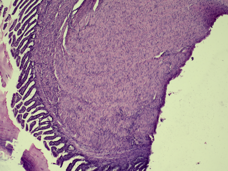
Figure 1: Small Intestine. Muscular tunic with dense, non-encapsulated erosion of smooth muscle fibers. Mucosa without histological alterations. H-E. 20x
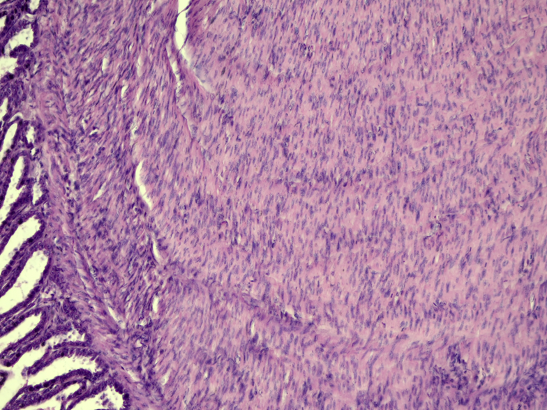
Figure 2: Small Intestine. Longitudinal muscular tunic with proliferation of smooth muscle fibers and compression of the submucosal tunic. H-E. 40x
Focally extensive areas of hemorrhage with scattered haemosiderin pigments were seen in serial sections of the liver. This area was evaluated compressing the hepatic parenchyma, with loss of architecture. Distended hepatocytes with intracytoplasmic vacuoles (interpreted as fat vacuoles). No inflammatory or neoplastic cells or microorganisms were evaluated. The histopathological diagnosis referred to focally extensive, moderate, acute hepatic hemorrhage.
Discussion
Leiomyomas are tumors that affect smooth muscle tissue (Uzal et al., 2016), and are mainly described in the digestive tract (Cusack et al., 2017; Oana et al., 2010; Van Wick et al., 2019) and the female reproductive system (Sahin et al., 2004; Urbina et al.., 2019). Macroscopically, these can be intraluminal, intramural, and extramural (Sagnotta et al., 2015), and usually show slow growth that may be associated with abdominal distention, obstruction of the gastrointestinal tract, or displacement of organs (Latimer, 1994).
Human case reports of intestinal leiomyoma state that most patients remain asymptomatic until the neoplasm reaches a large size, when the most common symptoms are pain associated with a palpable abdominal mass (Chen et al., 2008; Hatch et al., 2000; Sagnotta et al., 2015; Urbina et al., 2019). A study carried out with dogs mentioned that, of 29 cases of intestinal leiomyoma, 9 patients presented symptoms such as vomiting, palpable masses, diarrhea, weight loss and anorexia (Frost et al., 2003). The clinical signs observed in the patient were suggestive of pain (Murphy, 2009), coinciding with reports in humans and canines.
More frequent clinical evaluations, combined with increased longevity and a potential genetic predisposition associated with inbreeding, have resulted in increased oncological reports in companion birds and captive birds in general, when compared to free-living birds (Cusack et al., 2017).
Early diagnosis of intestinal leiomyomas is difficult because they are generally masses with an asymptomatic clinical course that are found under an intact or slightly affected mucosa (Uzal et al., 2016), added to diagnostic limitations in small patients (Robat et al., 2017). Diagnostic and treatment attempts remain challenging in many companion birds due to small patient size, difficulty in obtaining repeat intravascular access, increased anesthetic risks compared to other domestic animals, and lack of information on prognosis and efficacy of antineoplastic treatments in pet bird species (Robat et al., 2017). When available, samples of leiomyoma should be stained with Masson’s Trichrome, which stain positive for connective tissue and negative for myocytes. Therefore, Masson’s Trichrome should always be negative for smooth muscle tumors such as a leiomyoma. If diagnosed, the only treatment described for intestinal leiomyoma is surgical resection (Chen et al., 2008; Hatch et al., 2000; Sagnotta et al., 2015; Uzal et al., 2016).
Conclusion
The data obtained from the observable signs and physical examination, as well as necropsy findings and histopathological results, confirmed the diagnosis of intestinal leiomyoma. To the authors’ best knowledge, this is the first report of intestinal leiomyoma in a Budgerigar (Melopsittacus undulatus).

