1688-93391688-9339S1688-9339201400020000200112014001120141624412Estudio morfológico y morfométrico del agujero mentoniano mediante evaluación por tomografía computarizada Cone Beam en pacientes adultos dentados
Morphological and morphometric study of the mental foramen using cone-beam CT in dentate adult patients
Cabanillas Padilla, Juan *, Quea Cahuana, Eduardo **
* Docente de la Facultad de Odontología, Universidad de San Martín de Porres (USMP), Lima, Perú. Cirujano Dentista Juancabanillaspadilla@gmail.com
** Docente de la Facultad de Odontología, Universidad de San ]]>
Resumen ]]>
Objetivo. Estudiar la morfología y morfometría del agujero mentoniano mediante tomografía ConeBeam en pacientes adultos dentados Metodología. Estudio descriptivo transversal. Se estudiaron 180 tomografías ConeBeam analizando la distancia de la cortical superior e inferior del agujero mentoniano hasta la cresta alveolar y la basal mandibular respectivamente, así como la ubicación, forma, tamaño y presencia de agujeros accesorios. Resultados. Se encontró que la media respecto de la ]]>
Abstract ]]>
Objective. To study the morphology and morphometry of the mental foramen using cone-beam CT in dentate adult patients. Methods. Transversal descriptive study in which 180 cone-beam CTs were studied to analyze the distance between the upper and lower cortical areas of the mental foramen to the alveolar crest and the mandibular basal bone respectively, as well as the location, shape, size and presence of accessory holes. Results. It was found that the mean of the upper cortical area in relation to the alveolar crest was 15.00 mm and the ]]>
]]>
Fecha recibido: 09.03.14 - Fecha aceptado: 27.06.14
]]>
Introducción
La mandíbula tiene su origen en el primer arco braquial, presenta una osificación mixta y su crecimiento se dirige hacia ]]>
1-2).
El crecimiento mandibular cambia la dirección del agujero mentoniano. Al nacer, el haz neurovascular emerge por el agujero dirigido hacia adelante y en el adulto se encuentra dirigido hacia ]]>
(3).
El agujero mentoniano se define como una apertura en la superficie lateral de la mandíbula. En este punto el nervio alveolar inferior se bifurca dando origen al nervio mentoniano y al nervio incisivo, las cuales son ramas terminales, y responsables de la ]]>
1, 4, 5-7, 18).
Utilizando la tomografía computarizada Cone Beam se puede conocer con exactitud la ubicación, la forma, el tamaño del agujero mentoniano, y la presencia de agujeros accesorios. Esto permite realizar análisis morfométricos exactos, para ]]>
6, 24). De la misma manera esta tecnología brinda una imagen en escala real, en la cual se pueden realizar trazos y mediciones exactas, debido a que los voxeles presentan una configuración isotrópica. Asimismo, se puede obtener imágenes en los tres planos del espacio (8-10).
]]>
Materiales y métodos
Se analizaron 180 tomografías de pacientes adultos dentados de ambos sexos (74 hombres y 106 mujeres) entre 20 y 50 años, recolectadas del Centro de Diagnóstico por Imágenes, Lima – Perú, las tomografías fueron realizadas con un equipo marca Vatech, modelo Picasso Master, con el software Easy Dent, un campo de visión (FOV) de 20 X 19 mm, con 70 Kv, 8 Ma, con tiempo de exposición de 25 segundos y un punto focal de 0.5 mm. Las variables estudiadas en el plano transaxial se realizaron a través de cortes de 1 mm e intervalos de 0,5 mm.
Se realizó el análisis morfológico y morfométrico de los agujeros mentonianos de ambos lados. Las tomografías evaluadas presentaron todas las piezas dentarias y la cresta alveolar conservada. Se excluyeron tomografías con presencia de lesiones periapicales, dientes supernumerarios, pacientes con tratamiento ortodóntico y con ausencia bilateral del agujero mentoniano.
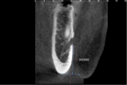
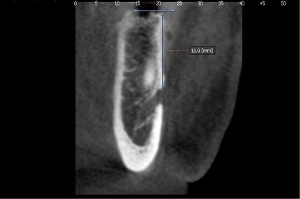
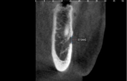
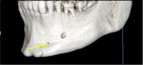
Se analizaron las distancias de las corticales superior e inferior del agujero mentoniano hacia la cresta alveolar y basal mandibular respectivamente y el tamaño del mismo en el corte transaxial, en este último se consideró la distancia entre las corticales superior e inferior de dicha estructura y las medidas se agruparon en rangos (Fig. 1 y 2).
Fig. 1: Ubicación del agujero mentoniano en el plano transaxial ]]>
Fig. 2 Tamaño del agujero mentoniano
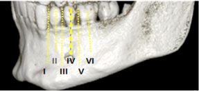
Para determinar la ubicación, forma y presencia de agujeros accesorios se utilizó la reconstrucción en 3D, para la ubicación se tomó como referencia los ejes longitudinales de las piezas dentarias según la clasificación de Al Jasser & Nwoku (21). Posición 1: Situado anterior del primer premolar, posición 2: En línea con el primer premolar, posición 3: Entre el primer y segundo premolar, posición 4: En línea con el segundo premolar, posición 5: Entre el segundo premolar y el primer molar, posición 6: En línea con el primer molar. (Fig. 3).
Se consideró la forma oval y circular como criterios de evaluación y se evaluó la presencia de agujeros accesorios (Fig. 4).
Fig. 3: Ubicación del agujero mentoniano según la clasificación de Al Jaser & Nwoku
Fig. 4 Presencia de agujeros mentonianos accesorios
El procesamiento y análisis de datos; se realizó a través del programa estadístico SPSS versión 15. Las variables cuantitativas se presentaron en valores mínimos, máximos, medias y desviaciones estándar. Se aplicó la prueba U de Mann-Whitney, para comparar las diferencias entre el lado derecho e izquierdo.
Las variables cualitativas se presentaron a través de tablas de distribución de frecuencias y se aplicaron las pruebas Chi cuadrado de Pearson y exacta de Fisher para comparar la diferencia entre el lado derecho e izquierdo. Todas las pruebas se trabajaron a un nivel de significancia de 5%. ]]>
Resultados
Las tomografías fueron evaluadas por tres especialistas calibrados (Kappa 1.00).
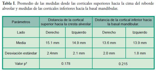
Se midió la distancia desde la cortical superior e inferior del agujero mentoniano hacia la cresta alveolar y basal mandibular respectivamente, no evidenciándose diferencias estadísticamente significativas entre los lados derecho e izquierdo de ambas mediciones. (p*0.178), (p*0.215) (Tabla I)
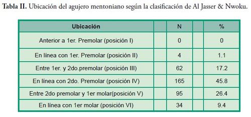
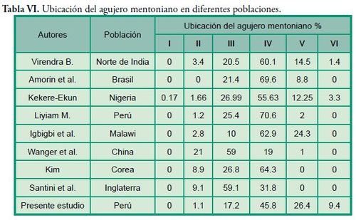
La ubicación más frecuente del agujero mentoniano se evidenció en el eje longitudinal del segundo premolar en ambos lados (posición IV) seguido de la posición V, III, VI, y II, no se observó ningún agujero mentoniano en la posición I, tampoco se evidenció diferencias significativas comparado con su lado contralateral. ( p*0.764) (Tabla II)
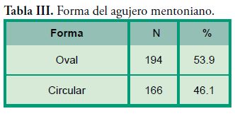
La forma oval se presentó con mayor frecuencia, no evidenciándose diferencia significativa comparado con su lado contralateral (p*0.057) (Tabla III).
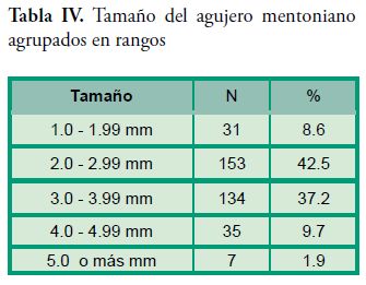
El tamaño del agujero mentoniano se agrupó en cinco rangos de medición, ubicándose la mayor cantidad de ellos (n= 153; 42.5 %) en el rango de 2 mm a 2.99 mm en ambos lados. No se evidenció diferencia significativa comparado con su lado contralateral (p*0.623) (Tabla IV)
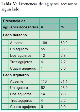
Se encontró que los agujeros accesorios estuvieron presentes en 100 casos (55.5 %) de las 180 tomografías analizadas. (Tabla V)
Discusión
]]>
4, 11) ; asimismo, otras investigaciones (6, 12) en donde se realizaron mediciones en mandíbulas secas, se encontraron resultados menores, los cuales difieren con el presente estudio.
La medida de la distancia de la cortical inferior del agujero mentoniano hacia la basal mandibular fue de (13.6 mm ± 2.0 mm) y ]]>
4, 13, 14).
Hay un debate considerable en la literatura respecto a la ubicación del agujero mentoniano en diferentes grupos étnicos. En este estudio se evidenció que dicha estructura se encontró ubicada en todas las muestras entre la raíz del primer premolar y la raíz del primer molar. ]]>
Los resultados obtenidos fueron semejantes a lo reportado por otras investigaciones (4, 6, 11, 13-17, 24) quienes observaron el agujero mentoniano en el eje longitudinal del segundo premolar. (Posición IV). Otros estudios (7, 12, 18, 19) evidenciaron la ubicación más frecuente del agujero mentoniano en la posición III, lo que discrepa con lo encontrado en el presente estudio, pues la posición V y III se ubicaron en el segundo y tercer lugar de frecuencia respectivamente. ]]>
Diversos autores han estudiado la ubicación del agujero mentoniano utilizando como referencia los ejes longitudinales de las piezas dentarias, sin embargo, se reportaron variabilidades en los resultados que se podría atribuir a un componente étnico (Tabla VI).
]]>
Respecto a la forma del agujero mentoniano existen diversas investigaciones cuyos resultados no guardan consenso en la clasificación de la misma, sin embargo, otros estudios consideran la forma oval y circular como criterio de evaluación, dichas investigaciones (4, 6, 11, 12, 15, 22 ) evidencian, en la ]]>
14, 16, 20, 24) discrepan con nuestros resultados, al reportar la forma circular como la más frecuente.
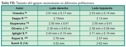
El tamaño del agujero mentoniano fue agrupado en 5 rangos. En la mayoría de los casos se encontraron en el rango de 2 mm a 2.99 mm en ambos lados, presentándose una frecuencia de 75 casos (41.7%) en el lado derecho y 78 casos (43.3%) en el lado izquierdo. ]]>
Distintos autores realizaron la evaluación del tamaño del agujero mentoniano en mandíbulas secas, encontrando similitud con los resultados obtenidos en el presente estudio en el rango de 2 mm a 2 .99 mm. (Tabla VII)
]]>
Los agujeros mentonianos accesorios estuvieron presentes en el 55.5 % de todos los casos, lo que difiere con la mayoría de estudios ]]>
6, 14, 15, 20 y 23). Singh R & Srivastav evidenció la presencia de agujeros accesorios en el 13% del total de la población evaluada, otros estudios (6, 14 ,15 y 23) evidenciaron una prevalencia de 6.67%, 6.6%, 3.92% y 6.5% respectivamente.
Todas las variables estudiadas coinciden con lo reportado por Igbigbi ]]>
Conclusión
Se concluye que el agujero mentoniano en una población adulta dentada se ubica en promedio en 13.75 mm por encima de la basal mandibular; la ubicación más frecuente se evidenció debajo del eje longitudinal del segundo premolar, la forma predominante fue oval, el tamaño se ubicó en el rango de 2 mm a 2.99 mm y se evidenció que los agujeros mentonianos accesorios estuvieron presentes en más de la mitad de los casos estudiados. (55.5 %)
Referencias
1. Velayos JL. Anatomía de la Cabeza para Odontólogos. 4a ed. Buenos aires: Editorial Panamericana, 2007.
2. Solano Reina JE, Mendoza Mendoza A. Crecimiento Craneofacial y Desarrollo de las Arcadas Dentarias. En: Odontopediatria.Barcelona: Masson. 2004. p37–53.
3. Ries Centeno GA. Cirugía bucal: Patología, Clínica y Terapéutica. 9a ed. Buenos Aires: Editorial el Ateneo. 1987. ]]>
4. Igbigbi PS, Lesbona S. The Position and Dimensions of the Mental Foramen in Adult Malawian Mandibles. West African J Med. 2005; 24 (3): 184-189.
5. Hassan T, Fauzi M, Hassan D. Bilateral Absence of Mental Foramen: A Rare Variation. Int J Anatomic Variations [en línea] 2010; 3: 187-189. Citado el 8 de Enero 2014. Disponible en: http://www.ijav.org/2010/ijav_10_167-169.pdf
6. Budhiraja V, Rastogi R, Lalwani R, Goel P, Chandra S. Study of Position, Shape, and Size of Mental Foramen Utilizing Various Parameters in Dry Adult Human Mandibles From North India. Int Scholarly Res Notices [en línea] 2012; 1-5. Citado el 27 de Noviembre 2013. Disponible en: http://dx.doi.org/10.5402/2013/961429
9. Oviedo P. Tomografía Cone Beam Aplicado a la Endodoncia [tesis] Lima- Perú. Universidad Peruana Cayetano Heredia: 2010.
10. Ige M. Tomografía Computarizada Volumétrica: Cone Beam” [tesis] Lima- Perú. Universidad Peruana Cayetano Heredia: 2010.
11. Agarwal D, Gupta S. Morphometric Analysis of Mental Foramen in Human Mandibles of South Gujarat. People’s Journal of Scientific Research 2011; 15.4(1).
14. Gupta S, Soni JS. Study of Anatomical Variations and Incidence of Mental Foramen and Accessory Mental Foramen in Dry Human Mandibles. Natl J Med Res 2012; 2 (1): 28-30.
16. Córdova L. Características Radiográficas del Foramen Mentoniano en Pacientes del Instituto de Salud Oral de la FAP del 2000 al 2008 [tesis] Lima- Perú. Universidad Nacional Federico Villarreal: 2009.
18. Rupesh S, Winnier J, Sherin A, Tatu J, Prasad A, venugopal R. Radiographic Study of the Location of Mental Foramen in a Randomly Selected Asian Indian Population on Digital Panoramic Radiographs. J Med Sci [en línea] 2011; 11 (2): 90-95. Citado el 20 de Diciembre 2013. Disponible en: http://scialert.net/fulltext/?doi=jms.2011.90.95
19. Gungor K, Ozturk M, Semiz M, Brooks SL. Location of Mental Foramen in Turkish Population, Coll Antropol 2006; 30 (4): 801–805.
24. Sekerci A, Sahman H, Sisman Y, Aksu Y. Morphometric analysis of the mental foramen in a Turkish population based on multi-slice computed tomography. J Oral and Maxillofacial Radio [en línea] 2013; 1: 1-7. Citado el 28 de Abril 2014. Disponible en: http://www.joomr.org/temp/JOralMaxillofacRadiol112-2975328_081553. pdf ]]>