<?xml version="1.0" encoding="ISO-8859-1"?><article xmlns:mml="http://www.w3.org/1998/Math/MathML" xmlns:xlink="http://www.w3.org/1999/xlink" xmlns:xsi="http://www.w3.org/2001/XMLSchema-instance">
<front>
<journal-meta>
<journal-id>1688-423X</journal-id>
<journal-title><![CDATA[Archivos de Medicina Interna]]></journal-title>
<abbrev-journal-title><![CDATA[Arch Med Int]]></abbrev-journal-title>
<issn>1688-423X</issn>
<publisher>
<publisher-name><![CDATA[Sociedad de Medicina Interna del Uruguay]]></publisher-name>
</publisher>
</journal-meta>
<article-meta>
<article-id>S1688-423X2015000200010</article-id>
<title-group>
<article-title xml:lang="es"><![CDATA[Absceso intramedular cervical en paciente inmunodeprimido]]></article-title>
<article-title xml:lang="en"><![CDATA[Intramedullary abscess of the cervical spinal cord in an immunodepressed patient]]></article-title>
</title-group>
<contrib-group>
<contrib contrib-type="author">
<name>
<surname><![CDATA[Cabrera]]></surname>
<given-names><![CDATA[Viviana]]></given-names>
</name>
<xref ref-type="aff" rid="A01"/>
</contrib>
<contrib contrib-type="author">
<name>
<surname><![CDATA[Spagnuolo]]></surname>
<given-names><![CDATA[Edgardo]]></given-names>
</name>
<xref ref-type="aff" rid="A01"/>
</contrib>
<contrib contrib-type="author">
<name>
<surname><![CDATA[Nicoli]]></surname>
<given-names><![CDATA[Edgar]]></given-names>
</name>
<xref ref-type="aff" rid="A01"/>
</contrib>
</contrib-group>
<aff id="A01">
<institution><![CDATA[,Universidad de la República (UdelaR) Facultad de Medicina Hospital Maciel]]></institution>
<addr-line><![CDATA[Montevideo ]]></addr-line>
<country>Uruguay</country>
</aff>
<pub-date pub-type="pub">
<day>00</day>
<month>07</month>
<year>2015</year>
</pub-date>
<pub-date pub-type="epub">
<day>00</day>
<month>07</month>
<year>2015</year>
</pub-date>
<volume>37</volume>
<numero>2</numero>
<fpage>98</fpage>
<lpage>100</lpage>
<copyright-statement/>
<copyright-year/>
<self-uri xlink:href="http://www.scielo.edu.uy/scielo.php?script=sci_arttext&amp;pid=S1688-423X2015000200010&amp;lng=en&amp;nrm=iso"></self-uri><self-uri xlink:href="http://www.scielo.edu.uy/scielo.php?script=sci_abstract&amp;pid=S1688-423X2015000200010&amp;lng=en&amp;nrm=iso"></self-uri><self-uri xlink:href="http://www.scielo.edu.uy/scielo.php?script=sci_pdf&amp;pid=S1688-423X2015000200010&amp;lng=en&amp;nrm=iso"></self-uri><abstract abstract-type="short" xml:lang="es"><p><![CDATA[Los abscesos intramedulares son lesiones poco frecuentes. Su incidencia es muy baja, lo que determina que su diagnóstico sea dificultoso. Se caracterizan por síndromes neurológicos variados, que están dominados por dolor vertebral y déficit neurológico, por lo general en presencia de fiebre. Se los vincula a focos infecciosos de vecindad y otras veces a diseminación hematógena. Son mas frecuentes en pacientes que presentan alguna inmunodepresión o afecciones sistémicas que favorezcan las infecciones en topografías poco habituales. Los autores presentan un caso de un paciente inmunodeprimido, con historia de dolor vertebral y posteriormente un severo déficit neurológico y en quien se diagnosticó un absceso intramedular, asociado a una espondilodiscitis. No se comprobó por los estudios ni en la cirugía continuidad entre las lesiones. Se hace un análisis del caso clínico y de la escasa bibliografía publicada al respecto.]]></p></abstract>
<abstract abstract-type="short" xml:lang="en"><p><![CDATA[Intramedullary spinal cord abscesses are infrequent lesions. Its incidence is very low, what determines that its hand held is a difficult one. Clinically they are characterized by varied neurological syndromes, which are dominated by vertebral pain and neurological deficits, associated with fever. The origin of these abscesses is usually related to neighbourhood foci, or to hematogenous dissemination from a distant focus. They are more frequent in patients qho are immunosuppressed or systemic conditions that promote infection in unusual topographies. The authors report a case of an inmunocompromised patient in whom an intramedullary cervical spinal cord abscess associated to spondylodiskitis was found. Neither in the image studies nor in surgery, was a continuity between both lesions confirmed. There is an analysis of the clinical case and the little literature published on the subject.]]></p></abstract>
<kwd-group>
<kwd lng="es"><![CDATA[Absceso intramedular]]></kwd>
<kwd lng="es"><![CDATA[Espondilodiscitis]]></kwd>
<kwd lng="en"><![CDATA[spinal cord abscess]]></kwd>
<kwd lng="en"><![CDATA[Surgery]]></kwd>
</kwd-group>
</article-meta>
</front><body><![CDATA[ <div class="Section1">      <p class="Seccin" style="text-align: left; line-height: normal;" align="left"><span style="font-size: 10pt; font-family: Verdana;" lang="ES-TRAD">ARCHIVOS DEL INSTITUTO DE NEUROLOG&Iacute;A<o:p></o:p></span></p>          <p class="Seccin" style="text-align: left; line-height: normal;" align="left"><span style="font-size: 10pt; font-family: Verdana;" lang="ES-TRAD"><o:p>&nbsp;</o:p></span></p>          <p class="Seccin" style="text-align: left; line-height: normal;" align="left"><span style="font-size: 10pt; font-family: Verdana;" lang="ES-TRAD"><o:p>&nbsp;</o:p></span></p>          <p class="Titarial18" style="text-align: left; line-height: normal;" align="left"><span style="font-size: 14pt; font-family: Verdana; font-style: normal;" lang="ES-TRAD">Absceso intramedular cervical en paciente inmunodeprimido<o:p></o:p></span></p>          <p class="Titarial14" style="text-align: left; line-height: normal;" align="left"><span style="font-size: 12pt; font-family: Verdana;" lang="EN-US">Intramedullary abscess of the cervical spinal cord in an     <br>     immunodepressed patient<o:p></o:p></span></p>          <p class="txt" style="text-align: left; text-indent: 0cm; line-height: normal;" align="left"><span style="font-size: 10pt; font-family: Verdana;" lang="EN-US"><o:p>&nbsp;</o:p></span></p>          <p class="autor" style="text-align: left; line-height: normal;" align="left"><span style="font-size: 10pt; font-family: Verdana; font-weight: normal;" lang="ES-TRAD">Dra. Viviana Cabrera<o:p></o:p></span></p>          <p class="autor" style="text-align: left; line-height: normal;" align="left"><span style="font-size: 10pt; font-family: Verdana; font-weight: normal;" lang="ES-TRAD">Residente de Neurocirug&iacute;a del Hospital Maciel. Facultad de Medicina. UdelaR. Montevideo.</span><span style="font-size: 10pt; font-family: Verdana; font-weight: normal;" lang="ES-TRAD"><o:p></o:p></span></p>          ]]></body>
<body><![CDATA[<p class="autor" style="text-align: left; line-height: normal;" align="left"><span style="font-size: 10pt; font-family: Verdana; font-weight: normal;" lang="ES-TRAD"><o:p>&nbsp;</o:p></span></p>          <p class="autor" style="text-align: left; line-height: normal;" align="left"><span style="font-size: 10pt; font-family: Verdana; font-weight: normal;" lang="ES-TRAD">Dr. Edgardo Spagnuolo<o:p></o:p></span></p>          <p class="autor" style="text-align: left; line-height: normal;" align="left"><span style="font-size: 10pt; font-family: Verdana; font-weight: normal;" lang="ES-TRAD">Neurocirujano, Jefe de Servicio de Neurocirug&iacute;a del Hospital Maciel. Facultad de Medicina. UdelaR. Montevideo.</span><span style="font-size: 10pt; font-family: Verdana; font-weight: normal;" lang="ES-TRAD"><o:p></o:p></span></p>          <p class="autor" style="text-align: left; line-height: normal;" align="left"><span style="font-size: 10pt; font-family: Verdana; font-weight: normal;" lang="ES-TRAD"><o:p>&nbsp;</o:p></span></p>          <p class="autor" style="text-align: left; line-height: normal;" align="left"><span style="font-size: 10pt; font-family: Verdana; font-weight: normal;" lang="ES-TRAD">Dr. Edgar Nicoli<o:p></o:p></span></p>          <p class="autor" style="text-align: left; line-height: normal;" align="left"><span style="font-size: 10pt; font-family: Verdana; font-weight: normal;" lang="ES-TRAD">Neurocirujano. Servicio de Neurocirug&iacute;a del Hospital Maciel. Facultad de Medicina. UdelaR. Montevideo.<o:p></o:p></span></p>          <p class="autor" style="text-align: left; line-height: normal;" align="left"><span style="font-size: 10pt; font-family: Verdana; font-weight: normal;" lang="ES-TRAD"><o:p>&nbsp;</o:p></span></p>          <p class="Helve7" style="text-align: left; line-height: normal;" align="left"><span style="font-size: 10pt; font-family: Verdana;" lang="ES-TRAD">Recibido: 17/04/15 &ndash; Aceptado: 07/07/15<o:p></o:p></span></p>          <p class="Helve7" style="text-align: left; line-height: normal;" align="left"><span style="font-size: 10pt; font-family: Verdana;" lang="ES-TRAD"><o:p>&nbsp;</o:p></span></p>          <p class="Helve7" style="text-align: left; line-height: normal;" align="left"><span style="font-size: 10pt; font-family: Verdana;" lang="ES-TRAD">Trabajo del Servicio de Neurocirug&iacute;a del Hospital Maciel (ASSE). Unidad Docente Asociada, Escuela de Graduados, Facultad de Medicina (UdelaR). Montevideo, Uruguay<o:p></o:p></span></p>          ]]></body>
<body><![CDATA[<p class="Helve7" style="text-align: left; line-height: normal;" align="left"><span style="font-size: 10pt; font-family: Verdana;" lang="ES-TRAD"><o:p>&nbsp;</o:p></span></p>          <p class="Helve7" style="text-align: left; line-height: normal;" align="left"><span style="font-size: 10pt; font-family: Verdana;" lang="ES-TRAD">Correspondencia: Dr. Edgardo Spagnulo. Servicio de Neurocirug&iacute;a del Hospital Maciel (ASSE). 25 de mayo 174, Montevideo, Uruguay. Correo electr&oacute;nico: <a href="mailto:spagjohn@yahoo.com">spagjohn@yahoo.com</a> <o:p></o:p></span></p>          <p class="Helve7" style="text-align: left; line-height: normal;" align="left"><span style="font-size: 10pt; font-family: Verdana;" lang="ES-TRAD"><o:p>&nbsp;</o:p></span></p>          <p class="MsoNormal" style="margin-bottom: 0.0001pt;"><span style="font-size: 10pt; font-family: Verdana;" lang="ES-TRAD">RESUMEN: Arch Med Interna 37(2): 98-100<o:p></o:p></span></p>          <p class="copete" style="text-align: left; line-height: normal;" align="left"><span style="font-size: 10pt; font-family: Verdana; font-weight: normal;" lang="ES-TRAD">Los abscesos intramedulares son lesiones poco frecuentes. Su incidencia es muy baja, lo que determina que su diagn&oacute;stico sea dificultoso. Se caracterizan por s&iacute;ndromes neurol&oacute;gicos variados, que est&aacute;n dominados por dolor vertebral y d&eacute;ficit neurol&oacute;gico, por lo general en presencia de fiebre. Se los vincula a focos infecciosos de vecindad y otras veces a diseminaci&oacute;n hemat&oacute;gena. Son mas frecuentes en pacientes que presentan alguna inmunodepresi&oacute;n o afecciones sist&eacute;micas que favorezcan las infecciones en topograf&iacute;as poco habituales. Los autores presentan un caso de un paciente inmunodeprimido, con historia de dolor vertebral y posteriormente un severo d&eacute;ficit neurol&oacute;gico y en quien se diagnostic&oacute; un absceso intramedular, asociado a una espondilodiscitis. No se comprob&oacute; por los estudios ni en la cirug&iacute;a continuidad entre las lesiones. Se hace un an&aacute;lisis del caso cl&iacute;nico y de la escasa bibliograf&iacute;a publicada al respecto<o:p></o:p></span></p>          <p class="copete" style="text-align: left; line-height: normal;" align="left"><span style="font-size: 10pt; font-family: Verdana; font-weight: normal;" lang="ES-TRAD">Palabras clave: Absceso intramedular. Espondilodiscitis.<o:p></o:p></span></p>          <p class="txt" style="text-align: left; text-indent: 0cm; line-height: normal;" align="left"><span style="font-size: 10pt; font-family: Verdana;" lang="ES-TRAD"><o:p>&nbsp;</o:p></span></p>          <p class="MsoNormal" style="margin-bottom: 0.0001pt;"><span style="font-size: 10pt; font-family: Verdana;" lang="EN-US">ABSTRACT: Arch Med Interna 37(2): 98-100<o:p></o:p></span></p>          <p class="copete" style="text-align: left; line-height: normal;" align="left"><span style="font-size: 10pt; font-family: Verdana; font-weight: normal;" lang="EN-US">Intramedullary spinal cord abscesses are infrequent lesions. Its incidence is very low, what determines that its hand held is a difficult one. Clinically they are characterized by varied neurological syndromes, which are dominated by vertebral<span style="">&nbsp; </span>pain and neurological deficits, associated with fever. The origin of these abscesses is usually related to neighbourhood foci, or to hematogenous dissemination from a distant focus. They are more frequent in patients qho are immunosuppressed or systemic conditions that promote infection in unusual topographies. The authors report a case of an inmunocompromised patient in whom an intramedullary cervical spinal cord abscess associated to spondylodiskitis was found. Neither in the image studies nor in surgery, was a continuity between both lesions confirmed. There is an analysis of the clinical case and the little literature published on the subject.<o:p></o:p></span></p>          <p class="copete" style="text-align: left; line-height: normal;" align="left"><span style="font-size: 10pt; font-family: Verdana; font-weight: normal;" lang="EN-US">Key words: spinal cord abscess. </span><span style="font-size: 10pt; font-family: Verdana; font-weight: normal;" lang="ES-TRAD">Surgery.<o:p></o:p></span></p>          ]]></body>
<body><![CDATA[<p class="copete" style="text-align: left; line-height: normal;" align="left"><span style="font-size: 10pt; font-family: Verdana; font-weight: normal;" lang="ES-TRAD"><o:p>&nbsp;</o:p></span></p>          <p class="txt" style="text-align: left; text-indent: 0cm; line-height: normal;" align="left"><span style="font-size: 10pt; font-family: Verdana;" lang="ES-TRAD"><o:p>&nbsp;</o:p></span></p>          <p class="Subt1" style="line-height: normal;"><span style="font-size: 10pt; font-family: Verdana; font-weight: normal;" lang="ES-TRAD">INTRODUCCI&Oacute;N<o:p></o:p></span></p>          <p class="txt" style="text-align: left; text-indent: 0cm; line-height: normal;" align="left"><span style="font-size: 10pt; font-family: Verdana;" lang="ES-TRAD"><o:p>&nbsp;</o:p></span></p>          <p class="txt" style="text-align: left; text-indent: 0cm; line-height: normal;" align="left"><span style="font-size: 10pt; font-family: Verdana;" lang="ES-TRAD">Las infecciones que comprometen el Sistema Nervioso Central (SNC) son poco frecuentes, si no se tienen en cuenta las infecciones extra-axiales. Predominan ampliamente los abscesos cerebrales, pero su incidencia ha descendido en las &uacute;ltimas d&eacute;cadas debido a la detecci&oacute;n y tratamiento precoz de focos infecciosos (sobre todo de la esfera otorrinolaringol&oacute;gica). Dentro de las infecciones del SNC, las infecciones intramedulares tienen una incidencia muy baja. Pero mucho m&aacute;s infrecuentes son las infecciones intramedulares<sup><a name="-1"></a><a name="-2"></a><a name="-3"></a><a name="-4"></a><a name="-5"></a><a name="-6"></a><a name="-7"></a><a name="-8"></a><a name="-9"></a>(<a href="#1">1-9</a>)</sup>. Se plantea que las infecciones medulares se dan en forma secundaria a un foco infeccioso de vecindad, como por ejemplo una infecci&oacute;n discovertebral (espondilodiscitis, osteomielitis vertebral) o que pueden ser secundarias a infecciones a distancia sembradas a nivel medular, por v&iacute;a hemat&oacute;gena<sup><a name="-10"></a><a name="-11"></a>(<a href="#10">10</a>,<a href="#11">11</a>)</sup>. Los abscesos intramedulares, son por lo tanto afecciones de muy baja incidencia, que se plantea deben darse en pacientes con inmunodepresi&oacute;n o con afecciones de fondo que favorezcan las infecciones y su diseminaci&oacute;n (por ejemplo diabetes de dif&iacute;cil control). De todas maneras, es poco frecuente que un paciente con una infecci&oacute;n a distancia, desarrolle exclusivamente como localizaci&oacute;n secundaria un absceso intramedular. Esto teniendo en cuenta la mala y escasa irrigaci&oacute;n medular. Por esto, el mecanismo que m&aacute;s se acepta es el secundario a infecciones vecinas. Lo raro en estos casos es que generalmente se da una colecci&oacute;n supurada intramedular y sin abscesos epidurales o meningitis asociados o como &uacute;nica lesi&oacute;n. Cabe tambi&eacute;n destacar que las anteriores son infecciones m&aacute;s frecuentes, pero no se acompa&ntilde;an de una diseminaci&oacute;n medular. Se pueden ver extensas colecciones epidurales, pero la duramadre es siempre una barrera infranqueable para la infecci&oacute;n. Tambi&eacute;n son muy escasas las publicaciones de infecciones intradurales por el mismo motivo. <o:p></o:p></span></p>          <p class="txt" style="text-align: left; text-indent: 0cm; line-height: normal;" align="left"><span style="font-size: 10pt; font-family: Verdana;" lang="ES-TRAD">Los autores presentan el caso de un paciente joven, portador de un absceso intramedular. Se hace un an&aacute;lisis del caso cl&iacute;nico, su imagenolog&iacute;a, su resoluci&oacute;n quir&uacute;rgica y se hace una revisi&oacute;n de la escasa bibliograf&iacute;a existente al respecto.<o:p></o:p></span></p>          <p class="txt" style="text-align: left; text-indent: 0cm; line-height: normal;" align="left"><span style="font-size: 10pt; font-family: Verdana;" lang="ES-TRAD"><o:p>&nbsp;</o:p></span></p>          <p class="Subt1" style="line-height: normal;"><span style="font-size: 10pt; font-family: Verdana; font-weight: normal;" lang="ES-TRAD">CASO CL&Iacute;NICO<o:p></o:p></span></p>          <p class="txt" style="text-align: left; text-indent: 0cm; line-height: normal;" align="left"><span style="font-size: 10pt; font-family: Verdana;" lang="ES-TRAD"><o:p>&nbsp;</o:p></span></p>          <p class="txt" style="text-align: left; text-indent: 0cm; line-height: normal;" align="left"><span style="font-size: 10pt; font-family: Verdana;" lang="ES-TRAD">Paciente de 20 a&ntilde;os, sexo masculino, HIV positivo de 2 a&ntilde;os de diagn&oacute;stico, en control m&eacute;dico irregular. Comienza 1 mes antes del ingreso con dolor cervical sin irradiaci&oacute;n que fue haci&eacute;ndose progresivamente m&aacute;s intenso. Dos semanas despu&eacute;s agrega fiebre y odinofagia. En la semana previa al ingreso nota parestesias y disestesias en ambos miembros inferiores, agregando luego impotencia funcional la que progresivamente se transform&oacute; en una paraparesia, que el d&iacute;a del ingreso le impide mantenerse de pie. Dos d&iacute;as previos hab&iacute;a agregado parestesias con disestesias en ambos miembros superiores y p&eacute;rdida de fuerzas en ambas manos. El d&iacute;a del ingreso refiere &ldquo;adormecimiento&rdquo; desde el cuello hacia abajo. No orin&oacute; en las &uacute;ltimas 36 hs. Al examen del ingreso: mal aspecto general. Adelgazado, palidez cut&aacute;neo mucosa. Febril, 39&ordm;C axilar. Mala perfusi&oacute;n perif&eacute;rica, hipotensi&oacute;n arterial, taquicardia. En lo neurol&oacute;gico: cuadriparesia severa a predominio crural, donde tiene fuerzas M1 en la escala del British Medical Council. Reflejos vivos con Babinski bilateral. Hipoestesia con nivel desde T1 y anestesia desde T3. Globo vesical que obliga a colocaci&oacute;n de sonda vesical. <o:p></o:p></span></p>          ]]></body>
<body><![CDATA[<p class="txt" style="text-align: left; text-indent: 0cm; line-height: normal;" align="left"><span style="font-size: 10pt; font-family: Verdana;" lang="ES-TRAD">Se estudia primero con RX y Tomograf&iacute;a Computada de raquis cervical, que confirmaron lesi&oacute;n compatible con espondilodiscitis C5-C6. Posteriormente se complet&oacute; con una Resonancia Magn&eacute;tica (RM) de todo el raquis, que evidencia a nivel cervical: lesi&oacute;n compatible con espondilodiscitis C5-C6 sin listesis y colecci&oacute;n prevertebral compatible con absceso. A nivel medular se evidencia un proceso centrado en la zona de la espondilodiscitis, con ensanchamiento medular, con &aacute;reas hiperintensas y otras hipointensas, compatibles con un extenso absceso acompa&ntilde;ado de edema (<a href="#fig_1">Figura 1</a>).</span></p>        <p class="txt" style="text-align: left; text-indent: 0cm; line-height: normal;" align="left"><span style="font-size: 10pt; font-family: Verdana;" lang="ES-TRAD"> &nbsp; &nbsp; &nbsp; &nbsp; &nbsp; &nbsp; &nbsp; &nbsp; &nbsp; &nbsp; &nbsp; &nbsp; &nbsp; &nbsp; &nbsp; &nbsp; &nbsp; &nbsp; &nbsp; &nbsp; &nbsp; &nbsp; &nbsp; &nbsp; &nbsp; &nbsp; &nbsp; &nbsp; &nbsp; &nbsp; &nbsp; &nbsp; &nbsp; &nbsp; &nbsp; &nbsp; &nbsp; &nbsp; &nbsp; &nbsp; &nbsp; &nbsp; &nbsp; &nbsp; &nbsp; &nbsp; &nbsp; &nbsp; &nbsp; &nbsp; &nbsp; &nbsp; &nbsp; &nbsp; &nbsp; &nbsp; &nbsp; &nbsp; &nbsp; &nbsp; &nbsp; &nbsp; &nbsp; &nbsp; &nbsp; &nbsp; &nbsp; &nbsp; &nbsp; &nbsp; &nbsp; &nbsp; &nbsp;&nbsp;</span></p>        <p class="txt" style="text-align: left; text-indent: 0cm; line-height: normal;" align="left"><a name="fig_1"></a><img style="width: 409px; height: 436px;" alt="" src="/img/revistas/ami/v37n2/2a10f1.jpg"></p>        <p class="txt" style="text-align: left; text-indent: 0cm; line-height: normal;" align="left"><span style="font-size: 10pt; font-family: Verdana;" lang="ES-TRAD">Se decide la intervenci&oacute;n de emergencia para evacuar el absceso intramedular. Se realiza una m&iacute;nima hemilaminectom&iacute;a a nivel de C5, con apertura dural y punci&oacute;n. Se realiza peque&ntilde;a apertura de duramadre y punci&oacute;n con trocar fino evacu&aacute;ndose varios cent&iacute;metros c&uacute;bicos de pus f&eacute;tido. A posteriori y luego de 48 hs. de intenso tratamiento para estabilizaci&oacute;n hemodin&aacute;mica se realiz&oacute; abordaje anterior, se evacu&oacute; el absceso prevertebral, posteriormente se sigui&oacute; con corporectom&iacute;a C5 y C6, con discectom&iacute;a y estabilizaci&oacute;n raqu&iacute;dea con instrumentaci&oacute;n anterior. Desde el ingreso se comenz&oacute; con tratamiento antibi&oacute;tico emp&iacute;rico. A las 72 hs. de la segunda cirug&iacute;a fallece en shock s&eacute;ptico. En el material purulento evacuado se encontraron abundantes leucocitos polimorfonucleares, pero el cultivo fue negativo para identificar germen. <o:p></o:p></span></p>          <p class="txt" style="text-align: left; text-indent: 0cm; line-height: normal;" align="left"><span style="font-size: 10pt; font-family: Verdana;" lang="ES-TRAD"><o:p>&nbsp;</o:p></span></p>          <p class="Subt1" style="line-height: normal;"><span style="font-size: 10pt; font-family: Verdana; font-weight: normal;" lang="ES-TRAD">DISCUSI&Oacute;N y comentarios<o:p></o:p></span></p>          <p class="txt" style="text-align: left; text-indent: 0cm; line-height: normal;" align="left"><span style="font-size: 10pt; font-family: Verdana;" lang="ES-TRAD"><o:p>&nbsp;</o:p></span></p>          <p class="txt" style="text-align: left; text-indent: 0cm; line-height: normal;" align="left"><span style="font-size: 10pt; font-family: Verdana;" lang="ES-TRAD">Desde la primera descripci&oacute;n de un absceso intramedular, hecha por Hart en 1830, son escasas las publicaciones sobre el tema<sup>(<a href="#9">9</a>)</sup>. Por ejemplo, en una revisi&oacute;n de 2009, Kurita y cols. encuentran unos 120 casos publicados de abscesos intramedulares<sup><a name="-12"></a><a name="-13"></a><a name="-14"></a>(<a href="#9">9</a>,<a href="#12">12-14</a>)</sup>. La mayor&iacute;a de las publicaciones son reportes de caso, salvo una serie con tres casos<sup>(<a href="#12">12</a>)</sup>. El mismo autor tambi&eacute;n destaca la extrema rareza de la patolog&iacute;a y de la importancia de hacer un diagn&oacute;stico precoz, ya que tienen una alta incidencia de mortalidad y de secuelas invalidantes entre los sobrevivientes. Le da gran importancia al uso de antibi&oacute;ticos adecuados, y que los mismos han incidido y en forma significativa en la evoluci&oacute;n posterior de estos pacientes.<o:p></o:p></span></p>          <p class="txt" style="text-align: left; text-indent: 0cm; line-height: normal;" align="left"><span style="font-size: 10pt; font-family: Verdana;" lang="ES-TRAD">En general se hace hincapi&eacute; en la rareza de la patolog&iacute;a, de su mal pron&oacute;stico vital y funcional si no se diagnostica precozmente y se trata adecuadamente<sup>(<a href="#1">1</a>,<a href="#3">3</a>,<a href="#4">4</a>,<a href="#13">13</a>)</sup>. Se discuten seg&uacute;n los diferentes autores las distintas t&eacute;cnicas quir&uacute;rgicas para evacuar el absceso, e incluso el tratamiento emp&iacute;rico con antibi&oacute;ticos sin cirug&iacute;a<sup>(<a href="#4">4</a>,<a href="#9">9</a>,<a href="#10">10</a>,<a href="#14">14</a>)</sup>. La mayor&iacute;a de las publicaciones hacen referencia a que el absceso intramedular es secundario a una lesi&oacute;n previa del SNC y sobre todo de la m&eacute;dula: abscesos secundarios a heridas de arma de fuego, a cirug&iacute;as intramedulares por otra causa, a senos d&eacute;rmicos, etc<sup>(<a href="#5">5-7</a>,<a href="#10">10</a>,<a href="#11">11</a>,<a href="#14">14</a>)</sup>. Vora publica en el 2004 un caso secundario a una toracotom&iacute;a por un carcinoma de es&oacute;fago<sup>(<a href="#13">13</a>)</sup>. <o:p></o:p></span></p>          <p class="txt" style="text-align: left; text-indent: 0cm; line-height: normal;" align="left"><span style="font-size: 10pt; font-family: Verdana;" lang="ES-TRAD">Otros autores relacionan la aparici&oacute;n de un absceso intramedular a la existencia de un tumor intramedular, como por ejemplo un ependimoma, mientras que hay tambi&eacute;n quienes han publicado casos relacionados con afecciones hematol&oacute;gicas, que determinan una isquemia cr&oacute;nica medular, con estasis circulatoria, que en presencia de un foco infeccioso a distancia pueden favorecer la formaci&oacute;n de un absceso intramedular<sup>(<a href="#2">2</a>,<a href="#5">5</a>,<a href="#7">7</a>,<a href="#8">8</a>)</sup>. <o:p></o:p></span></p>          ]]></body>
<body><![CDATA[<p class="txt" style="text-align: left; text-indent: 0cm; line-height: normal;" align="left"><span style="font-size: 10pt; font-family: Verdana;" lang="ES-TRAD">El absceso intramedular es una afecci&oacute;n grave y con una elevada morbimortalidad que est&aacute; entre 9 y 90% seg&uacute;n diferentes autores<sup>(<a href="#2">2</a>,<a href="#6">6</a>,<a href="#9">9</a>,<a href="#10">10</a>,<a href="#13">13</a>)</sup>. La mortalidad ha ido bajando en forma dram&aacute;tica en los &uacute;ltimos 20 a&ntilde;os, gracias a la permanente aparici&oacute;n de nuevos antibi&oacute;ticos<sup>(<a href="#2">2</a>,<a href="#6">6</a>)</sup>. Algunos autores plantean que las cifras de mortalidad son altas si al absceso medular se asocia espondilodiscitis con severa destrucci&oacute;n &oacute;sea<sup>(<a href="#4">4</a>,<a href="#13">13</a>)</sup>. En estos casos la muerte se debe a diseminaci&oacute;n sist&eacute;mica del proceso infeccioso.<o:p></o:p></span></p>          <p class="txt" style="text-align: left; text-indent: 0cm; line-height: normal;" align="left"><span style="font-size: 10pt; font-family: Verdana;" lang="ES-TRAD">El diagn&oacute;stico de absceso intramedular se plantea por la cl&iacute;nica y se confirma con la imagenolog&iacute;a. La cl&iacute;nica de nuestro paciente es comparable con lo mencionado en la revisi&oacute;n realizada por Kurita de que la fiebre, el s&iacute;ndrome neurol&oacute;gico deficitario y los trastornos esfinterianos dominaron la cl&iacute;nica<sup>(<a href="#9">9</a>)</sup>. Tambi&eacute;n es compatible en que en un elevado n&uacute;mero de casos hab&iacute;a antecedentes de un foco infeccioso de vecindad.<o:p></o:p></span></p>          <p class="txt" style="text-align: left; text-indent: 0cm; line-height: normal;" align="left"><span style="font-size: 10pt; font-family: Verdana;" lang="ES-TRAD">En cuanto a la imagenolog&iacute;a, el estudio que permite el diagnostico de absceso intramedular es la RM. Se puede ver la lesi&oacute;n y su vinculaci&oacute;n o no con focos de vecindad<sup>(<a href="#7">7</a>)</sup>. La confirmaci&oacute;n del germen se obtiene con la cirug&iacute;a, aunque los cultivos pueden ser negativos, ya que no se recomienda esperar el cultivo para comenzar el tratamiento antibi&oacute;tico dada la gravedad que supone este tipo de lesi&oacute;n. <o:p></o:p></span></p>          <p class="txt" style="text-align: left; text-indent: 0cm; line-height: normal;" align="left"><span style="font-size: 10pt; font-family: Verdana;" lang="ES-TRAD">
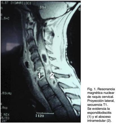
Si bien se reportan casos en los que no hab&iacute;a foco infeccioso demostrable ni de vecindad ni a distancia, en el caso analizado en esta presentaci&oacute;n hab&iacute;a un foco infeccioso de vecindad (espondilodiscitis). En el mismo, la imagenolog&iacute;a simple (RX de columna cervical) y la Tomograf&iacute;a Computada de raquis fueron positivas en demostrar una lesi&oacute;n discovertebral, con las caracter&iacute;sticas de una espondilodiscitis C5-C6, con alteraciones vertebrales, secundarias a osteomielitis. Estas lesiones explican el cuadro doloroso de semanas de evoluci&oacute;n, tipificado como dolor raqu&iacute;deo y el s&iacute;ndrome toxi-infeccioso de los d&iacute;as previos al ingreso. Por el d&eacute;ficit neurol&oacute;gico, que evolucion&oacute; r&aacute;pidamente, se solicit&oacute; una RM de raquis, que evidenci&oacute; una extensa lesi&oacute;n medular, con aspecto de un absceso multilobulado asociado a edema y ensanchamiento medular cervical. Se destaca que no hab&iacute;a compromiso epidural ni subdural. El proceso patol&oacute;gico medular estaba centrado en la topograf&iacute;a de la lesi&oacute;n discovertebral, pero no hab&iacute;a evidencia imagenol&oacute;gica de continuidad. La terap&eacute;utica quir&uacute;rgica requiri&oacute; dos intervenciones, una de emergencia para evacuar el absceso, dado lo severo del d&eacute;ficit neurol&oacute;gico y una segunda a las 48 hs y ya estabilizado el paciente, donde se realiz&oacute; la corporectom&iacute;a y posterior instrumentaci&oacute;n raqu&iacute;dea. A pesar del tratamiento quir&uacute;rgico y del tratamiento emp&iacute;rico con antibi&oacute;ticos instituido desde el ingreso, la evoluci&oacute;n no fue buena. A las 72 hs. de la segunda cirug&iacute;a fallece, dado que hab&iacute;a evolucionado a un cuadro infeccioso generalizado con repercusi&oacute;n multisist&eacute;mica (shock s&eacute;ptico).<o:p></o:p></span></p>          <p class="txt" style="text-align: left; text-indent: 0cm; line-height: normal;" align="left"><span style="font-size: 10pt; font-family: Verdana;" lang="ES-TRAD">Respecto a los g&eacute;rmenes, el </span><span style="font-size: 10pt; font-family: Verdana;" lang="ES-TRAD">Staphilococcus aureus</span><span style="font-size: 10pt; font-family: Verdana;" lang="ES-TRAD"> es el que predomina, pero se han encontrado a g&eacute;rmenes agresivos poco comunes, hongos, micobacterias, etc. Tambi&eacute;n hay casos de cultivo negativo<sup>(<a href="#13">13</a>)</sup>.<o:p></o:p></span></p>          <p class="txt" style="text-align: left; text-indent: 0cm; line-height: normal;" align="left"><span style="font-size: 10pt; font-family: Verdana;" lang="ES-TRAD">Predominan en el sexo masculino y la topograf&iacute;a m&aacute;s frecuente en altura a nivel medular es dorsal o a nivel terminal medular. Menos frecuentes son las lesiones a nivel cervical<o:p></o:p></span></p>          <p class="txt" style="text-align: left; text-indent: 0cm; line-height: normal;" align="left"><span style="font-size: 10pt; font-family: Verdana;" lang="ES-TRAD">Por &uacute;ltimo se los divide tambi&eacute;n en cuanto al tiempo de evoluci&oacute;n de los s&iacute;ntomas en agudos, subagudos y cr&oacute;nicos. Los dos primeros por lo general se vinculan a g&eacute;rmenes comunes, mientras que los cr&oacute;nicos se ven m&aacute;s frecuentemente en infecciones espec&iacute;ficas como micobacterias.<o:p></o:p></span></p>          <p class="txt" style="text-align: left; text-indent: 0cm; line-height: normal;" align="left"><span style="font-size: 10pt; font-family: Verdana;" lang="ES-TRAD">El tratamiento de los abscesos medulares ha variado con los a&ntilde;os. Hasta no hace muchos a&ntilde;os se planteaba la necesidad de una extensa laminectom&iacute;a y evacuaci&oacute;n completa de la colecci&oacute;n supurada. En la actualidad y dependiendo de la situaci&oacute;n cl&iacute;nica se puede plantear desde una m&iacute;nima cirug&iacute;a, con punci&oacute;n y evacuaci&oacute;n de material purulento, hasta aquellos que plantean que si la situaci&oacute;n cl&iacute;nica lo permite y si la evoluci&oacute;n no muestra agravaci&oacute;n de la signolog&iacute;a, se podr&iacute;a hacer tratamiento exclusivamente antibi&oacute;tico. Hay reportes de curaci&oacute;n exclusivamente con este tratamiento<sup>(<a href="#1">1</a>,<a href="#2">2</a>,<a href="#4">4</a>,<a href="#9">9</a>,<a href="#11">11</a>,<a href="#14">14</a>)</sup>.<o:p></o:p></span></p>          <p class="txt" style="text-align: left; text-indent: 0cm; line-height: normal;" align="left"><sup><span style="font-size: 10pt; font-family: Verdana;" lang="ES-TRAD"><o:p>&nbsp;</o:p></span></sup></p>          <p class="txt" style="text-align: left; text-indent: 0cm; line-height: normal;" align="left"><span style="font-size: 10pt; font-family: Verdana;" lang="ES-TRAD"><o:p>&nbsp;</o:p></span></p>          ]]></body>
<body><![CDATA[<p class="Subt1" style="line-height: normal;"><span style="font-size: 10pt; font-family: Verdana; font-weight: normal;" lang="ES-TRAD">Bibliograf&iacute;a<o:p></o:p></span></p>          <p class="txt" style="text-align: left; text-indent: 0cm; line-height: normal;" align="left"><span style="font-size: 10pt; font-family: Verdana;" lang="ES-TRAD"><o:p>&nbsp;</o:p></span></p>          <!-- ref --><p class="Bibliog" style="margin-left: 36pt; text-align: left; text-indent: -36pt; line-height: normal;" align="left"><span style="font-size: 10pt; font-family: Verdana;" lang="EN-US"><a name="1"></a><a href="#-1">1</a>.<span style="">&nbsp;&nbsp;&nbsp;&nbsp;&nbsp;&nbsp; </span>Blacklock B, Hood T, Maxwell, R. Intramedullary spinal cord abscess. Case Report. J Neurosurg 1982;57:270-273.    <o:p></o:p></span></p>          <!-- ref --><p class="Bibliog" style="margin-left: 36pt; text-align: left; text-indent: -36pt; line-height: normal;" align="left"><span style="font-size: 10pt; font-family: Verdana;" lang="EN-US"><a name="2"></a><a href="#-2">2</a>.<span style="">&nbsp;&nbsp;&nbsp;&nbsp;&nbsp;&nbsp; </span>Brasme C, Peruzzi P, Bertault R. Intramedullary abscess of the spinal cord in a patient with a right tolift shunt. Case Report. Clin Infect Dis 1997;24:89-90.    <o:p></o:p></span></p>          <!-- ref --><p class="Bibliog" style="margin-left: 36pt; text-align: left; text-indent: -36pt; line-height: normal;" align="left"><span style="font-size: 10pt; font-family: Verdana;" lang="EN-US"><a name="3"></a><a href="#-3">3</a>.<span style="">&nbsp;&nbsp;&nbsp;&nbsp;&nbsp;&nbsp; </span>Byrne R, Van Roem K, Whisler W. Intramedullary abscess: a report of two cases. Neurosurgery 1994;35:321-326.    <o:p></o:p></span></p>          <!-- ref --><p class="Bibliog" style="margin-left: 36pt; text-align: left; text-indent: -36pt; line-height: normal;" align="left"><span style="font-size: 10pt; font-family: Verdana;" lang="EN-US"><a name="4"></a><a href="#-4">4</a>.<span style="">&nbsp;&nbsp;&nbsp;&nbsp;&nbsp;&nbsp; </span>Chan C, Gold W. Intramedullary abscess of the spinal cord in the antibiotic era. Clin Infect Dis 1998;27:619-623.    <o:p></o:p></span></p>          ]]></body>
<body><![CDATA[<!-- ref --><p class="Bibliog" style="margin-left: 36pt; text-align: left; text-indent: -36pt; line-height: normal;" align="left"><span style="font-size: 10pt; font-family: Verdana;" lang="EN-US"><a name="5"></a><a href="#-5">5</a>.<span style="">&nbsp;&nbsp;&nbsp;&nbsp;&nbsp;&nbsp; </span>Desai K, Muzundar D, Goel A. Holocord intramedullary abscess. Spinal Cord 1999;37:866-870.    <o:p></o:p></span></p>          <!-- ref --><p class="Bibliog" style="margin-left: 36pt; text-align: left; text-indent: -36pt; line-height: normal;" align="left"><span style="font-size: 10pt; font-family: Verdana;" lang="EN-US"><a name="6"></a><a href="#-6">6</a>.<span style="">&nbsp;&nbsp;&nbsp;&nbsp;&nbsp;&nbsp; </span>Gerlach R, Zimmermann M, Hermann E, Kieslich M, Weidauer S, Seifert V. Large intramedullary abscess of the spinal cord associated with an epidermoid cyst without dermal sinus. Case report. J Neurosurg Spine 2007;7:357-361.    <o:p></o:p></span></p>          <!-- ref --><p class="Bibliog" style="margin-left: 36pt; text-align: left; text-indent: -36pt; line-height: normal;" align="left"><span style="font-size: 10pt; font-family: Verdana;" lang="EN-US"><a name="7"></a><a href="#-7">7</a>.<span style="">&nbsp;&nbsp;&nbsp;&nbsp;&nbsp;&nbsp; </span>Jungreis C, Rothfus W, Latchaw R. Tumors and infections of the spine and spine cord. In: Latchaw R, editor. MRI and CT imaging of the head, neck and spine. <st1:city w:st="on"><st1:place w:st="on">St. Louis</st1:place></st1:city>: Mosby; 1991. pp. 1183-1223.    <o:p></o:p></span></p>          <!-- ref --><p class="Bibliog" style="margin-left: 36pt; text-align: left; text-indent: -36pt; line-height: normal;" align="left"><span style="font-size: 10pt; font-family: Verdana;" lang="EN-US"><a name="8"></a><a href="#-8">8</a>.<span style="">&nbsp;&nbsp;&nbsp;&nbsp;&nbsp;&nbsp; </span>Koppel B, Daras M, Duffy K. Intramedullary spinal cord abscess. Neurosurgery 1990;26:1441-1446.    <o:p></o:p></span></p>          <!-- ref --><p class="Bibliog" style="margin-left: 36pt; text-align: left; text-indent: -36pt; line-height: normal;" align="left"><span style="font-size: 10pt; font-family: Verdana;" lang="EN-US"><a name="9"></a><a href="#-9">9</a>.<span style="">&nbsp;&nbsp;&nbsp;&nbsp;&nbsp;&nbsp; </span>Kurita N, Sakuray Y, Tanguchi M, Terao T, Takahashi H, Mannen T. Intramedullary spinal cord abscess treated with antibiotic. </span><span style="font-size: 10pt; font-family: Verdana;">Case report. Neurol Med Chir (Tokyo) 2009;49: 262-268.    <o:p></o:p></span></p>          ]]></body>
<body><![CDATA[<!-- ref --><p class="Bibliog" style="margin-left: 36pt; text-align: left; text-indent: -36pt; line-height: normal;" align="left"><span style="font-size: 10pt; font-family: Verdana;"><a name="10"></a><a href="#-10">10</a>. <span style="">&nbsp;&nbsp;&nbsp; </span><span style="">&nbsp;</span>Maldonado L, Soloaga E, Veltri M. Absceso intramedular en paciente con Enfermedad de Rendu Osler Weber. </span><span style="font-size: 10pt; font-family: Verdana;" lang="EN-US">Medicina (B. Aires) 2007;67:2-6.    <o:p></o:p></span></p>          <!-- ref --><p class="Bibliog" style="margin-left: 36pt; text-align: left; text-indent: -36pt; line-height: normal;" align="left"><span style="font-size: 10pt; font-family: Verdana;" lang="EN-US"><a name="11"></a><a href="#-11">11</a>.<span style="">&nbsp;&nbsp;&nbsp;&nbsp; </span>Murphy K, Brunberg J, Quint D. Spinal cord infection: myelitis and abscess formation. AJNR 1998;19:341-348.    <o:p></o:p></span></p>          <!-- ref --><p class="Bibliog" style="margin-left: 36pt; text-align: left; text-indent: -36pt; line-height: normal;" align="left"><span style="font-size: 10pt; font-family: Verdana;" lang="EN-US"><a name="12"></a><a href="#-12">12</a>.<span style="">&nbsp;&nbsp;&nbsp;&nbsp; </span>Tacconi L, Johnston F, Thomas D. Intramedullary spinal cord abscess. Neurosurgery 1995;37:817-819.    <o:p></o:p></span></p>          <!-- ref --><p class="Bibliog" style="margin-left: 36pt; text-align: left; text-indent: -36pt; line-height: normal;" align="left"><span style="font-size: 10pt; font-family: Verdana;" lang="EN-US"><a name="13"></a><a href="#-13">13</a>.<span style="">&nbsp;&nbsp;&nbsp;&nbsp; </span>Vora A , Raad I, McCutcheon E. Intramedullary abscess from group F Streptococcus. Surg Infect (Larchmt) 2004;5(2):200-204.    <o:p></o:p></span></p>          <!-- ref --><p class="Bibliog" style="margin-left: 36pt; text-align: left; text-indent: -36pt; line-height: normal;" align="left"><span style="font-size: 10pt; font-family: Verdana;" lang="EN-US"><a name="14"></a><a href="#-14">14</a>.<span style="">&nbsp;&nbsp;&nbsp;&nbsp; </span>Wright RL. Intramedullary spinal cord abscess. Report a case secondary to stab wound with good recovery following operation. J Neurosurg 1965;23: 208-210.    <o:p></o:p></span></p>          ]]></body>
<body><![CDATA[<p class="txt" style="text-align: left; text-indent: 0cm; line-height: normal;" align="left"><span style="font-size: 10pt; font-family: Verdana;" lang="EN-US"><o:p>&nbsp;</o:p></span></p>          <p class="MsoNormal"><span style="font-size: 10pt; font-family: Verdana;" lang="ES-TRAD"><o:p>&nbsp;</o:p></span></p>      </div>           ]]></body><back>
<ref-list>
<ref id="B1">
<label>1</label><nlm-citation citation-type="journal">
<person-group person-group-type="author">
<name>
<surname><![CDATA[Blacklock]]></surname>
<given-names><![CDATA[B]]></given-names>
</name>
<name>
<surname><![CDATA[Hood]]></surname>
<given-names><![CDATA[T]]></given-names>
</name>
<name>
<surname><![CDATA[Maxwell]]></surname>
<given-names><![CDATA[R]]></given-names>
</name>
</person-group>
<article-title xml:lang="en"><![CDATA[Intramedullary spinal cord abscess: Case Report]]></article-title>
<source><![CDATA[J Neurosurg]]></source>
<year>1982</year>
<volume>57</volume>
<page-range>270-273</page-range></nlm-citation>
</ref>
<ref id="B2">
<label>2</label><nlm-citation citation-type="journal">
<person-group person-group-type="author">
<name>
<surname><![CDATA[Brasme]]></surname>
<given-names><![CDATA[C]]></given-names>
</name>
<name>
<surname><![CDATA[Peruzzi]]></surname>
<given-names><![CDATA[P]]></given-names>
</name>
<name>
<surname><![CDATA[Bertault]]></surname>
<given-names><![CDATA[R]]></given-names>
</name>
</person-group>
<article-title xml:lang="en"><![CDATA[Intramedullary abscess of the spinal cord in a patient with a right tolift shunt: Case Report]]></article-title>
<source><![CDATA[Clin Infect Dis]]></source>
<year>1997</year>
<volume>24</volume>
<page-range>89-90</page-range></nlm-citation>
</ref>
<ref id="B3">
<label>3</label><nlm-citation citation-type="journal">
<person-group person-group-type="author">
<name>
<surname><![CDATA[Byrne]]></surname>
<given-names><![CDATA[R]]></given-names>
</name>
<name>
<surname><![CDATA[Van Roem]]></surname>
<given-names><![CDATA[K]]></given-names>
</name>
<name>
<surname><![CDATA[Whisler]]></surname>
<given-names><![CDATA[W]]></given-names>
</name>
</person-group>
<article-title xml:lang="en"><![CDATA[Intramedullary abscess: a report of two cases]]></article-title>
<source><![CDATA[Neurosurgery]]></source>
<year>1994</year>
<volume>35</volume>
<page-range>321-326</page-range></nlm-citation>
</ref>
<ref id="B4">
<label>4</label><nlm-citation citation-type="journal">
<person-group person-group-type="author">
<name>
<surname><![CDATA[Chan]]></surname>
<given-names><![CDATA[C]]></given-names>
</name>
<name>
<surname><![CDATA[Gold]]></surname>
<given-names><![CDATA[W]]></given-names>
</name>
</person-group>
<article-title xml:lang="en"><![CDATA[Intramedullary abscess of the spinal cord in the antibiotic era]]></article-title>
<source><![CDATA[Clin Infect Dis]]></source>
<year>1998</year>
<volume>27</volume>
<page-range>619-623</page-range></nlm-citation>
</ref>
<ref id="B5">
<label>5</label><nlm-citation citation-type="journal">
<person-group person-group-type="author">
<name>
<surname><![CDATA[Desai]]></surname>
<given-names><![CDATA[K]]></given-names>
</name>
<name>
<surname><![CDATA[Muzundar]]></surname>
<given-names><![CDATA[D]]></given-names>
</name>
<name>
<surname><![CDATA[Goel]]></surname>
<given-names><![CDATA[A]]></given-names>
</name>
</person-group>
<article-title xml:lang="en"><![CDATA[Holocord intramedullary abscess]]></article-title>
<source><![CDATA[Spinal Cord]]></source>
<year>1999</year>
<volume>37</volume>
<page-range>866-870</page-range></nlm-citation>
</ref>
<ref id="B6">
<label>6</label><nlm-citation citation-type="journal">
<person-group person-group-type="author">
<name>
<surname><![CDATA[Gerlach]]></surname>
<given-names><![CDATA[R]]></given-names>
</name>
<name>
<surname><![CDATA[Zimmermann]]></surname>
<given-names><![CDATA[M]]></given-names>
</name>
<name>
<surname><![CDATA[Hermann]]></surname>
<given-names><![CDATA[E]]></given-names>
</name>
<name>
<surname><![CDATA[Kieslich]]></surname>
<given-names><![CDATA[M]]></given-names>
</name>
<name>
<surname><![CDATA[Weidauer]]></surname>
<given-names><![CDATA[S]]></given-names>
</name>
<name>
<surname><![CDATA[Seifert]]></surname>
<given-names><![CDATA[V]]></given-names>
</name>
</person-group>
<article-title xml:lang="en"><![CDATA[Large intramedullary abscess of the spinal cord associated with an epidermoid cyst without dermal sinus: Case report]]></article-title>
<source><![CDATA[J Neurosurg Spine]]></source>
<year>2007</year>
<volume>7</volume>
<page-range>357-361</page-range></nlm-citation>
</ref>
<ref id="B7">
<label>7</label><nlm-citation citation-type="book">
<person-group person-group-type="author">
<name>
<surname><![CDATA[Jungreis]]></surname>
<given-names><![CDATA[C]]></given-names>
</name>
<name>
<surname><![CDATA[Rothfus]]></surname>
<given-names><![CDATA[W]]></given-names>
</name>
<name>
<surname><![CDATA[Latchaw]]></surname>
<given-names><![CDATA[R]]></given-names>
</name>
</person-group>
<article-title xml:lang="en"><![CDATA[Tumors and infections of the spine and spine cord]]></article-title>
<person-group person-group-type="editor">
<name>
<surname><![CDATA[Latchaw]]></surname>
<given-names><![CDATA[R]]></given-names>
</name>
</person-group>
<source><![CDATA[MRI and CT imaging of the head, neck and spine]]></source>
<year>1991</year>
<page-range>1183-1223</page-range><publisher-loc><![CDATA[St. Louis ]]></publisher-loc>
<publisher-name><![CDATA[Mosby]]></publisher-name>
</nlm-citation>
</ref>
<ref id="B8">
<label>8</label><nlm-citation citation-type="journal">
<person-group person-group-type="author">
<name>
<surname><![CDATA[Koppel]]></surname>
<given-names><![CDATA[B]]></given-names>
</name>
<name>
<surname><![CDATA[Daras]]></surname>
<given-names><![CDATA[M]]></given-names>
</name>
<name>
<surname><![CDATA[Duffy]]></surname>
<given-names><![CDATA[K]]></given-names>
</name>
</person-group>
<article-title xml:lang="en"><![CDATA[Intramedullary spinal cord abscess]]></article-title>
<source><![CDATA[Neurosurgery]]></source>
<year>1990</year>
<volume>26</volume>
<page-range>1441-1446</page-range></nlm-citation>
</ref>
<ref id="B9">
<label>9</label><nlm-citation citation-type="journal">
<person-group person-group-type="author">
<name>
<surname><![CDATA[Kurita]]></surname>
<given-names><![CDATA[N]]></given-names>
</name>
<name>
<surname><![CDATA[Sakuray]]></surname>
<given-names><![CDATA[Y]]></given-names>
</name>
<name>
<surname><![CDATA[Tanguchi]]></surname>
<given-names><![CDATA[M]]></given-names>
</name>
<name>
<surname><![CDATA[Terao]]></surname>
<given-names><![CDATA[T]]></given-names>
</name>
<name>
<surname><![CDATA[Takahashi]]></surname>
<given-names><![CDATA[H]]></given-names>
</name>
<name>
<surname><![CDATA[Mannen]]></surname>
<given-names><![CDATA[T]]></given-names>
</name>
</person-group>
<article-title xml:lang="en"><![CDATA[Intramedullary spinal cord abscess treated with antibiotic: Case report]]></article-title>
<source><![CDATA[Neurol Med Chir]]></source>
<year>2009</year>
<volume>49</volume>
<page-range>262-268</page-range></nlm-citation>
</ref>
<ref id="B10">
<label>10</label><nlm-citation citation-type="journal">
<person-group person-group-type="author">
<name>
<surname><![CDATA[Maldonado]]></surname>
<given-names><![CDATA[L]]></given-names>
</name>
<name>
<surname><![CDATA[Soloaga]]></surname>
<given-names><![CDATA[E]]></given-names>
</name>
<name>
<surname><![CDATA[Veltri]]></surname>
<given-names><![CDATA[M]]></given-names>
</name>
</person-group>
<article-title xml:lang="es"><![CDATA[Absceso intramedular en paciente con Enfermedad de Rendu Osler Weber]]></article-title>
<source><![CDATA[Medicina]]></source>
<year>2007</year>
<volume>67</volume>
<page-range>2-6</page-range><publisher-loc><![CDATA[B. Aires ]]></publisher-loc>
</nlm-citation>
</ref>
<ref id="B11">
<label>11</label><nlm-citation citation-type="journal">
<person-group person-group-type="author">
<name>
<surname><![CDATA[Murphy]]></surname>
<given-names><![CDATA[K]]></given-names>
</name>
<name>
<surname><![CDATA[Brunberg]]></surname>
<given-names><![CDATA[J]]></given-names>
</name>
<name>
<surname><![CDATA[Quint]]></surname>
<given-names><![CDATA[D]]></given-names>
</name>
</person-group>
<article-title xml:lang="en"><![CDATA[Spinal cord infection: myelitis and abscess formation]]></article-title>
<source><![CDATA[AJNR]]></source>
<year>1998</year>
<volume>19</volume>
<page-range>341-348</page-range></nlm-citation>
</ref>
<ref id="B12">
<label>12</label><nlm-citation citation-type="journal">
<person-group person-group-type="author">
<name>
<surname><![CDATA[Tacconi]]></surname>
<given-names><![CDATA[L]]></given-names>
</name>
<name>
<surname><![CDATA[Johnston]]></surname>
<given-names><![CDATA[F]]></given-names>
</name>
<name>
<surname><![CDATA[Thomas]]></surname>
<given-names><![CDATA[D]]></given-names>
</name>
</person-group>
<article-title xml:lang="en"><![CDATA[Intramedullary spinal cord abscess]]></article-title>
<source><![CDATA[Neurosurgery]]></source>
<year>1995</year>
<volume>37</volume>
<page-range>817-819</page-range></nlm-citation>
</ref>
<ref id="B13">
<label>13</label><nlm-citation citation-type="journal">
<person-group person-group-type="author">
<name>
<surname><![CDATA[Vora]]></surname>
<given-names><![CDATA[A]]></given-names>
</name>
<name>
<surname><![CDATA[Raad]]></surname>
<given-names><![CDATA[I]]></given-names>
</name>
<name>
<surname><![CDATA[McCutcheon]]></surname>
<given-names><![CDATA[E]]></given-names>
</name>
</person-group>
<article-title xml:lang="en"><![CDATA[Intramedullary abscess from group F Streptococcus]]></article-title>
<source><![CDATA[Surg Infect]]></source>
<year>2004</year>
<volume>5</volume>
<numero>2</numero>
<issue>2</issue>
<page-range>200-204</page-range><publisher-loc><![CDATA[Larchmt ]]></publisher-loc>
</nlm-citation>
</ref>
<ref id="B14">
<label>14</label><nlm-citation citation-type="journal">
<person-group person-group-type="author">
<name>
<surname><![CDATA[Wright]]></surname>
<given-names><![CDATA[RL]]></given-names>
</name>
</person-group>
<article-title xml:lang="en"><![CDATA[Intramedullary spinal cord abscess: Report a case secondary to stab wound with good recovery following operation]]></article-title>
<source><![CDATA[J Neurosurg]]></source>
<year>1965</year>
<volume>23</volume>
<page-range>208-210</page-range></nlm-citation>
</ref>
</ref-list>
</back>
</article>
