<?xml version="1.0" encoding="ISO-8859-1"?><article xmlns:mml="http://www.w3.org/1998/Math/MathML" xmlns:xlink="http://www.w3.org/1999/xlink" xmlns:xsi="http://www.w3.org/2001/XMLSchema-instance">
<front>
<journal-meta>
<journal-id>1688-423X</journal-id>
<journal-title><![CDATA[Archivos de Medicina Interna]]></journal-title>
<abbrev-journal-title><![CDATA[Arch Med Int]]></abbrev-journal-title>
<issn>1688-423X</issn>
<publisher>
<publisher-name><![CDATA[Sociedad de Medicina Interna del Uruguay]]></publisher-name>
</publisher>
</journal-meta>
<article-meta>
<article-id>S1688-423X2013000300004</article-id>
<title-group>
<article-title xml:lang="es"><![CDATA[Elastometría hepática de transición.: Nueva técnica de diagnóstico en hepatología.]]></article-title>
<article-title xml:lang="en"><![CDATA[Transient elastometry of the liver.: New diagnostic technique in hepatology.]]></article-title>
</title-group>
<aff id="A01">
<institution><![CDATA[,UdelaR Facultad de Medicina ]]></institution>
<addr-line><![CDATA[Montevideo ]]></addr-line>
</aff>
<pub-date pub-type="pub">
<day>00</day>
<month>12</month>
<year>2013</year>
</pub-date>
<pub-date pub-type="epub">
<day>00</day>
<month>12</month>
<year>2013</year>
</pub-date>
<volume>35</volume>
<numero>3</numero>
<fpage>80</fpage>
<lpage>84</lpage>
<copyright-statement/>
<copyright-year/>
<self-uri xlink:href="http://www.scielo.edu.uy/scielo.php?script=sci_arttext&amp;pid=S1688-423X2013000300004&amp;lng=en&amp;nrm=iso"></self-uri><self-uri xlink:href="http://www.scielo.edu.uy/scielo.php?script=sci_abstract&amp;pid=S1688-423X2013000300004&amp;lng=en&amp;nrm=iso"></self-uri><self-uri xlink:href="http://www.scielo.edu.uy/scielo.php?script=sci_pdf&amp;pid=S1688-423X2013000300004&amp;lng=en&amp;nrm=iso"></self-uri><abstract abstract-type="short" xml:lang="es"><p><![CDATA[Es conocida la importancia que reviste el grado de fibrosis en el manejo de las enfermedades hepáticas crónicas. Desde hace muchos años el estudio de elección para evaluar la misma es la biopsia hepática. En la actualidad se busca un método alternativo y de carácter no invasivo, para su diagnóstico y evaluación. Es así que han surgido pruebas tanto serológicas como imagenológicas, las que solas o en combinación, han logrado sustituir en muchos casos a la punción biópsica hepática. Este trabajo tiene como objetivo conocer una nueva técnica de diagnóstico por imagen que evalúa la fibrosis del hígado de una forma segura, precisa y de bajo costo. La elastometría de transición es actualmente un método ampliamente usado alrededor del mundo para la valoración de los pacientes portadores de enfermedades hepáticas crónicas, en particular las virales, y en especial la hepatitis crónica por virus C. La misma estará próximamente disponible en el país con posibilidad de acceso de todo el sistema sanitario.]]></p></abstract>
<abstract abstract-type="short" xml:lang="en"><p><![CDATA[The importance of the degree of fibrosis in the management of chronic liver diseases is well recognized. For many years, the liver biopsy has been the study of choice to evaluate it. More recently, efforts have been geared to finding an alternative non invasive method for its diagnosis and assessment. Hence, there is evidence that both combined or alone, new serology and imaging testing have succeeded in replacing the liver biopsy punction. This work is intended to discuss a new imaging diagnostic technique to evaluate the liver in a harmless, precise and inexpensive way. Transient elastometry is a broadly used method currently available around the world for the work-up of patients with chronic liver diseases -particularly viral infections- and more specifically, chronic hepatitis caused by virus C. The technique will be available in the country soon, and the entire health system will have access to it.]]></p></abstract>
<kwd-group>
<kwd lng="es"><![CDATA[Fibrosis hepática]]></kwd>
<kwd lng="es"><![CDATA[Enfermedad hepática crónica]]></kwd>
<kwd lng="es"><![CDATA[Elastometría hepática]]></kwd>
<kwd lng="en"><![CDATA[Liver fibrosis]]></kwd>
<kwd lng="en"><![CDATA[Chronic liver disease]]></kwd>
<kwd lng="en"><![CDATA[Liver elastometry]]></kwd>
</kwd-group>
</article-meta>
</front><body><![CDATA[ <div class="Section1">      <p style="margin-right: 42.25pt; text-align: justify; line-height: 14pt;"> <font face="Verdana" size="2">Revisi&oacute;n</font><o:p></o:p></p>        <p style="margin-right: 42.25pt; text-align: center; line-height: 18pt;" align="center"><b><i> <span style="font-size: 13pt; "><font face="Verdana">Elastometr&iacute;a hep&aacute;tica de transici&oacute;n. Nueva t&eacute;cnica de diagn&oacute;stico en hepatolog&iacute;a.</font></span><o:p></o:p></i></b></p>        <p style="margin: 5.65pt 42.25pt 0.0001pt 0cm; text-align: center; line-height: 14pt;" align="center"><b> <span style="font-family: Verdana; letter-spacing: -0.15pt">Transient elastometry of the liver. New diagnostic technique in hepatology.</span></b><span style="font-size: 9pt; font-family: Helvetica;"><o:p></o:p></span></p>        <p style="margin-right: 42.25pt; text-align: center; line-height: 120%;" align="center">&nbsp;</p>        <p style="margin-right: 42.25pt; text-align: center; line-height: 120%;" align="center">&nbsp;</p>        <p style="margin-right: 42.25pt; text-align: center; line-height: 120%;" align="center"><b> <font face="Verdana"><span style="font-size: 10pt; line-height: 120%; ">Dr. Marcos Pintos</span></font><o:p></o:p></b></p>        <p style="margin-right: 42.25pt; text-align: center; line-height: 120%;" align="center"> <font face="Verdana"><span style="font-size: 10pt; line-height: 120%; ">Ex Asistente Cl&iacute;nica M&eacute;dica. Facultad de Medicina. UdelaR. Montevideo.</span></font><o:p></o:p></p>        <p style="margin-right: 42.25pt; text-align: center; line-height: 120%;" align="center"> <span style="font-size: 10pt; line-height: 120%; font-family: Verdana;"><o:p>&nbsp;</o:p></span></p>        <p style="margin-right: 42.25pt; text-align: center; line-height: 120%;" align="center"><b> <span style="font-size: 10pt; line-height: 120%; "><span style=""> <font face="Verdana">&nbsp;</font></span><font face="Verdana">Dra. Mercedes Perendones</font></span><o:p></o:p></b></p>        ]]></body>
<body><![CDATA[<p style="margin-right: 42.25pt; text-align: center; line-height: 120%;" align="center"> <span style="font-size: 10pt; line-height: 120%; "><font face="Verdana">Profesor Adjunto Cl&iacute;nica M&eacute;dica. Facultad de Medicina. UdelaR. Montevideo.</font></span><b><o:p></o:p></b></p>        <p style="margin-right: 42.25pt; text-align: center; line-height: 120%;" align="center"><b> <span style="font-size: 10pt; line-height: 120%; font-family: Verdana;"><o:p>&nbsp;</o:p></span></b></p>        <p style="margin-right: 42.25pt; text-align: center; line-height: 120%;" align="center"><b> <span style="font-size: 10pt; line-height: 120%; "><font face="Verdana">Dra. Mabel Go&ntilde;i</font></span><o:p></o:p></b></p>        <p style="margin-right: 42.25pt; text-align: center; line-height: 120%;" align="center"> <span style="font-size: 10pt; line-height: 120%; "><font face="Verdana">Profesor Agregado Cl&iacute;nica M&eacute;dica. Facultad de Medicina. UdelaR. Montevideo.</font></span></p>        <p style="margin-right: 42.25pt; text-align: center; line-height: 120%;" align="center">&nbsp;</p>        <p style="margin-right: 42.25pt; text-align: justify; text-indent: 17pt; line-height: 120%;"> <span style="font-size: 10pt; line-height: 120%; font-family: Verdana;"><o:p>&nbsp;</o:p></span></p>        <p style="margin-right: 42.25pt; text-align: justify; line-height: 7.5pt;"> <span style="font-size: 10pt; "><font face="Verdana">Recibido: 30/07/13 - Aceptado: 28/10/13</font></span><o:p></o:p></p>        <p style="margin-right: 42.25pt; text-align: justify; line-height: 7.5pt;"> <span style="font-size: 10pt; "><font face="Verdana">Correspondencia: Dra. Mercedes Perendones, Larravide s/n esquina Cabrera - Hospital Pasteur. Montevideo. Tel&eacute;fax: 25088131 interno 196. Correo electr&oacute;nico: <a href="mailto:draperendones@adinet.com.uy">draperendones@adinet.com.uy</a></font></span></p>        <p style="margin-right: 42.25pt; text-align: justify; line-height: 7.5pt;"> &nbsp;</p>        <p style="margin: 0cm 42.25pt 0.0001pt 5.65pt; text-align: justify; line-height: 120%;"><b> <span style="font-size: 10pt; line-height: 120%; "><font face="Verdana">RESUMEN: Arch Med Interna 2013 - 35(3):80-84</font></span><o:p></o:p></b></p>        ]]></body>
<body><![CDATA[<p style="margin-right: 42.25pt; text-align: justify; line-height: 120%;"> <font face="Verdana"><span style="font-size: 10pt; line-height: 120%; ">Es conocida la importancia que reviste el grado de fibrosis en el manejo de las enfermedades hep&aacute;ticas cr&oacute;nicas. Desde hace muchos a&ntilde;os el estudio de elecci&oacute;n para evaluar la misma es la biopsia hep&aacute;tica. En la actualidad se busca un m&eacute;todo alternativo y de car&aacute;cter no invasivo, para su diagn&oacute;stico y evaluaci&oacute;n. Es as&iacute; que han surgido pruebas tanto serol&oacute;gicas como imagenol&oacute;gicas, las que solas o en combinaci&oacute;n, han logrado sustituir en muchos casos a la punci&oacute;n bi&oacute;psica hep&aacute;tica. Este trabajo tiene como objetivo conocer una nueva t&eacute;cnica de diagn&oacute;stico por imagen que eval&uacute;a la fibrosis del h&iacute;gado de una forma segura, precisa y de bajo costo. La elastometr&iacute;a de transici&oacute;n es actualmente un m&eacute;todo ampliamente usado alrededor del mundo para la valoraci&oacute;n de los pacientes portadores de enfermedades hep&aacute;ticas cr&oacute;nicas, en particular las virales, y en especial la hepatitis cr&oacute;nica por virus C. La misma estar&aacute; pr&oacute;ximamente disponible en el pa&iacute;s con posibilidad de acceso de todo el sistema sanitario.</span></font><o:p></o:p></p>        <p style="margin-right: 42.25pt; text-align: justify; line-height: 120%;"><b> <span style="font-size: 10pt; line-height: 120%; font-family: Verdana;"><o:p>&nbsp;</o:p></span></b></p>        <p style="margin-right: 42.25pt; text-align: justify; line-height: 120%;"><b> <span style="font-size: 10pt; line-height: 120%; "><font face="Verdana">Palabras clave: Fibrosis hep&aacute;tica, Enfermedad hep&aacute;tica cr&oacute;nica, Elastometr&iacute;a hep&aacute;tica.</font></span><o:p></o:p></b></p>        <p style="margin-right: 42.25pt; text-align: justify; line-height: 120%;"><b> <span style="font-size: 10pt; line-height: 120%; font-family: Verdana;"><o:p>&nbsp;</o:p></span></b></p>        <p style="margin: 0cm 42.25pt 0.0001pt 5.65pt; text-align: justify; line-height: 120%;"><b> <font face="Verdana"><span style="font-size: 10pt; line-height: 120%; ">ABSTRACT: Arch Med Interna 2013 - 35(3):80-84</span></font><o:p></o:p></b></p>        <p style="margin-right: 42.25pt; text-align: justify; line-height: 120%;"> <font face="Verdana"><span style="font-size: 10pt; line-height: 120%; ">The importance of the degree of fibrosis in the management of chronic liver diseases is well recognized. For many years, the liver biopsy has been the study of choice to evaluate it. More recently, efforts have been geared to finding an alternative non invasive method for its diagnosis and assessment.<span style="">&nbsp; </span>Hence, there is evidence that both combined or alone, new serology and imaging testing have succeeded in replacing the liver biopsy punction. This work is intended to discuss a new imaging diagnostic technique to evaluate the liver in a harmless, precise and inexpensive way. Transient elastometry is a broadly used method currently available around the world for the work-up of patients with chronic liver diseases &ndash;particularly viral infections&ndash; and more specifically, chronic hepatitis caused by virus C. The technique will be available in the country soon, and the entire health system will have access to it.</span></font><o:p></o:p></p>        <p style="margin-right: 42.25pt; text-align: justify; line-height: 120%;"><b> <span style="font-size: 10pt; line-height: 120%; font-family: Verdana;"><o:p>&nbsp;</o:p></span></b></p>        <p style="margin-right: 42.25pt; text-align: justify; line-height: 120%;"><b> <span style="font-size: 10pt; line-height: 120%; font-family: Verdana;">Keywords: Liver fibrosis, Chronic liver disease, Liver elastometry.</span></b></p>        <p style="margin-right: 42.25pt; text-align: justify; line-height: 120%;">&nbsp;</p>        <p style="margin-right: 42.25pt; text-align: justify; line-height: 120%;">&nbsp;</p>        ]]></body>
<body><![CDATA[<p style="margin-right: 42.25pt; line-height: 120%;"><b> <span style="font-size: 10pt; line-height: 120%; "><font face="Verdana">INTRODUCCI&Oacute;N</font></span><o:p></o:p></b></p>        <p style="margin-right: 42.25pt; text-align: justify; text-indent: 17pt; line-height: 120%;"> <span style="font-size: 10pt; line-height: 120%; font-family: Verdana;"><o:p>&nbsp;</o:p></span></p>        <p style="margin-right: 42.25pt; text-align: justify; text-indent: 17pt; line-height: 120%;"> <font face="Verdana"><span style="font-size: 10pt; line-height: 120%; ">Las enfermedades hep&aacute;ticas cr&oacute;nicas tienen como consecuencia la activaci&oacute;n de mecanismos inflamatorios, provocando la sustituci&oacute;n progresiva de los hepatocitos por tejido fibroso, lo que conduce en &uacute;ltimo t&eacute;rmino a la cirrosis. Este es el estadio final com&uacute;n de las hepatopat&iacute;as cr&oacute;nicas y entra&ntilde;a un grave problema de salud asociado a una importante morbimortalidad. </span></font> <o:p></o:p></p>        <p style="margin-right: 42.25pt; text-align: justify; text-indent: 17pt; line-height: 120%;"> <font face="Verdana"><span style="font-size: 10pt; line-height: 120%; ">Las enfermedades mas prevalentes que ocasionan fibrosis hep&aacute;tica son la hepatitis v&iacute;rica C (VHC) y el consumo excesivo de alcohol, seguidas de otras etiolog&iacute;as como la hepatitis B (VHB), la cirrosis biliar primaria (CBP), la colangitis esclerosante primaria, la hemocromatosis, la hepatitis autoinmune, la enfermedad de h&iacute;gado graso no alcoh&oacute;lico (EHGNA) y la enfermedad de Wilson<sup> (<a href="#1">1</a>)</sup>.<a name="-1"></a></span></font><o:p></o:p></p>        <p style="margin-right: 42.25pt; text-align: justify; text-indent: 17pt; line-height: 120%;"> <font face="Verdana"><span style="font-size: 10pt; line-height: 120%; ">En cualquier hepatopat&iacute;a cr&oacute;nica el pron&oacute;stico del paciente est&aacute; determinado fundamentalmente, por el desarrollo y la extensi&oacute;n de la fibrosis. </span></font> <o:p></o:p></p>        <p style="margin-right: 42.25pt; text-align: justify; text-indent: 17pt; line-height: 120%;"> <font face="Verdana"><span style="font-size: 10pt; line-height: 120%; ">El proceso suele ser progresivo, pero tambi&eacute;n puede permanecer estable, o incluso, y en contra de lo que se pensaba, puede revertir, tanto desde fibrosis avanzada como desde cirrosis, hecho que esta relacionado con la posibilidad de realizar tratamiento espec&iacute;fico a la etiolog&iacute;a<sup> (<a href="#1">1</a>,<a href="#2">2</a>)</sup>.<a name="-2"></a> El diagnostico de la fibrosis hep&aacute;tica y su estadio tiene fundamental importancia para la indicaci&oacute;n y el inicio del tratamiento de algunas de estas patolog&iacute;as, as&iacute; como para su posterior control y seguimiento. La misi&oacute;n del m&eacute;dico tratante radica en la realizaci&oacute;n de un diagn&oacute;stico preciso, tanto para prevenir la progresi&oacute;n de la fibrosis utilizando la terap&eacute;utica adecuada, como para minimizar o retrasar la aparici&oacute;n de las complicaciones relacionadas con el estadio de cirrosis<sup> (<a href="#3">3</a>)</sup>.<a name="-3"></a></span></font><o:p></o:p></p>        <p style="margin-right: 42.25pt; text-align: justify; text-indent: 17pt; line-height: 120%;"> <span style="font-size: 10pt; line-height: 120%; font-family: Verdana;"><o:p>&nbsp;</o:p></span></p>        <p style="margin-right: 42.25pt; line-height: 120%;"><b> <span style="font-size: 10pt; line-height: 120%; "><font face="Verdana">METODOLOG&Iacute;A </font></span> <o:p></o:p></b></p>        <p style="margin-right: 42.25pt; text-align: justify; text-indent: 17pt; line-height: 120%;"> <span style="font-size: 10pt; line-height: 120%; font-family: Verdana;"><o:p>&nbsp;</o:p></span></p>        <p style="margin-right: 42.25pt; text-align: justify; text-indent: 17pt; line-height: 120%;"> <font face="Verdana"><span style="font-size: 10pt; line-height: 120%; ">El objetivo de la presente revisi&oacute;n es describir la utilidad de la elastometr&iacute;a de transici&oacute;n para la detecci&oacute;n de fibrosis en las enfermedades hep&aacute;ticas cr&oacute;nicas, sus fortalezas y debilidades. </span> </font> <o:p></o:p></p>        ]]></body>
<body><![CDATA[<p style="margin-right: 42.25pt; text-align: justify; text-indent: 17pt; line-height: 120%;"> <font face="Verdana"><span style="font-size: 10pt; line-height: 120%; ">Se realiz&oacute; una revisi&oacute;n del tema en base a la literatura publicada desde el a&ntilde;o 2005 al 2013 en revistas cient&iacute;fico-m&eacute;dicas arbitradas. Los criterios de b&uacute;squeda fueron estudios cl&iacute;nicos y/o metaan&aacute;lisis en pacientes de ambos sexos, mayores de 18 a&ntilde;os y portadores de enfermedad hep&aacute;tica cr&oacute;nica, en idioma ingl&eacute;s o espa&ntilde;ol. </span> </font> <o:p></o:p></p>        <p style="margin-right: 42.25pt; text-align: justify; text-indent: 17pt; line-height: 120%;"> <font face="Verdana"><span style="font-size: 10pt; line-height: 120%; ">Se utiliz&oacute; el portal Timb&oacute; como forma de acceso a las principales bases de datos en l&iacute;nea como son PubMed, Medline, Lilacs, The Cochrane Library, Springer, Scopus, Science Direct, OvidSP, Jstor, entre otras. </span></font> <o:p></o:p></p>        <p style="margin-right: 42.25pt; text-align: justify; text-indent: 17pt; line-height: 120%;"> <span style="font-size: 10pt; line-height: 120%; font-family: Verdana;"><o:p>&nbsp;</o:p></span></p>        <p style="margin-right: 42.25pt; line-height: 120%;"><b> <span style="font-size: 10pt; line-height: 120%; "><span style=""> <font face="Verdana">&nbsp;</font></span><font face="Verdana">MARCO TE&Oacute;RICO </font></span> <o:p></o:p></b></p>        <p style="margin-right: 42.25pt; text-align: justify; text-indent: 17pt; line-height: 120%;"> <span style="font-size: 10pt; line-height: 120%; font-family: Verdana;"><o:p>&nbsp;</o:p></span></p>        <p style="margin-right: 42.25pt; line-height: 120%;"><b> <span style="font-size: 10pt; line-height: 120%; "><font face="Verdana">Biopsia hep&aacute;tica </font></span> <o:p></o:p></b></p>        <p style="margin-right: 42.25pt; text-align: justify; text-indent: 17pt; line-height: 120%;"> <span style="font-size: 10pt; line-height: 120%; "><font face="Verdana">Actualmente la biopsia hep&aacute;tica constituye la t&eacute;cnica diagn&oacute;stica de elecci&oacute;n para determinar el grado de fibrosis hep&aacute;tica y es considerada el gold standard de diagnostico de la enfermedad<sup> (<a href="#1">1</a>)</sup>.</font></span><o:p></o:p></p>        <p style="margin-right: 42.25pt; text-align: justify; text-indent: 17pt; line-height: 120%;"> <font face="Verdana"><span style="font-size: 10pt; line-height: 120%; ">La forma m&aacute;s frecuente actual de obtenci&oacute;n de la muestra es por punci&oacute;n transparietal a ciegas o guiada por ecograf&iacute;a o tomograf&iacute;a. El tama&ntilde;o de la muestra oscila entre 1 y 4 cm de longitud y 1,2 a 2 mm de di&aacute;metro, lo que tan solo representa el 1/50.000 de la masa total del h&iacute;gado. </span></font> <o:p></o:p></p>        <p style="margin-right: 42.25pt; text-align: justify; text-indent: 17pt; line-height: 120%;"> <font face="Verdana"><span style="font-size: 10pt; line-height: 120%; ">El an&aacute;lisis histopatol&oacute;gico de la biopsia informa no solo el grado de fibrosis hep&aacute;tica, sino tambi&eacute;n la actividad necroinflamatoria, el grado de esteatosis, a la vez que puede aportar informaci&oacute;n sobre la etiolog&iacute;a del trastorno hep&aacute;tico<sup> (<a href="#1">1</a>)</sup>. </span> </font> <o:p></o:p></p>        <p style="margin-right: 42.25pt; text-align: justify; text-indent: 17pt; line-height: 120%;"> <span style="font-size: 10pt; line-height: 120%; "><font face="Verdana">Existen diferentes scores para diferenciar el estadio de fibro</font></span><span style="letter-spacing: -0.05pt;"><font face="Verdana"><span style="font-size: 10pt; line-height: 120%; ">sis hep&aacute;tica. Una de las m&aacute;s utilizadas es el METAVIR scoring system que mide la fibrosis en una escala de 0-4, donde F0 corresponde a su ausencia; F1 a fibrosis portal sin septos; F2 a fibrosis portal con algunos septos; F3 cuando presenta numerosos septos sin cirrosis; y F4 a cirrosis. Se considera fibrosis significativa cuando el estadio de fibrosis es F2 y fibrosis avanzada cuando se informa un estadio F3-F4. </span></font> <o:p></o:p></span></p>        ]]></body>
<body><![CDATA[<p style="margin-right: 42.25pt; text-align: justify; text-indent: 17pt; line-height: 120%;"> <font face="Verdana"><span style="font-size: 10pt; line-height: 120%; ">La detecci&oacute;n de pacientes en estadio F2 o superior conlleva importantes implicaciones cl&iacute;nicas pues, en la mayor&iacute;a de los casos, se considera el umbral para iniciar un tratamiento espec&iacute;fico etiol&oacute;gico<sup> (<a href="#1">1</a>)</sup>. </span></font> <o:p></o:p></p>        <p style="margin-right: 42.25pt; text-align: justify; text-indent: 17pt; line-height: 120%;"> <span style="font-size: 10pt; line-height: 120%; "><font face="Verdana">Adem&aacute;s del METAVIR, existen otros m&eacute;todos como la escala de clasificaci&oacute;n de Knodell o &iacute;ndice de actividad histol&oacute;gica, la de Ishak o la de Brunt que son especificas para fibrosis debida a EHGNA<sup> (<a href="#1">1</a>)</sup>.</font></span><o:p></o:p></p>        <p style="margin-right: 42.25pt; text-align: justify; text-indent: 17pt; line-height: 120%;"> <font face="Verdana"><span style="font-size: 10pt; line-height: 120%; ">A pesar de que la biopsia es considerada el patr&oacute;n oro para valorar el estadio de fibrosis hep&aacute;tica, su exactitud y reproducibilidad han sido cuestionadas por varios motivos. Por un lado, los posibles errores de muestreo (por tama&ntilde;o inadecuado, fragmentaci&oacute;n tisular, o por la propia afectaci&oacute;n heterog&eacute;nea del par&eacute;nquima hep&aacute;tico). Estudios laparosc&oacute;picos han mostrado que el diagn&oacute;stico de cirrosis a trav&eacute;s de la biopsia hep&aacute;tica percut&aacute;nea no se realiza correctamente en un 10 a 30% de los casos<sup> (<a href="#1">1</a>,<a href="#2">2</a>)</sup>. Por otro lado las diferencias existentes inter e intraobservador en la interpretaci&oacute;n de las biopsias, que puede llegar al 20% a&uacute;n en pat&oacute;logos expertos<sup>&nbsp;(<a href="#4">4</a>,<a href="#5">5</a>)</sup>.<a name="-4"></a><a name="-5"></a> Existen adem&aacute;s contraindicaciones que imposibilitan su realizaci&oacute;n como las alteraciones de la coagulaci&oacute;n sangu&iacute;nea o la falta de colaboraci&oacute;n del paciente<sup> (<a href="#1">1</a>)</sup>. Se suma a lo anterior que por su car&aacute;cter invasivo, este procedimiento tampoco es el adecuado para el seguimiento seriado de la patolog&iacute;a hep&aacute;tica, as&iacute; como su respuesta terap&eacute;utica<sup> (<a href="#1">1</a>,<a href="#4">4</a>)</sup>. </span></font> <o:p></o:p></p>        <p style="margin-right: 42.25pt; text-align: justify; text-indent: 17pt; line-height: 120%;"> <span style="font-size: 10pt; line-height: 120%; "><font face="Verdana">Otras limitaciones no menos importantes son su elevado costo y una baja aceptaci&oacute;n por parte de los pacientes, que en ocasiones conlleva demoras en el inicio del tratamiento. </font></span> <o:p></o:p></p>        <p style="margin-right: 42.25pt; text-align: justify; text-indent: 17pt; line-height: 120%;"> <span style="font-size: 10pt; line-height: 120%; "><font face="Verdana">Adem&aacute;s, la biopsia no est&aacute; exenta de riesgos ya que tiene una morbilidad de 1 cada 1.000 pacientes. Si bien la mayor&iacute;a de las complicaciones son menores y asociadas a la punci&oacute;n percut&aacute;nea como dolor, hemorragia leve que no requiere transfusi&oacute;n o hipotensi&oacute;n transitoria; se han descrito complicaciones mayores como hemorragia que requiere transfusi&oacute;n o tratamiento quir&uacute;rgico, perforaci&oacute;n de &oacute;rganos adyacentes, neumo-hemotorax, peritonitis biliar, sepsis y reacci&oacute;n al&eacute;rgica a anest&eacute;sicos. En un porcentaje muy peque&ntilde;o (1:10.000 pacientes) puede ocasionar la muerte<sup> (<a href="#1">1</a>,<a href="#3">3</a>)</sup>.</font></span><o:p></o:p></p>        <p style="margin-right: 42.25pt; text-align: justify; text-indent: 17pt; line-height: 120%;"> <font face="Verdana"><span style="font-size: 10pt; line-height: 120%; ">Es por este motivo que los cient&iacute;ficos se han preocupado en desarrollar una t&eacute;cnica diagn&oacute;stica no invasiva y de bajo riesgo para el diagn&oacute;stico y estadificaci&oacute;n de los pacientes con fibrosis hep&aacute;tica. Existiendo actualmente m&eacute;todos serol&oacute;gicos e imagenol&oacute;gicos con gran aceptaci&oacute;n en la pr&aacute;ctica m&eacute;dica. </span></font> <o:p></o:p></p>        <p style="margin-right: 42.25pt; text-align: justify; text-indent: 17pt; line-height: 120%;"> <span style="font-size: 10pt; line-height: 120%; font-family: Verdana;"><o:p>&nbsp;</o:p></span></p>        <p style="margin-right: 42.25pt; line-height: 120%;"><b> <span style="font-size: 10pt; line-height: 120%; "><font face="Verdana">T&eacute;cnicas no invasivas para la valoraci&oacute;n de fibrosis hep&aacute;tica </font></span> <o:p></o:p></b></p>        <p style="margin-right: 42.25pt; text-align: justify; text-indent: 17pt; line-height: 120%;"> <span style="font-size: 10pt; line-height: 120%; font-family: Verdana;"><o:p>&nbsp;</o:p></span></p>        <p style="margin-right: 42.25pt; line-height: 120%;"><b> <span style="font-size: 10pt; line-height: 120%; "><font face="Verdana">Ex&aacute;menes serol&oacute;gicos </font></span> <o:p></o:p></b></p>        ]]></body>
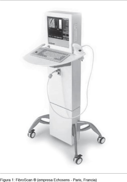
<body><![CDATA[<p style="margin-right: 42.25pt; text-align: justify; text-indent: 17pt; line-height: 120%;"> <span style="font-size: 10pt; line-height: 120%; "><font face="Verdana">Dentro de los ex&aacute;menes serol&oacute;gicos, unos son de c&aacute;lculo sencillo, como el APRI, el &iacute;ndice de Forns, el FIB-4 o el Fibroindex, mientras que otros suponen una formulaci&oacute;n m&aacute;s compleja. </font></span> <o:p></o:p></p>        <p style="margin-right: 42.25pt; text-align: justify; text-indent: 17pt; line-height: 120%;"> <span style="font-size: 10pt; line-height: 120%; "><font face="Verdana">Entre los biomarcadores directos, podemos diferenciar los que se asocian al dep&oacute;sito de mol&eacute;culas en la matriz extracelular (laminina, procolageno, acido hialuronico); los relacionados con la degradaci&oacute;n de dicha matriz extracelular (prop&eacute;ptido aminoterminal tipo III procolageno, inhibidores tisulares de metaloproteinasas TIMP-1 y TIMP-2, y metaloproteinasas); y las citoquinas asociadas a fibrosis hep&aacute;tica (factores de crecimiento tisular TGF-&alpha;, TGF-&beta;, y el factor beta de crecimiento derivado de plaquetas PDGF-BB)<sup> (<a href="#1">1</a>,<a href="#6">6</a>)</sup>.<a name="-6"></a> </font></span> <o:p></o:p></p>        <p style="margin-right: 42.25pt; text-align: justify; text-indent: 17pt; line-height: 120%;"> <font face="Verdana"><span style="font-size: 10pt; line-height: 120%; ">La principal desventaja de estos marcadores es su falta de especificidad pues realmente reflejan los procesos de fibrog&eacute;nesis y fibrin&oacute;lisis, y no directamente la presencia de fibrosis, y algunos se ven influidos por enfermedades extra hep&aacute;ticas.</span></font><o:p></o:p></p>        <p style="margin-right: 42.25pt; text-align: justify; text-indent: 17pt; line-height: 120%;"> <span style="font-size: 10pt; line-height: 120%; font-family: Verdana;"><o:p>&nbsp;</o:p></span></p>        <p style="margin-right: 42.25pt; line-height: 120%;"><b> <span style="font-size: 10pt; line-height: 120%; "><font face="Verdana">T&eacute;cnicas de imagen </font></span> <o:p></o:p></b></p>        <p style="margin-right: 42.25pt; text-align: justify; text-indent: 17pt; line-height: 120%;"> <font face="Verdana"><span style="font-size: 10pt; line-height: 120%; ">La ecograf&iacute;a, la tomograf&iacute;a computarizada (TC) o la resonancia magn&eacute;tica (RM) son algunos de los m&eacute;todos de imagen empleados como t&eacute;cnicas no invasivas en el estudio de fibrosis hep&aacute;tica. En la mayor&iacute;a de los casos, han demostrado ser capaces de detectar cambios en el par&eacute;nquima hep&aacute;tico cuando existe una fibrosis significativa, pero no resultan &uacute;tiles para detectar estadios inferiores ni para realizar la estatificaci&oacute;n de fibrosis. </span> </font> <o:p></o:p></p>        <p style="margin-right: 42.25pt; text-align: justify; text-indent: 17pt; line-height: 120%;"> <span style="font-size: 10pt; line-height: 120%; font-family: Verdana; letter-spacing: -0.1pt">En general, son procedimientos costosos, con ciertos riesgos porque en algunos pacientes es necesario administrar contraste y en otros se expone al paciente a radiacio</span><font face="Verdana"><span style="font-size: 10pt; line-height: 120%; ">nes<sup>&nbsp;(<a href="#1">1</a>)</sup>.</span></font><o:p></o:p></p>        <p style="margin-right: 42.25pt; text-align: justify; text-indent: 17pt; line-height: 120%;"> <font face="Verdana"><span style="font-size: 10pt; line-height: 120%; ">La elastometr&iacute;a o elastograf&iacute;a de transici&oacute;n (ET) es una nueva t&eacute;cnica diagn&oacute;stica que ha revolucionado el campo del diagnostico no invasivo para la fibrosis hep&aacute;tica. Descrita por primera vez en la literatura m&eacute;dica en el a&ntilde;o 1999, es hasta el momento comercializada con el nombre de FibroScan<sup>&reg;</sup> (FS) por la empresa Echosens (Paris, Francia) (<a href="#fig1">Figura 1</a>).<a name="-fig1"></a> </span></font> <o:p></o:p></p>        <p style="margin-right: 42.25pt; text-align: justify; text-indent: 17pt; line-height: 120%;"> <span style="font-size: 10pt; line-height: 120%; font-family: Verdana;">El uso de la ET en pacientes con enfermedad hep&aacute;tica se basa en la relaci&oacute;n existente entre la rigidez del h&iacute;gado y el grado de fibrosis hep&aacute;tica<sup> (<a href="#1">1</a>)</sup>.</span></p>      <p style="margin-right: 42.25pt; text-align: justify; text-indent: 17pt; line-height: 120%;"></p>      ]]></body>
<body><![CDATA[<p style="margin-right: 42.25pt; text-align: justify; text-indent: 17pt; line-height: 120%;"> <font face="Verdana" size="2"><a name="fig1"></a><a href="#-fig1"><img style="border: 0px solid ; width: 505px; height: 721px;" alt="" src="/img/revistas/ami/v35n3/3a04f1.jpg"></a></font><span style="font-size: 9pt; line-height: 120%; font-family: Helvetica;"><o:p></o:p></span></p>        <p style="margin-right: 42.25pt; text-align: justify; text-indent: 17pt; line-height: 120%;"> <span style="font-size: 10pt; line-height: 120%; font-family: Verdana;"><o:p>&nbsp;</o:p></span></p>        <p style="margin-right: 42.25pt; line-height: 120%;"><b> <span style="font-size: 10pt; line-height: 120%; "><font face="Verdana">ELASTOGRAF&Iacute;A DE TRANSICI&Oacute;N </font></span> <o:p></o:p></b></p>        <p style="margin-right: 42.25pt; text-align: justify; text-indent: 17pt; line-height: 120%;"> <span style="font-size: 10pt; line-height: 120%; font-family: Verdana;"><o:p>&nbsp;</o:p></span></p>        <p style="margin-right: 42.25pt; line-height: 120%;"><b> <span style="font-size: 10pt; line-height: 120%; "><font face="Verdana">Descripci&oacute;n de la t&eacute;cnica y ventajas de la misma </font></span> <o:p></o:p></b></p>        <p style="margin-right: 42.25pt; text-align: justify; text-indent: 17pt; line-height: 120%;"> <font face="Verdana"><span style="font-size: 10pt; line-height: 120%; ">El FibroScan<sup>&reg;</sup> consta de un transductor de ultrasonidos (de 5 MHz de frecuencia) acoplado sobre el eje de un vibrador. La sonda del transductor de ultrasonidos se coloca sobre la piel, en un espacio intercostal a la altura del l&oacute;bulo derecho hep&aacute;tico. Con la ayuda de una imagen ecogr&aacute;fica en modo tiempo-movimiento se selecciona el &aacute;rea a estudiar, que debe tener un espesor determinado y estar libre de estructuras vasculares importantes. Una vez localizada el &aacute;rea m&aacute;s adecuada, se inicia la exploraci&oacute;n presionando el bot&oacute;n de la sonda para generar una vibraci&oacute;n de baja frecuencia (50 Hz) y moderada amplitud. Esta vibraci&oacute;n induce una onda el&aacute;stica que se propagara a trav&eacute;s de los tejidos, siendo analizada mediante la adquisici&oacute;n de se&ntilde;ales de ultrasonido con las que se genera el mapa de rastreo de dicha onda el&aacute;stica, de acuerdo a los par&aacute;metros de profundidad y tiempo. De esta manera, se determina la velocidad de propagaci&oacute;n de la onda el&aacute;stica que est&aacute; relacionada con la elasticidad tisular hep&aacute;tica: a menor elasticidad del tejido, mayor velocidad de propagaci&oacute;n de la onda<sup> (<a href="#1">1</a>,<a href="#3">3</a>,<a href="#7">7</a>)</sup>.<a name="-7"></a> </span> </font> <o:p></o:p></p>        <p style="margin-right: 42.25pt; text-align: justify; text-indent: 17pt; line-height: 120%;"> <font face="Verdana"><span style="font-size: 10pt; line-height: 120%; ">El FibroScan<sup>&reg;</sup> mide la elasticidad de un cilindro de par&eacute;nquima hep&aacute;tico de aproximadamente 1 cm de di&aacute;metro y 2-4 cm de longitud, lo que representa un volumen 100 veces mayor que el explorado con la biopsia y por esto se considera que es m&aacute;s representativo de la totalidad del par&eacute;nquima hep&aacute;tico. Aporta una informaci&oacute;n que se aproxima 1:500 en relaci&oacute;n a la masa total del h&iacute;gado, comparado con 1:50.000 que es lo que aporta la punci&oacute;n bi&oacute;psica hep&aacute;tica<sup> (1,3)</sup>. La profundidad de las mediciones oscila entre 25 y 45 mm, distancia que evita la interferencia en la medici&oacute;n del tejido celular subcut&aacute;neo y la capsula hep&aacute;tica, en la mayor&iacute;a de los adultos. Para cada paciente, generalmente se realizan diez mediciones de la elasticidad y el resultado final que ofrece el FibroScan&reg; es la mediana de todas las mediciones realizadas. Los resultados se expresan en kilopascales (kPa), y el rango de valores de elasticidad va de 2,5 a 75 kPa. Cuando la medici&oacute;n no es v&aacute;lida, el FibroScan&reg; no da ning&uacute;n resultado. Es el software el encargado de determinar cu&aacute;ndo una medici&oacute;n es v&aacute;lida o no. Se considera que la exploraci&oacute;n es correcta si la proporci&oacute;n de mediciones validas respecto al total de mediciones realizadas es de al menos el 60% y el rango intercuartilico no excede el 30% del valor de la mediana, para evitar excesiva variabilidad entre los resultados<sup> (<a href="#1">1</a>,<a href="#4">4</a>)</sup>.</span></font><o:p></o:p></p>        <p style="margin-right: 42.25pt; text-align: justify; text-indent: 17pt; line-height: 120%;"> <span style="font-size: 10pt; line-height: 120%; "><font face="Verdana">Esta tecnolog&iacute;a tiene otras ventajas como su car&aacute;cter no invasivo, no doloroso, de f&aacute;cil realizaci&oacute;n, un tiempo de exploraci&oacute;n corto (puede tardar menos de 5 minutos) y la obtenci&oacute;n inmediata de resultados, y que &eacute;stos son operador independientes<sup> (<a href="#3">3</a>,<a href="#4">4</a>)</sup>. </font></span> <o:p></o:p></p>        <p style="margin-right: 42.25pt; text-align: justify; text-indent: 17pt; line-height: 120%;"> <span style="font-size: 10pt; line-height: 120%; font-family: Verdana;"><o:p>&nbsp;</o:p></span></p>        <p style="margin-right: 42.25pt; line-height: 120%;"><b> <span style="font-size: 10pt; line-height: 120%; "><font face="Verdana">Efectividad diagn&oacute;stica y seguridad </font></span> <o:p></o:p></b></p>        ]]></body>
<body><![CDATA[<p style="margin-right: 42.25pt; text-align: justify; text-indent: 17pt; line-height: 120%;"> <font face="Verdana"><span style="font-size: 10pt; line-height: 120%; ">La valoraci&oacute;n est&aacute;ndar de la efectividad de un test se realiza mirando el &aacute;rea bajo la curva (AUROC), teniendo como medida perfecta un score de 1,0<sup> (<a href="#4">4</a>)</sup>. Se acepta que una prueba diagn&oacute;stica es excelente cuando su AUROC es superior a 90% y buena si es superior al 80%. </span> </font> <o:p></o:p></p>        <p style="margin-right: 42.25pt; text-align: justify; text-indent: 17pt; line-height: 120%;"> <font face="Verdana"><span style="font-size: 10pt; line-height: 120%; ">La ET presenta un AUROC global del 92 al 98%, consider&aacute;ndose por tanto una t&eacute;cnica v&aacute;lida y de gran utilidad cl&iacute;nica para detectar cirrosis, al punto de que en esta situaci&oacute;n podr&iacute;a suprimirse la realizaci&oacute;n de la biopsia<sup> (<a href="#1">1</a>)</sup>.</span></font><o:p></o:p></p>        <p style="margin-right: 42.25pt; text-align: justify; text-indent: 17pt; line-height: 120%;"> <span style="font-size: 10pt; line-height: 120%; "><font face="Verdana">Apoya esto, que la correlaci&oacute;n entre la elasticidad media con ET y la fibrosis diagn&oacute;stica mediante biopsia fue analizada en 25 estudios, describi&eacute;ndose en 21 de ellos una relaci&oacute;n estad&iacute;sticamente significativa (p &le; 0,05)<sup> (<a href="#1">1</a>)</sup>.</font></span><o:p></o:p></p>        <p style="margin-right: 42.25pt; text-align: justify; text-indent: 17pt; line-height: 120%;"> <font face="Verdana"><span style="font-size: 10pt; line-height: 120%; ">La capacidad diagn&oacute;stica de la ET ser&iacute;a mayor en la identificaci&oacute;n de la fibrosis avanzada o cirrosis, y resultar&iacute;a menos exacta en el diagnostico de los estadios iniciales e intermedios, existiendo gran heterogeneidad en los estudios para estos grados de fibrosis.</span></font><o:p></o:p></p>        <p style="margin-right: 42.25pt; text-align: justify; text-indent: 17pt; line-height: 120%;"> <font face="Verdana"><span style="font-size: 10pt; line-height: 120%; ">Para la mayor&iacute;a de los autores lo m&aacute;s importante desde el punto de vista cl&iacute;nico es que la ET sea capaz de detectar los niveles de fibrosis superiores a F2 con el fin de iniciar el tratamiento<sup> (<a href="#1">1</a>)</sup>. </span></font> <o:p></o:p></p>        <p style="margin-right: 42.25pt; text-align: justify; text-indent: 17pt; line-height: 120%;"> <font face="Verdana"><span style="font-size: 10pt; line-height: 120%; ">Es &uacute;til adem&aacute;s en el seguimiento cl&iacute;nico de la fibrosis hep&aacute;tica, dado que se trata de un proceso en continua evoluci&oacute;n; y en la monitorizaci&oacute;n de pacientes en tratamiento<sup> (<a href="#1">1</a>) </sup>al tratarse de una t&eacute;cnica no invasiva, r&aacute;pida y de bajo costo. En relaci&oacute;n con la seguridad de la ET, existe acuerdo en que es una prueba segura, sin efectos adversos<sup> (<a href="#3">3</a>,<a href="#4">4</a>)</sup>.</span></font><o:p></o:p></p>        <p style="margin-right: 42.25pt; text-align: justify; text-indent: 17pt; line-height: 120%;"> <span style="font-size: 10pt; line-height: 120%; font-family: Verdana;"><o:p>&nbsp;</o:p></span></p>        <p style="margin-right: 42.25pt; line-height: 120%;"><b> <span style="font-size: 10pt; line-height: 120%; "><font face="Verdana">Limitaciones de uso </font></span> <o:p></o:p></b></p>        <p style="margin-right: 42.25pt; text-align: justify; text-indent: 17pt; line-height: 120%;"> <font face="Verdana"><span style="font-size: 10pt; line-height: 120%; ">La t&eacute;cnica tiene buena resoluci&oacute;n en el 95% de los pacientes. Dentro de las limitaciones para la aplicaci&oacute;n de esta se encuentran la ascitis, la obesidad y los espacios intercostales estrechos. </span> </font> <o:p></o:p></p>        <p style="margin-right: 42.25pt; text-align: justify; text-indent: 17pt; line-height: 120%;"> <font face="Verdana"><span style="font-size: 10pt; line-height: 120%; ">La ascitis impide su aplicaci&oacute;n dado que los l&iacute;quidos interfieren con la se&ntilde;al de onda, sin embargo visto que la misma es indicativa de cirrosis hep&aacute;tica, en estos casos no suele ser necesario valorar el nivel de fibrosis<sup> (<a href="#4">4</a>,<a href="#7">7</a>)</sup>.</span></font><o:p></o:p></p>        ]]></body>
<body><![CDATA[<p style="margin-right: 42.25pt; text-align: justify; text-indent: 17pt; line-height: 120%;"> <font face="Verdana"><span style="font-size: 10pt; line-height: 120%; ">En referencia a la obesidad, se considera que es el &uacute;nico factor independiente del fallo de medida con ET cuando el &iacute;ndice de masa corporal es mayor de 28<sup> (<a href="#3">3</a>,<a href="#4">4</a>,<a href="#7">7</a>)</sup>. </span> </font> <o:p></o:p></p>        <p style="margin-right: 42.25pt; text-align: justify; text-indent: 17pt; line-height: 120%;"> <font face="Verdana"><span style="font-size: 10pt; line-height: 120%; ">Se ha comprobado alg&uacute;n grado de discordancia con la biopsia hep&aacute;tica en uno de cada 8 pacientes obesos, y &eacute;sta est&aacute; dada fundamentalmente por una sobreestimaci&oacute;n de la fibrosis<sup> (<a href="#8">8</a>)</sup>.<a name="-8"></a></span></font><o:p></o:p></p>        <p style="margin-right: 42.25pt; text-align: justify; text-indent: 17pt; line-height: 120%;"> <font face="Verdana"><span style="font-size: 10pt; line-height: 120%; ">Con referencia a este inconveniente actualmente se han desarrollado nuevas sondas o transductores espec&iacute;ficos que mejoran la performance del estudio. </span></font> <o:p></o:p></p>        <p style="margin-right: 42.25pt; text-align: justify; text-indent: 17pt; line-height: 120%;"> <span style="font-size: 10pt; line-height: 120%; "><font face="Verdana">Recientemente se ha desarrollado una sonda espec&iacute;fica para pacientes obesos (sonda XL), que mejora el diagn&oacute;stico de la ET en estos pacientes. </font></span> <o:p></o:p></p>        <p style="margin-right: 42.25pt; text-align: justify; text-indent: 17pt; line-height: 120%;"> <span style="font-size: 10pt; line-height: 120%; "><font face="Verdana">Seg&uacute;n se ha comunicado recientemente, la mitad de los pacientes que no pueden ser valorados con el transductor est&aacute;ndar por su obesidad, son estudiados con &eacute;xito cuando se utiliza la nueva sonda XL<sup> (<a href="#9">9</a>)</sup>.<a name="-9"></a> La combinaci&oacute;n de ambas sondas permite obtener un 91,2% de &eacute;xitos en las mediciones de la ET. </font></span> <o:p></o:p></p>        <p style="margin-right: 42.25pt; text-align: justify; text-indent: 17pt; line-height: 120%;"> <span style="font-size: 10pt; line-height: 120%; font-family: Verdana;"><o:p>&nbsp;</o:p></span></p>        <p style="margin-right: 42.25pt; line-height: 120%;"><b> <span style="font-size: 10pt; line-height: 120%; "><font face="Verdana">Elastograf&iacute;a de transici&oacute;n en relaci&oacute;n a otros m&eacute;todos diagn&oacute;sticos no invasivos </font></span> <o:p></o:p></b></p>        <p style="margin-right: 42.25pt; text-align: justify; text-indent: 17pt; line-height: 120%;"> <font face="Verdana"><span style="font-size: 10pt; line-height: 120%; ">Se ha estudiado el uso combinado de la ET con marcadores serol&oacute;gicos, lo que parece aumentar la capacidad diagnostica. De esta forma se estudi&oacute; el comportamiento diagnostico del FibroScan&reg; junto con FibroTest y APRI y se encontr&oacute; que el mayor grado de concordancia contra la biopsia hep&aacute;tica se alcanzaba al utilizar de forma combinada FibroScan&reg; y FibroTest. Tanto es as&iacute;, que hay autores que plantean que el uso combinado de ambas t&eacute;cnicas sustituyen totalmente la biopsia hep&aacute;tica en los pacientes con VHC<sup>&nbsp;(<a href="#1">1</a>,<a href="#3">3</a>,<a href="#10">10</a>)</sup>.<a name="-10"></a> </span> </font> <o:p></o:p></p>        <p style="margin-right: 42.25pt; text-align: justify; text-indent: 17pt; line-height: 120%;"> <span style="font-size: 10pt; line-height: 120%; font-family: Verdana;"><o:p>&nbsp;</o:p></span></p>        <p style="margin-right: 42.25pt; line-height: 120%;"><b> <span style="font-size: 10pt; line-height: 120%; "><font face="Verdana">Aplicaci&oacute;n </font></span> <o:p></o:p></b></p>        ]]></body>
<body><![CDATA[<p style="margin-right: 42.25pt; text-align: justify; text-indent: 17pt; line-height: 120%;"> <font face="Verdana"><span style="font-size: 10pt; line-height: 120%; ">Las patolog&iacute;as hep&aacute;ticas cr&oacute;nicas secundarias a infecciones virales, han sido las m&aacute;s estudiadas, sobre todo por VHC. </span> </font> <o:p></o:p></p>        <p style="margin-right: 42.25pt; text-align: justify; text-indent: 17pt; line-height: 120%;"> <span style="font-size: 10pt; line-height: 120%; "><font face="Verdana">Numerosos estudios han demostrado la correlaci&oacute;n significativa que existe en el diagn&oacute;stico de fibrosis por ET vs biopsia hep&aacute;tica en pacientes con VHC, con un AUROC de 0,77 a 0,90 para fibrosis moderada (F2 o m&aacute;s) y de 0,90 a 0,97 para fibrosis severa (estadio de cirrosis), con puntos de corte de 6.2 a 8,7 kPa y de 9,6 a 14,8 kPa respectivamente<sup>&nbsp;(<a href="#1">1</a>,<a href="#11">11</a>-<a href="#13">13</a>)</sup>.<a name="-11"></a><a name="-12"></a><a name="-13"></a> Tambi&eacute;n se ha demostrado la utilidad de la ET para valorar la disminuci&oacute;n de la fibrosis en los pacientes respondedores al tratamiento con Interfer&oacute;n-pegilado m&aacute;s Ribavirina. La monitorizaci&oacute;n de este grupo de pacientes parece ser una de las m&aacute;s prometedoras para esta t&eacute;cnica<sup> (<a href="#11">11</a>,<a href="#14">14</a>-<a href="#17">17</a>)</sup>.<a name="-14"></a><a name="-15"></a><a name="-16"></a><a name="-17"></a> Por &uacute;ltimo la ET ha sido utilizada en determinar la recurrencia de VHC luego del trasplante hep&aacute;tico, teniendo un AUROC de 0,90 a 0,98 para el punto de corte de 8,5 kPa<sup> (<a href="#12">12</a>)</sup>. </font></span> <o:p></o:p></p>        <p style="margin-right: 42.25pt; text-align: justify; text-indent: 17pt; line-height: 120%;"> <span style="font-size: 10pt; line-height: 120%; "><font face="Verdana">Estudios recientes, sobre todo en poblaciones de origen asi&aacute;tico, han demostrado igual rendimiento diagn&oacute;stico de la ET para pacientes con VHB. Tiene un AUROC de 0,81 a 0,95 para fibrosis significativa y de 0,80 a 0,98 para cirrosis con puntos de corte de 6,3 a 7,9 kPa y de 9,0 a 13,8 kPa respectivamente. El punto de corte es menor que en los pacientes con VHC, lo que puede explicarse porque la fibrosis ocasionada por el VHB es de menor entidad, visto que es una patolog&iacute;a que tiende a generar mayor nodularidad<sup> (<a href="#12">12</a>)</sup>. </font></span> <o:p></o:p></p>        <p style="margin-right: 42.25pt; text-align: justify; text-indent: 17pt; line-height: 120%;"> <font face="Verdana"><span style="font-size: 10pt; line-height: 120%; ">Se ha comenzado a estudiar el valor diagn&oacute;stico de la ET para otras etiolog&iacute;as hep&aacute;ticas cr&oacute;nicas no virales con resultados alentadores, pero que a&uacute;n requieren validaci&oacute;n para su punto de corte<sup> (<a href="#12">12</a>,<a href="#18">18</a>)</sup><a name="-18"></a>. En los casos de EHGNA donde el problema fundamental es diferenciar los pacientes con esteatosis simple de aquellos con esteatohepatitis, el Fibroscan&reg; es igualmente &uacute;til sobre todo cuando se lo asocia a pruebas serol&oacute;gicas<sup> (<a href="#8">8</a>,<a href="#19">19</a>, <a href="#20">20</a>)</sup>.<a name="-19"></a><a name="-20"></a> </span> </font> <o:p></o:p></p>        <p style="margin-right: 42.25pt; text-align: justify; text-indent: 17pt; line-height: 120%;"> <span style="font-size: 10pt; line-height: 120%; "><font face="Verdana">Pero adem&aacute;s se ha utilizado la ET en la valoraci&oacute;n de la presi&oacute;n portal y la presencia de hepatocarcinoma. Vizzutti et al han demostrado que la ET es &uacute;til para la determinaci&oacute;n de la progresi&oacute;n de la presi&oacute;n portal, lo que se traduce en la predicci&oacute;n del desarrollo de v&aacute;rices esof&aacute;gicas grandes con un AUROC de 0,76 a 0,85<sup> (<a href="#12">12</a>)</sup>. Kazemi et al plantean que con un punto de corte de 20 kPa es posible descartar la presencia de v&aacute;rices esof&aacute;gicas de grado 2/3 y de v&aacute;rices en fundus g&aacute;strico con un VPN del 90%, evitando la realizaci&oacute;n de endoscop&iacute;as en un 50% de los pacientes<sup> (<a href="#1">1</a>,<a href="#13">13</a>)</sup>. </font></span> <o:p></o:p></p>        <p style="margin-right: 42.25pt; text-align: justify; text-indent: 17pt; line-height: 120%;"> <span style="font-size: 10pt; line-height: 120%; "><font face="Verdana">Foucher et al han correlacionado distintos puntos de corte del Fibroscan&reg; con la presencia de v&aacute;rices esof&aacute;gicas en distintos tipos de situaciones como son la clasificaci&oacute;n de Child-Pugh, la presencia de ascitis o sangrado digestivo previo<sup> (<a href="#12">12</a>)</sup>. Se ha estudiado la capacidad de la ET en predecir la presencia de hepatocarcinoma, as&iacute; como la recurrencia del mismo en pacientes sometidos a tratamientos curativos, encontrando valores de corte con resultados estad&iacute;sticamente significativos<sup> (<a href="#12">12</a>)</sup>. </font></span> <o:p></o:p></p>        <p style="margin-right: 42.25pt; text-align: justify; text-indent: 17pt; line-height: 120%;"> <span style="font-size: 10pt; line-height: 120%; font-family: Verdana;"><o:p>&nbsp;</o:p></span></p>        <p style="margin-right: 42.25pt; line-height: 120%;"><b> <span style="font-size: 10pt; line-height: 120%; "><font face="Verdana">Impacto </font></span> <o:p></o:p></b></p>        <p style="margin-right: 42.25pt; text-align: justify; text-indent: 17pt; line-height: 120%;"> <font face="Verdana"><span style="font-size: 10pt; line-height: 120%; ">La introducci&oacute;n del FibroScan<sup>&reg;</sup> en los sistemas de salud no requiere una infraestructura especial, tan solo serian necesarias instancias de capacitaci&oacute;n para el personal encargado de la exploraci&oacute;n<sup> (<a href="#1">1</a>)</sup>. </span></font> <o:p></o:p></p>        <p style="margin-right: 42.25pt; text-align: justify; text-indent: 17pt; line-height: 120%;"> <span style="font-size: 10pt; line-height: 120%; "><font face="Verdana">Resulta evidente, adem&aacute;s, que la ET permitir&iacute;a estudiar un mayor volumen de pacientes que la biopsia, dando, con ello, la oportunidad de acceder al tratamiento a m&aacute;s pacientes y de no retrasar ni el diagnostico ni el inicio del tratamiento cuando este sea necesario. </font></span> <o:p></o:p></p>        ]]></body>
<body><![CDATA[<p style="margin-right: 42.25pt; text-align: justify; text-indent: 17pt; line-height: 120%;"> <font face="Verdana"><span style="font-size: 10pt; line-height: 120%; ">La incorporaci&oacute;n de la ET implicar&iacute;a un gasto de inversi&oacute;n inicial pero su utilizaci&oacute;n posiblemente llevar&iacute;a a un considerable ahorro a medio y largo plazo. Como se ha mencionado, se espera que el uso de esta tecnolog&iacute;a genere una reducci&oacute;n del n&uacute;mero de biopsias, con la consiguiente reducci&oacute;n de las complicaciones asociadas a su pr&aacute;ctica, que se traducir&iacute;a en potenciales ahorros para el sistema sanitario<sup> (<a href="#1">1</a>)</sup>. </span> </font> <o:p></o:p></p>        <p style="margin-right: 42.25pt; text-align: justify; text-indent: 17pt; line-height: 120%;"> <font face="Verdana"><span style="font-size: 10pt; line-height: 120%; ">Se han publicado algoritmos de pr&aacute;ctica cl&iacute;nica para el abordaje de distintas enfermedades hep&aacute;ticas, tomando como referencia los hallazgos de la ET y marcadores serol&oacute;gicos de fibrosis, con la finalidad de suprimir al m&aacute;ximo la necesidad de punci&oacute;n bi&oacute;psica y minimizar los riesgos<sup> (<a href="#3">3</a>, <a href="#8">8</a>,<a href="#19">19</a>)</sup>.</span></font><o:p></o:p></p>        <p style="margin-right: 42.25pt; text-align: justify; text-indent: 17pt; line-height: 120%;"> <font face="Verdana"><span style="font-size: 10pt; line-height: 120%; ">Las autoridades de salud francesas han aprobado desde hace ya varios a&ntilde;os un algoritmo de estudio basado en ET y test serol&oacute;gicos no invasivos, para los pacientes con VHC no complicada y su respuesta al tratamiento, lo que posteriormente se ha confirmado en estudios cl&iacute;nicos<sup> (<a href="#7">7</a>,<a href="#21">21</a>)</sup>.<a name="-21"></a></span></font><o:p></o:p></p>        <p style="margin-right: 42.25pt; text-align: justify; text-indent: 17pt; line-height: 120%;"> <span style="font-size: 10pt; line-height: 120%; font-family: Verdana;"><o:p>&nbsp;</o:p></span></p>        <p style="margin-right: 42.25pt; line-height: 120%;"><b> <span style="font-size: 10pt; line-height: 120%; "><font face="Verdana">Disponibilidad en Uruguay </font></span> <o:p></o:p></b></p>        <p style="margin-right: 42.25pt; text-align: justify; text-indent: 17pt; line-height: 120%;"> <font face="Verdana"><span style="font-size: 10pt; line-height: 120%; ">La Agencia Nacional de Investigaci&oacute;n e Innovaci&oacute;n (ANII) aprob&oacute; este a&ntilde;o un proyecto presentado por dos grupos m&eacute;dicos (UDA Enfermedades He&aacute;to-Bilio-Pancre&aacute;ticas del Hospital Central de las Fuerzas Armadas y la Policl&iacute;nica de Hepatolog&iacute;a del Hospital Pasteur) para la adquisici&oacute;n del Fibroscan<sup>&reg;</sup> en el marco de los programas para la adquisici&oacute;n de tecnolog&iacute;a de punta no existente en el pa&iacute;s. Actualmente se est&aacute; en la etapa de autorizar su importaci&oacute;n, por parte del Departamento de Tecnolog&iacute;a M&eacute;dica del Ministerio de Salud P&uacute;blica. Se considera que para inicios del a&ntilde;o 2014 estar&aacute; en condiciones de uso. El proyecto comprende actividades asistenciales, docentes y de investigaci&oacute;n, lo que permitir&aacute; en un futuro pr&oacute;ximo contar con experiencia nacional.</span></font><o:p></o:p></p>        <p style="margin-right: 42.25pt; text-align: justify; text-indent: 17pt; line-height: 120%;"> <span style="font-size: 10pt; line-height: 120%; font-family: Verdana;"><o:p>&nbsp;</o:p></span></p>        <p style="margin-right: 42.25pt; line-height: 120%;"><b> <span style="font-size: 10pt; line-height: 120%; "><font face="Verdana">CONCLUSI&Oacute;N </font></span> <o:p></o:p></b></p>        <p style="margin-right: 42.25pt; text-align: justify; text-indent: 17pt; line-height: 120%;"> <span style="font-size: 10pt; line-height: 120%; font-family: Verdana;"><o:p>&nbsp;</o:p></span></p>        <p style="margin-right: 42.25pt; text-align: justify; text-indent: 17pt; line-height: 120%;"> <font face="Verdana"><span style="font-size: 10pt; line-height: 120%; ">La Elastograf&iacute;a de Transici&oacute;n, aparece como una nueva tecnolog&iacute;a sanitaria no invasiva, r&aacute;pida y efectiva para el diagnostico de fibrosis hep&aacute;tica, sin efectos secundarios para el paciente. Esta ampliamente aceptada su utilidad para el diagn&oacute;stico y estatificaci&oacute;n en los pacientes portadores de enfermedades hep&aacute;ticas cr&oacute;nicas virales, sobre todo por VHC. En otras etiolog&iacute;as y/o situaciones cl&iacute;nicas su uso est&aacute; a&uacute;n en v&iacute;as de desarrollo, siendo sin duda un campo promisorio de investigaci&oacute;n y asistencia cl&iacute;nica. </span></font> <o:p></o:p></p>        ]]></body>
<body><![CDATA[<p style="margin-right: 42.25pt; text-align: justify; text-indent: 17pt; line-height: 120%;"> <span style="font-size: 10pt; line-height: 120%; font-family: Verdana;"><o:p>&nbsp;</o:p></span></p>        <p style="margin-right: 42.25pt; line-height: 120%;"><b> <span style="font-size: 10pt; line-height: 120%; "><font face="Verdana">BIBLIOGRAF&Iacute;A</font></span><o:p></o:p></b></p>        <p style="margin-right: 42.25pt; text-align: justify; text-indent: 17pt; line-height: 120%;"> <span style="font-size: 10pt; line-height: 120%; font-family: Verdana;"><o:p>&nbsp;</o:p></span></p>        <p style="margin: 0cm 42.25pt 0.0001pt 17pt; text-align: justify; text-indent: -17pt; line-height: 120%;"> <span style="font-size: 10pt; line-height: 120%; "><font face="Verdana"><a href="#-1">1</a>.<span style="">&nbsp;&nbsp;&nbsp;&nbsp; </span>Asensio del Barrio C, Polo de Santos M, Luengo Matos S, Sanchez Gomez L, Alcazar Alcazar R. &ldquo;Elastografia de Transici&oacute;n (FibroScan&reg;) en el diagnostico de fibrosis hep&aacute;tica: revisi&oacute;n sistem&aacute;tica y meta-an&aacute;lisis&rdquo;. [en l&iacute;nea]. Madrid: Agencia de Evaluaci&oacute;n de Tecnolog&iacute;as Sanitarias (AETS), [acceso octubre 2013] 2009. Disponible en: <a href="http://gesdoc.isciii.es/gesdoccontroller?action=download&amp;id=14/09/2012-eab16a6bf7">http://gesdoc.isciii.es/gesdoccontroller?action=download&amp;id=14/09/2012-eab16a6bf7</a></font></span><o:p></o:p></p>        <!-- ref --><p style="margin: 0cm 42.25pt 0.0001pt 17pt; text-align: justify; text-indent: -17pt; line-height: 120%;"> <span style="font-size: 10pt; line-height: 120%; "><font face="Verdana"><a href="#-2">2</a>.<span style="">&nbsp;&nbsp;&nbsp;&nbsp; </span>Zois CD, Baltayiannis GH, Karayiannis P, Tsianos EV. Systematic review: hepatic fibrosis -regression with therapy. Aliment Pharmacol Ther 2008; 28(10):1175-1187.    </font></span><o:p></o:p></p>        <!-- ref --><p style="margin: 0cm 42.25pt 0.0001pt 17pt; text-align: justify; text-indent: -17pt; line-height: 120%;"> <span style="font-size: 10pt; line-height: 120%; "><font face="Verdana"><a href="#-3">3</a>.<span style="">&nbsp;&nbsp;&nbsp;&nbsp; </span>Nguyen-Khac E, Capron D. Noninvasive diagnosis of liver fibrosis by ultrasonic transient elastography (Fibroscan). Eur J Gastroenterol Hepatol ; 2006; 18:1321-1325.     </font></span> <o:p></o:p></p>        <!-- ref --><p style="margin: 0cm 42.25pt 0.0001pt 17pt; text-align: justify; text-indent: -17pt; line-height: 120%;"> <span style="font-size: 10pt; line-height: 120%; "><font face="Verdana"><a href="#-4">4</a>.<span style="">&nbsp;&nbsp;&nbsp;&nbsp; </span>Castera L, Forns X, Alberti A. Non-invasive evaluation of liver fibrosis using transient elastography. Journal of Hepatology 48 (2008) 835-847.    </font></span><o:p></o:p></p>        ]]></body>
<body><![CDATA[<!-- ref --><p style="margin: 0cm 42.25pt 0.0001pt 17pt; text-align: justify; text-indent: -17pt; line-height: 120%;"> <span style="font-size: 10pt; line-height: 120%; "><font face="Verdana"><a href="#-5">5</a>.<span style="">&nbsp;&nbsp;&nbsp;&nbsp; </span>Malekzadeh R, Poustchi H. Fibroscan for assessing liver fibrosis: An acceptable alternative for liver biopsy. Hepat Mon. 2011; 11:157-158.     </font></span> <o:p></o:p></p>        <!-- ref --><p style="margin: 0cm 42.25pt 0.0001pt 17pt; text-align: justify; text-indent: -17pt; line-height: 120%;"> <span style="font-size: 10pt; line-height: 120%; "><font face="Verdana"><a href="#-6">6</a>.<span style="">&nbsp;&nbsp;&nbsp;&nbsp; </span>Anastasiou J, Alisa A, Virtue S, Portmann B, Murray-Lyon I, Williams R. Noninvasive markers of fibrosis and inflammation in clinical practice: prospective comparison with liver biopsy. Eur J Gastroenterol Hepatol, 2010 22:474-480.     </font></span> <o:p></o:p></p>        <!-- ref --><p style="margin: 0cm 42.25pt 0.0001pt 17pt; text-align: justify; text-indent: -17pt; line-height: 120%;"> <span style="font-size: 10pt; line-height: 120%; "><font face="Verdana"><a href="#-7">7</a>.<span style="">&nbsp;&nbsp;&nbsp;&nbsp; </span>Cast&eacute;ra L, Foucher J, Bernard PH, Carvalho F, Allaix D, Merrouche W. Pitfalls of Liver Stiffness Measurement: A 5-Year Prospective Study of 13,369 Examinations. Hepatology. 2010;51(3):828-35.    </font></span><o:p></o:p></p>        <!-- ref --><p style="margin: 0cm 42.25pt 0.0001pt 17pt; text-align: justify; text-indent: -17pt; line-height: 120%;"> <span style="font-size: 10pt; line-height: 120%; "><font face="Verdana"><a href="#-8">8</a>.<span style="">&nbsp;&nbsp;&nbsp;&nbsp; </span>Myers RP, Pomier-Layrargues G, Kirsch R, Pollett A, Beaton M, Levstik M, et al. Discordance in fibrosis staging between liver biopsy and transient elastography using the FibroScan XL probe. J Hepatol. 2012 Mar;56(3):564-70.    </font></span><o:p></o:p></p>        <!-- ref --><p style="margin: 0cm 42.25pt 0.0001pt 17pt; text-align: justify; text-indent: -17pt; line-height: 120%;"> <span style="font-size: 10pt; line-height: 120%; "><font face="Verdana"><a href="#-9">9</a>.<span style="">&nbsp;&nbsp;&nbsp;&nbsp; </span>de L&eacute;dinghen V, Wong VW, Vergniol J, Wong GL, Foucher J, Chu SH, et al. Diagnosis of liver fibrosis and cirrhosis using liver stiffness measurement: Comparison between M and XL probe of FibroScan. J Hepatol. 2012;56:833-9.    </font></span><o:p></o:p></p>        ]]></body>
<body><![CDATA[<!-- ref --><p style="margin: 0cm 42.25pt 0.0001pt 17pt; text-align: justify; text-indent: -17pt; line-height: 120%;"> <span style="font-size: 10pt; line-height: 120%; "><font face="Verdana"><a href="#-10">10</a>.<span style="">&nbsp;&nbsp; </span>Cast&eacute;ra L, Vergniol J, Foucher J, Le Bail B, Chanteloup E, Haaser M, et al. Prospective comparison of transient elastography, Fibrotest, APRI, and liver biopsy for the assessment of fibrosis in chronic hepatitis C. Gastroenterology. 2005; 128: 343-50.    </font></span><o:p></o:p></p>        <!-- ref --><p style="margin: 0cm 42.25pt 0.0001pt 17pt; text-align: justify; text-indent: -17pt; line-height: 120%;"> <span style="font-size: 10pt; line-height: 120%; "><font face="Verdana"><a href="#-11">11</a>.<span style="">&nbsp;&nbsp; </span>Takemoto R, Nakamuta M, Aoyagi Y, Fujino T, Yasutake K, Koga K, et al. Validity of FibroScan values for predicting hepatic fibrosis stage in patients with chronic HCV infection. J Dig Dis. 2009;10: 145-8.    </font></span><o:p></o:p></p>        <!-- ref --><p style="margin: 0cm 42.25pt 0.0001pt 17pt; text-align: justify; text-indent: -17pt; line-height: 120%;"> <span style="font-size: 10pt; line-height: 120%; "><font face="Verdana"><a href="#-12">12</a>.<span style="">&nbsp;&nbsp; </span>Jung KS, Kim SU. Clinical applications of transient elastography. Clin Mol Hepatol. 2012;18: 163-73.    </font></span><o:p></o:p></p>        <!-- ref --><p style="margin: 0cm 42.25pt 0.0001pt 17pt; text-align: justify; text-indent: -17pt; line-height: 120%;"> <span style="font-size: 10pt; line-height: 120%; "><font face="Verdana"><a href="#-13">13</a>.<span style="">&nbsp;&nbsp; </span>Carri&oacute;n JA. Utility of Fibroscan in the evaluation of liver fibrosis. Gastroenterol Hepatol 2009; 320: 415-23.    </font></span><o:p></o:p></p>        <!-- ref --><p style="margin: 0cm 42.25pt 0.0001pt 17pt; text-align: justify; text-indent: -17pt; line-height: 120%;"> <span style="font-size: 10pt; line-height: 120%; "><font face="Verdana"><a href="#-14">14</a>.<span style="">&nbsp;&nbsp; </span>Vergniol, J Foucher J, Cast&eacute;ra L, Bernard PH, Tournan R, Terrebonne E. Changes of non-invasive markers and FibroScan values during HCV treatment. J Viral Hepat. 2009 Feb;16(2):132-40.     </font></span> <o:p></o:p></p>        ]]></body>
<body><![CDATA[<!-- ref --><p style="margin: 0cm 42.25pt 0.0001pt 17pt; text-align: justify; text-indent: -17pt; line-height: 120%;"> <span style="font-size: 10pt; line-height: 120%; "><font face="Verdana"><a href="#-15">15</a>.<span style="">&nbsp;&nbsp; </span>Wang JH, Changchien CS, Hung CH, Tung WC, Kee KM, Chen CH et al. Liver stiffness decrease after effective antiviral therapy in patients with chronic hepatitis C: Longitudinal&ordm; study using FibroScan. J Gastroenterol Hepatol. 2010; 25:964-9.    </font></span><o:p></o:p></p>        <!-- ref --><p style="margin: 0cm 42.25pt 0.0001pt 17pt; text-align: justify; text-indent: -17pt; line-height: 120%;"> <span style="font-size: 10pt; line-height: 120%; "><font face="Verdana"><a href="#-16">16</a>.<span style="">&nbsp;&nbsp; </span>Scott DR, Levy MT. Liver transient elastography (Fibroscan): a place in the management algorithms of chronic viral hepatitis. Antivir Ther, 2010; 15: 1-11.    </font></span><o:p></o:p></p>        <!-- ref --><p style="margin: 0cm 42.25pt 0.0001pt 17pt; text-align: justify; text-indent: -17pt; line-height: 120%;"> <span style="font-size: 10pt; line-height: 120%; "><font face="Verdana"><a href="#-17">17</a>.<span style="">&nbsp;&nbsp; </span>Fransen van de Putte D, Blom R, van Soest H, Mundt M, Verveer C, Arends J, et al. Impact of Fibroscan on management of chronic viral hepatitis in clinical practice. Ann Hepatol 2011; 4: 469-476.    </font></span><o:p></o:p></p>        <!-- ref --><p style="margin: 0cm 42.25pt 0.0001pt 17pt; text-align: justify; text-indent: -17pt; line-height: 120%;"> <span style="font-size: 10pt; line-height: 120%; "><font face="Verdana"><a href="#-18">18</a>.<span style="">&nbsp;&nbsp; </span>Ganne-Carrie N, Ziol M, de Ledinghen V, Douvin C, Marcellin P, Castera L, et al. Accuracy of liver stiffness measurement for the diagnosis of cirrhosis in patients with chronic liver diseases. Hepatology 2006;44:1511-1517.    </font></span><o:p></o:p></p>        <!-- ref --><p style="margin: 0cm 42.25pt 0.0001pt 17pt; text-align: justify; text-indent: -17pt; line-height: 120%;"> <span style="font-size: 10pt; line-height: 120%; "><font face="Verdana"><a href="#-19">19</a>.<span style="">&nbsp;&nbsp; </span>Ter&aacute;n Lantar&oacute;n A, Crespo Garc&iacute;a J. Cribado de la enfermedad hep&aacute;tica por dep&oacute;sito de grasa: c&oacute;mo y a quien. Gastroenterol Hepatol. 2011; 34: 278-288.     </font></span> <o:p></o:p></p>        ]]></body>
<body><![CDATA[<!-- ref --><p style="margin: 0cm 42.25pt 0.0001pt 17pt; text-align: justify; text-indent: -17pt; line-height: 120%;"> <span style="font-size: 10pt; line-height: 120%; "><font face="Verdana"><a href="#-20">20</a>.<span style="">&nbsp;&nbsp; </span>Gaia S, Carenzi S, Barilli AL, Bugianesi E, Smedile A, Brunello F, et al. Reliability of transient elastography for the detection of fibrosis in Non-Alcoholic Fatty Liver Disease and chronic viral hepatitis. J Hepatol. 2011;54:64-71.    </font></span><o:p></o:p></p>        <!-- ref --><p style="margin: 0cm 42.25pt 0.0001pt 17pt; text-align: justify; text-indent: -17pt; line-height: 120%;"> <span style="font-size: 10pt; line-height: 120%; font-family: Verdana;"><a name="21"></a><a href="#-21">21</a>.<span style="">&nbsp;&nbsp; </span>Poynard T, de Ledinghen V, Zarski JP, Stanciu C, Munteanu M, Vergniol J, et al. Relative performances of FibroTest, Fibroscan, and biopsy for the assessment of the stage of liver fibrosis in patients with chronic hepatitis C: A step toward the truth in the absence of a gold standard. J Hepatol. 2012 Mar;56(3):541-8.    </span></p>    </div>         ]]></body><back>
<ref-list>
<ref id="B1">
<label>1</label><nlm-citation citation-type="">
<person-group person-group-type="author">
<name>
<surname><![CDATA[Asensio del Barrio]]></surname>
<given-names><![CDATA[C]]></given-names>
</name>
<name>
<surname><![CDATA[Polo de Santos]]></surname>
<given-names><![CDATA[M]]></given-names>
</name>
<name>
<surname><![CDATA[Luengo Matos]]></surname>
<given-names><![CDATA[S]]></given-names>
</name>
<name>
<surname><![CDATA[Sanchez Gomez]]></surname>
<given-names><![CDATA[L]]></given-names>
</name>
<name>
<surname><![CDATA[Alcazar Alcazar]]></surname>
<given-names><![CDATA[R]]></given-names>
</name>
</person-group>
<collab>Agencia de Evaluación de Tecnologías Sanitarias</collab>
<source><![CDATA[Elastografia de Transición (FibroScan®) en el diagnostico de fibrosis hepática: revisión sistemática y meta-análisis]]></source>
<year>2009</year>
<publisher-loc><![CDATA[Madrid ]]></publisher-loc>
</nlm-citation>
</ref>
<ref id="B2">
<label>2</label><nlm-citation citation-type="journal">
<person-group person-group-type="author">
<name>
<surname><![CDATA[Zois]]></surname>
<given-names><![CDATA[CD]]></given-names>
</name>
<name>
<surname><![CDATA[Baltayiannis]]></surname>
<given-names><![CDATA[GH]]></given-names>
</name>
<name>
<surname><![CDATA[Karayiannis]]></surname>
<given-names><![CDATA[P]]></given-names>
</name>
<name>
<surname><![CDATA[Tsianos]]></surname>
<given-names><![CDATA[EV]]></given-names>
</name>
</person-group>
<article-title xml:lang="en"><![CDATA[Systematic review: hepatic fibrosis -regression with therapy]]></article-title>
<source><![CDATA[Aliment Pharmacol Ther]]></source>
<year>2008</year>
<volume>28</volume>
<numero>10</numero>
<issue>10</issue>
<page-range>1175-1187</page-range></nlm-citation>
</ref>
<ref id="B3">
<label>3</label><nlm-citation citation-type="journal">
<person-group person-group-type="author">
<name>
<surname><![CDATA[Nguyen-Khac]]></surname>
<given-names><![CDATA[E]]></given-names>
</name>
<name>
<surname><![CDATA[Capron]]></surname>
<given-names><![CDATA[D]]></given-names>
</name>
</person-group>
<article-title xml:lang="en"><![CDATA[Noninvasive diagnosis of liver fibrosis by ultrasonic transient elastography (Fibroscan)]]></article-title>
<source><![CDATA[Eur J Gastroenterol Hepatol]]></source>
<year>2006</year>
<volume>18</volume>
<page-range>1321-1325</page-range></nlm-citation>
</ref>
<ref id="B4">
<label>4</label><nlm-citation citation-type="journal">
<person-group person-group-type="author">
<name>
<surname><![CDATA[Castera]]></surname>
<given-names><![CDATA[L]]></given-names>
</name>
<name>
<surname><![CDATA[Forns]]></surname>
<given-names><![CDATA[X]]></given-names>
</name>
<name>
<surname><![CDATA[Alberti]]></surname>
<given-names><![CDATA[A]]></given-names>
</name>
</person-group>
<article-title xml:lang="en"><![CDATA[Non-invasive evaluation of liver fibrosis using transient elastography]]></article-title>
<source><![CDATA[Journal of Hepatology]]></source>
<year>2008</year>
<volume>48</volume>
<page-range>835-847</page-range></nlm-citation>
</ref>
<ref id="B5">
<label>5</label><nlm-citation citation-type="journal">
<person-group person-group-type="author">
<name>
<surname><![CDATA[Malekzadeh]]></surname>
<given-names><![CDATA[R]]></given-names>
</name>
<name>
<surname><![CDATA[Poustchi]]></surname>
<given-names><![CDATA[H]]></given-names>
</name>
</person-group>
<article-title xml:lang="en"><![CDATA[Fibroscan for assessing liver fibrosis: An acceptable alternative for liver biopsy]]></article-title>
<source><![CDATA[Hepat Mon]]></source>
<year>2011</year>
<volume>11</volume>
<page-range>157-158</page-range></nlm-citation>
</ref>
<ref id="B6">
<label>6</label><nlm-citation citation-type="journal">
<person-group person-group-type="author">
<name>
<surname><![CDATA[Anastasiou]]></surname>
<given-names><![CDATA[J]]></given-names>
</name>
<name>
<surname><![CDATA[Alisa]]></surname>
<given-names><![CDATA[A]]></given-names>
</name>
<name>
<surname><![CDATA[Virtue]]></surname>
<given-names><![CDATA[S]]></given-names>
</name>
<name>
<surname><![CDATA[Portmann]]></surname>
<given-names><![CDATA[B]]></given-names>
</name>
<name>
<surname><![CDATA[Murray-Lyon]]></surname>
<given-names><![CDATA[I]]></given-names>
</name>
<name>
<surname><![CDATA[Williams]]></surname>
<given-names><![CDATA[R]]></given-names>
</name>
</person-group>
<article-title xml:lang="en"><![CDATA[Noninvasive markers of fibrosis and inflammation in clinical practice: prospective comparison with liver biopsy]]></article-title>
<source><![CDATA[Eur J Gastroenterol Hepatol]]></source>
<year>2010</year>
<volume>22</volume>
<page-range>474-480</page-range></nlm-citation>
</ref>
<ref id="B7">
<label>7</label><nlm-citation citation-type="journal">
<person-group person-group-type="author">
<name>
<surname><![CDATA[Castéra]]></surname>
<given-names><![CDATA[L]]></given-names>
</name>
<name>
<surname><![CDATA[Foucher]]></surname>
<given-names><![CDATA[J]]></given-names>
</name>
<name>
<surname><![CDATA[Bernard]]></surname>
<given-names><![CDATA[PH]]></given-names>
</name>
<name>
<surname><![CDATA[Carvalho]]></surname>
<given-names><![CDATA[F]]></given-names>
</name>
<name>
<surname><![CDATA[Allaix]]></surname>
<given-names><![CDATA[D]]></given-names>
</name>
<name>
<surname><![CDATA[Merrouche]]></surname>
<given-names><![CDATA[W]]></given-names>
</name>
</person-group>
<article-title xml:lang="en"><![CDATA[Pitfalls of Liver Stiffness Measurement: A 5-Year Prospective Study of 13,369 Examinations]]></article-title>
<source><![CDATA[Hepatology]]></source>
<year>2010</year>
<volume>51</volume>
<numero>3</numero>
<issue>3</issue>
<page-range>828-35</page-range></nlm-citation>
</ref>
<ref id="B8">
<label>8</label><nlm-citation citation-type="journal">
<person-group person-group-type="author">
<name>
<surname><![CDATA[Myers]]></surname>
<given-names><![CDATA[RP]]></given-names>
</name>
<name>
<surname><![CDATA[Pomier-Layrargues]]></surname>
<given-names><![CDATA[G]]></given-names>
</name>
<name>
<surname><![CDATA[Kirsch]]></surname>
<given-names><![CDATA[R]]></given-names>
</name>
<name>
<surname><![CDATA[Pollett]]></surname>
<given-names><![CDATA[A]]></given-names>
</name>
<name>
<surname><![CDATA[Beaton]]></surname>
<given-names><![CDATA[M]]></given-names>
</name>
<name>
<surname><![CDATA[Levstik]]></surname>
<given-names><![CDATA[M]]></given-names>
</name>
</person-group>
<article-title xml:lang="en"><![CDATA[Discordance in fibrosis staging between liver biopsy and transient elastography using the FibroScan XL probe]]></article-title>
<source><![CDATA[J Hepatol]]></source>
<year>2012</year>
<month> M</month>
<day>ar</day>
<volume>56</volume>
<numero>3</numero>
<issue>3</issue>
<page-range>564-70</page-range></nlm-citation>
</ref>
<ref id="B9">
<label>9</label><nlm-citation citation-type="journal">
<person-group person-group-type="author">
<name>
<surname><![CDATA[de Lédinghen]]></surname>
<given-names><![CDATA[V]]></given-names>
</name>
<name>
<surname><![CDATA[Wong]]></surname>
<given-names><![CDATA[VW]]></given-names>
</name>
<name>
<surname><![CDATA[Vergniol]]></surname>
<given-names><![CDATA[J]]></given-names>
</name>
<name>
<surname><![CDATA[Wong]]></surname>
<given-names><![CDATA[GL]]></given-names>
</name>
<name>
<surname><![CDATA[Foucher]]></surname>
<given-names><![CDATA[J]]></given-names>
</name>
<name>
<surname><![CDATA[Chu]]></surname>
<given-names><![CDATA[SH]]></given-names>
</name>
</person-group>
<article-title xml:lang="en"><![CDATA[Diagnosis of liver fibrosis and cirrhosis using liver stiffness measurement: Comparison between M and XL probe of FibroScan]]></article-title>
<source><![CDATA[J Hepatol]]></source>
<year>2012</year>
<volume>56</volume>
<page-range>833-9</page-range></nlm-citation>
</ref>
<ref id="B10">
<label>10</label><nlm-citation citation-type="journal">
<person-group person-group-type="author">
<name>
<surname><![CDATA[Castéra]]></surname>
<given-names><![CDATA[L]]></given-names>
</name>
<name>
<surname><![CDATA[Vergniol]]></surname>
<given-names><![CDATA[J]]></given-names>
</name>
<name>
<surname><![CDATA[Foucher]]></surname>
<given-names><![CDATA[J]]></given-names>
</name>
<name>
<surname><![CDATA[Le Bail]]></surname>
<given-names><![CDATA[B]]></given-names>
</name>
<name>
<surname><![CDATA[Chanteloup]]></surname>
<given-names><![CDATA[E]]></given-names>
</name>
<name>
<surname><![CDATA[Haaser]]></surname>
<given-names><![CDATA[M]]></given-names>
</name>
</person-group>
<article-title xml:lang="en"><![CDATA[Prospective comparison of transient elastography, Fibrotest, APRI, and liver biopsy for the assessment of fibrosis in chronic hepatitis C]]></article-title>
<source><![CDATA[Gastroenterology]]></source>
<year>2005</year>
<volume>128</volume>
<page-range>343-50</page-range></nlm-citation>
</ref>
<ref id="B11">
<label>11</label><nlm-citation citation-type="journal">
<person-group person-group-type="author">
<name>
<surname><![CDATA[Takemoto]]></surname>
<given-names><![CDATA[R]]></given-names>
</name>
<name>
<surname><![CDATA[Nakamuta]]></surname>
<given-names><![CDATA[M]]></given-names>
</name>
<name>
<surname><![CDATA[Aoyagi]]></surname>
<given-names><![CDATA[Y]]></given-names>
</name>
<name>
<surname><![CDATA[Fujino]]></surname>
<given-names><![CDATA[T]]></given-names>
</name>
<name>
<surname><![CDATA[Yasutake]]></surname>
<given-names><![CDATA[K]]></given-names>
</name>
<name>
<surname><![CDATA[Koga]]></surname>
<given-names><![CDATA[K]]></given-names>
</name>
</person-group>
<article-title xml:lang="en"><![CDATA[Validity of FibroScan values for predicting hepatic fibrosis stage in patients with chronic HCV infection]]></article-title>
<source><![CDATA[J Dig Dis]]></source>
<year>2009</year>
<volume>10</volume>
<page-range>145-8</page-range></nlm-citation>
</ref>
<ref id="B12">
<label>12</label><nlm-citation citation-type="journal">
<person-group person-group-type="author">
<name>
<surname><![CDATA[Jung]]></surname>
<given-names><![CDATA[KS]]></given-names>
</name>
<name>
<surname><![CDATA[Kim]]></surname>
<given-names><![CDATA[SU]]></given-names>
</name>
</person-group>
<article-title xml:lang="en"><![CDATA[Clinical applications of transient elastography]]></article-title>
<source><![CDATA[Clin Mol Hepatol]]></source>
<year>2012</year>
<volume>18</volume>
<page-range>163-73</page-range></nlm-citation>
</ref>
<ref id="B13">
<label>13</label><nlm-citation citation-type="journal">
<person-group person-group-type="author">
<name>
<surname><![CDATA[Carrión]]></surname>
<given-names><![CDATA[JA]]></given-names>
</name>
</person-group>
<article-title xml:lang="en"><![CDATA[Utility of Fibroscan in the evaluation of liver fibrosis]]></article-title>
<source><![CDATA[Gastroenterol Hepatol]]></source>
<year>2009</year>
<volume>320</volume>
<page-range>415-23</page-range></nlm-citation>
</ref>
<ref id="B14">
<label>14</label><nlm-citation citation-type="journal">
<person-group person-group-type="author">
<name>
<surname><![CDATA[Vergniol]]></surname>
<given-names><![CDATA[J]]></given-names>
</name>
<name>
<surname><![CDATA[Foucher]]></surname>
<given-names><![CDATA[J]]></given-names>
</name>
<name>
<surname><![CDATA[Castéra]]></surname>
<given-names><![CDATA[L]]></given-names>
</name>
<name>
<surname><![CDATA[Bernard]]></surname>
<given-names><![CDATA[PH]]></given-names>
</name>
<name>
<surname><![CDATA[Tournan]]></surname>
<given-names><![CDATA[R]]></given-names>
</name>
<name>
<surname><![CDATA[Terrebonne]]></surname>
<given-names><![CDATA[E]]></given-names>
</name>
</person-group>
<article-title xml:lang="en"><![CDATA[Changes of non-invasive markers and FibroScan values during HCV treatment]]></article-title>
<source><![CDATA[J Viral Hepat]]></source>
<year>2009</year>
<month> F</month>
<day>eb</day>
<volume>16</volume>
<numero>2</numero>
<issue>2</issue>
<page-range>132-40</page-range></nlm-citation>
</ref>
<ref id="B15">
<label>15</label><nlm-citation citation-type="journal">
<person-group person-group-type="author">
<name>
<surname><![CDATA[Wang]]></surname>
<given-names><![CDATA[JH]]></given-names>
</name>
<name>
<surname><![CDATA[Changchien]]></surname>
<given-names><![CDATA[CS]]></given-names>
</name>
<name>
<surname><![CDATA[Hung]]></surname>
<given-names><![CDATA[CH]]></given-names>
</name>
<name>
<surname><![CDATA[Tung]]></surname>
<given-names><![CDATA[WC]]></given-names>
</name>
<name>
<surname><![CDATA[Kee]]></surname>
<given-names><![CDATA[KM]]></given-names>
</name>
<name>
<surname><![CDATA[Chen]]></surname>
<given-names><![CDATA[CH]]></given-names>
</name>
</person-group>
<article-title xml:lang="en"><![CDATA[Liver stiffness decrease after effective antiviral therapy in patients with chronic hepatitis C: Longitudinal study using FibroScan]]></article-title>
<source><![CDATA[J Gastroenterol Hepatol]]></source>
<year>2010</year>
<volume>25</volume>
<page-range>964-9</page-range></nlm-citation>
</ref>
<ref id="B16">
<label>16</label><nlm-citation citation-type="journal">
<person-group person-group-type="author">
<name>
<surname><![CDATA[Scott]]></surname>
<given-names><![CDATA[DR]]></given-names>
</name>
<name>
<surname><![CDATA[Levy]]></surname>
<given-names><![CDATA[MT]]></given-names>
</name>
</person-group>
<article-title xml:lang="en"><![CDATA[Liver transient elastography (Fibroscan): a place in the management algorithms of chronic viral hepatitis]]></article-title>
<source><![CDATA[Antivir Ther]]></source>
<year>2010</year>
<volume>15</volume>
<page-range>1-11</page-range></nlm-citation>
</ref>
<ref id="B17">
<label>17</label><nlm-citation citation-type="journal">
<person-group person-group-type="author">
<name>
<surname><![CDATA[Fransen van de Putte]]></surname>
<given-names><![CDATA[D]]></given-names>
</name>
<name>
<surname><![CDATA[Blom]]></surname>
<given-names><![CDATA[R]]></given-names>
</name>
<name>
<surname><![CDATA[van Soest]]></surname>
<given-names><![CDATA[H]]></given-names>
</name>
<name>
<surname><![CDATA[Mundt]]></surname>
<given-names><![CDATA[M]]></given-names>
</name>
<name>
<surname><![CDATA[Verveer]]></surname>
<given-names><![CDATA[C]]></given-names>
</name>
<name>
<surname><![CDATA[Arends]]></surname>
<given-names><![CDATA[J]]></given-names>
</name>
</person-group>
<article-title xml:lang="en"><![CDATA[Impact of Fibroscan on management of chronic viral hepatitis in clinical practice]]></article-title>
<source><![CDATA[Ann Hepatol]]></source>
<year>2011</year>
<volume>4</volume>
<page-range>469-476</page-range></nlm-citation>
</ref>
<ref id="B18">
<label>18</label><nlm-citation citation-type="journal">
<person-group person-group-type="author">
<name>
<surname><![CDATA[Ganne-Carrie]]></surname>
<given-names><![CDATA[N]]></given-names>
</name>
<name>
<surname><![CDATA[Ziol]]></surname>
<given-names><![CDATA[M]]></given-names>
</name>
<name>
<surname><![CDATA[de Ledinghen]]></surname>
<given-names><![CDATA[V]]></given-names>
</name>
<name>
<surname><![CDATA[Douvin]]></surname>
<given-names><![CDATA[C]]></given-names>
</name>
<name>
<surname><![CDATA[Marcellin]]></surname>
<given-names><![CDATA[P]]></given-names>
</name>
<name>
<surname><![CDATA[Castera]]></surname>
<given-names><![CDATA[L]]></given-names>
</name>
</person-group>
<article-title xml:lang="en"><![CDATA[Accuracy of liver stiffness measurement for the diagnosis of cirrhosis in patients with chronic liver diseases]]></article-title>
<source><![CDATA[Hepatology]]></source>
<year>2006</year>
<volume>44</volume>
<page-range>1511-1517</page-range></nlm-citation>
</ref>
<ref id="B19">
<label>19</label><nlm-citation citation-type="journal">
<person-group person-group-type="author">
<name>
<surname><![CDATA[Terán Lantarón]]></surname>
<given-names><![CDATA[A]]></given-names>
</name>
<name>
<surname><![CDATA[Crespo García]]></surname>
<given-names><![CDATA[J]]></given-names>
</name>
</person-group>
<article-title xml:lang="es"><![CDATA[Cribado de la enfermedad hepática por depósito de grasa: cómo y a quien]]></article-title>
<source><![CDATA[Gastroenterol Hepatol]]></source>
<year>2011</year>
<volume>34</volume>
<page-range>278-288</page-range></nlm-citation>
</ref>
<ref id="B20">
<label>20</label><nlm-citation citation-type="journal">
<person-group person-group-type="author">
<name>
<surname><![CDATA[Gaia]]></surname>
<given-names><![CDATA[S]]></given-names>
</name>
<name>
<surname><![CDATA[Carenzi]]></surname>
<given-names><![CDATA[S]]></given-names>
</name>
<name>
<surname><![CDATA[Barilli]]></surname>
<given-names><![CDATA[AL]]></given-names>
</name>
<name>
<surname><![CDATA[Bugianesi]]></surname>
<given-names><![CDATA[E]]></given-names>
</name>
<name>
<surname><![CDATA[Smedile]]></surname>
<given-names><![CDATA[A]]></given-names>
</name>
<name>
<surname><![CDATA[Brunello]]></surname>
<given-names><![CDATA[F]]></given-names>
</name>
</person-group>
<article-title xml:lang="en"><![CDATA[Reliability of transient elastography for the detection of fibrosis in Non-Alcoholic Fatty Liver Disease and chronic viral hepatitis]]></article-title>
<source><![CDATA[J Hepatol]]></source>
<year>2011</year>
<volume>54</volume>
<page-range>64-71</page-range></nlm-citation>
</ref>
<ref id="B21">
<label>21</label><nlm-citation citation-type="journal">
<person-group person-group-type="author">
<name>
<surname><![CDATA[Poynard]]></surname>
<given-names><![CDATA[T]]></given-names>
</name>
<name>
<surname><![CDATA[de Ledinghen]]></surname>
<given-names><![CDATA[V]]></given-names>
</name>
<name>
<surname><![CDATA[Zarski]]></surname>
<given-names><![CDATA[JP]]></given-names>
</name>
<name>
<surname><![CDATA[Stanciu]]></surname>
<given-names><![CDATA[C]]></given-names>
</name>
<name>
<surname><![CDATA[Munteanu]]></surname>
<given-names><![CDATA[M]]></given-names>
</name>
<name>
<surname><![CDATA[Vergniol]]></surname>
<given-names><![CDATA[J]]></given-names>
</name>
</person-group>
<article-title xml:lang="en"><![CDATA[Relative performances of FibroTest, Fibroscan, and biopsy for the assessment of the stage of liver fibrosis in patients with chronic hepatitis C: A step toward the truth in the absence of a gold standard]]></article-title>
<source><![CDATA[J Hepatol]]></source>
<year>2012</year>
<month>Ma</month>
<day>r</day>
<volume>56</volume>
<numero>3</numero>
<issue>3</issue>
<page-range>541-8</page-range></nlm-citation>
</ref>
</ref-list>
</back>
</article>
