<?xml version="1.0" encoding="ISO-8859-1"?><article xmlns:mml="http://www.w3.org/1998/Math/MathML" xmlns:xlink="http://www.w3.org/1999/xlink" xmlns:xsi="http://www.w3.org/2001/XMLSchema-instance">
<front>
<journal-meta>
<journal-id>1688-0420</journal-id>
<journal-title><![CDATA[Revista Uruguaya de Cardiología]]></journal-title>
<abbrev-journal-title><![CDATA[Rev.Urug.Cardiol.]]></abbrev-journal-title>
<issn>1688-0420</issn>
<publisher>
<publisher-name><![CDATA[Sociedad Uruguaya de Cardiología]]></publisher-name>
</publisher>
</journal-meta>
<article-meta>
<article-id>S1688-04202016000200008</article-id>
<title-group>
<article-title xml:lang="es"><![CDATA[Reporte de valores normales de deformación miocárdica en una población sana uruguaya]]></article-title>
</title-group>
<contrib-group>
<contrib contrib-type="author">
<name>
<surname><![CDATA[Parma]]></surname>
<given-names><![CDATA[Gabriel]]></given-names>
</name>
<xref ref-type="aff" rid="A01"/>
</contrib>
<contrib contrib-type="author">
<name>
<surname><![CDATA[Américo]]></surname>
<given-names><![CDATA[Carlos]]></given-names>
</name>
<xref ref-type="aff" rid="A01"/>
</contrib>
<contrib contrib-type="author">
<name>
<surname><![CDATA[Dayan]]></surname>
<given-names><![CDATA[Víctor]]></given-names>
</name>
<xref ref-type="aff" rid="A01"/>
</contrib>
<contrib contrib-type="author">
<name>
<surname><![CDATA[Lluberas]]></surname>
<given-names><![CDATA[Natalia]]></given-names>
</name>
<xref ref-type="aff" rid="A01"/>
</contrib>
<contrib contrib-type="author">
<name>
<surname><![CDATA[Martínez]]></surname>
<given-names><![CDATA[Fabián]]></given-names>
</name>
<xref ref-type="aff" rid="A01"/>
</contrib>
<contrib contrib-type="author">
<name>
<surname><![CDATA[Fajardo]]></surname>
<given-names><![CDATA[Ana]]></given-names>
</name>
<xref ref-type="aff" rid="A01"/>
</contrib>
<contrib contrib-type="author">
<name>
<surname><![CDATA[Pazos]]></surname>
<given-names><![CDATA[Arturo]]></given-names>
</name>
<xref ref-type="aff" rid="A01"/>
</contrib>
<contrib contrib-type="author">
<name>
<surname><![CDATA[Lluberas]]></surname>
<given-names><![CDATA[Ricardo]]></given-names>
</name>
<xref ref-type="aff" rid="A01"/>
</contrib>
<contrib contrib-type="author">
<name>
<surname><![CDATA[Florio]]></surname>
<given-names><![CDATA[Lucía]]></given-names>
</name>
<xref ref-type="aff" rid="A01"/>
</contrib>
</contrib-group>
<aff id="A01">
<institution><![CDATA[,UdelaR Centro Cardiovascular Universitario ]]></institution>
<addr-line><![CDATA[ ]]></addr-line>
</aff>
<pub-date pub-type="pub">
<day>00</day>
<month>08</month>
<year>2016</year>
</pub-date>
<pub-date pub-type="epub">
<day>00</day>
<month>08</month>
<year>2016</year>
</pub-date>
<volume>31</volume>
<numero>2</numero>
<fpage>8</fpage>
<lpage>8</lpage>
<copyright-statement/>
<copyright-year/>
<self-uri xlink:href="http://www.scielo.edu.uy/scielo.php?script=sci_arttext&amp;pid=S1688-04202016000200008&amp;lng=en&amp;nrm=iso"></self-uri><self-uri xlink:href="http://www.scielo.edu.uy/scielo.php?script=sci_abstract&amp;pid=S1688-04202016000200008&amp;lng=en&amp;nrm=iso"></self-uri><self-uri xlink:href="http://www.scielo.edu.uy/scielo.php?script=sci_pdf&amp;pid=S1688-04202016000200008&amp;lng=en&amp;nrm=iso"></self-uri><abstract abstract-type="short" xml:lang="es"><p><![CDATA[Antecedentes: el strain longitudinal global (SLG) es una herramienta diagnóstica útil en definidos escenarios clínicos, como el de la valoración de disfunción sistólica precoz. Existen varios métodos para su cuantificación y escasas referencias del método Vector velocity imaging (VVI) que validen sus valores normales para el ventrículo izquierdo y el derecho (VI, VD). Objetivo: conocer cifras normales del SLG del VI y del VD por método VVI en población sana y compararlas con series internacionales por igual método. Valorar su correlación con otras medidas de función sistólica, medidas antropométricas y hemodinámicas. Material y métodos: se trata de un estudio prospectivo, observacional, utilizando el strain longitudinal por método VVI en voluntarios sanos. Se seleccionó una muestra de conveniencia de 57 sujetos sanos -edad ³18 años-, reclutados de manera prospectiva. Se realizó cuestionario y examen físico, ecocardiograma Doppler color y strain longitudinal regional y global del VI y del VD. Para el análisis de los datos se dividió la población por sexo, y se compararon los datos de SLGVI y SLGVD obtenidos, con los de otra población de referencia internacional. Resultado: se incluyeron 52 pacientes. Los valores del SLG VI: -19,8 ± 1,7%; SLG VD: -20,9% ± 2,9%. La comparación en cuanto a diferencias de medias entre nuestra población y la de referencia internacional para el VI (%) con igual método de análisis: 0,04 (95%IC -0,51 - 0,52; p: 0,98). Comparando con el trabajo de VD (%): -0,5 (95%IC -1,2 - 0,17; p: 0,13). Conclusión: evaluamos en forma detallada la deformación normal del miocardio en una cohorte amplia de voluntarios sanos y expusimos por vez primera para la región y por método de strain (VVI) valores de referencia de SLG y regional del VI y VD. No se encontraron diferencias con series internacionales en la media de SLG del VI y VD.]]></p></abstract>
<kwd-group>
<kwd lng="es"><![CDATA[ECOCARDIOGRAFÍA]]></kwd>
<kwd lng="es"><![CDATA[MIOCARDIO / anomalías]]></kwd>
<kwd lng="es"><![CDATA[VALORES DE REFERENCIA]]></kwd>
<kwd lng="es"><![CDATA[VOLUNTARIOS SANOS]]></kwd>
</kwd-group>
</article-meta>
</front><body><![CDATA[      <basefont size="3"> <multicol gutter="18" cols="2"></multicol>     <p><font color="#d02437" face="Candara" size="3"> <b>Art&iacute;culo original&nbsp;</b> </font>    <br>       <font color="#1f1a17" face="Candara" size="2">Rev Urug Cardiol 2016; 31</font></p>         <p>&nbsp;</p>         <p align="left"><font color="#1f1a17" face="CentSchbook BT" size="6"> Reporte de valores normales de deformaci&oacute;n mioc&aacute;rdica en una poblaci&oacute;n  sana uruguaya&nbsp; </font></p>         <br>         <p align="left"><font color="#da251d" face="Candara" size="4"><a name="1."></a> Dres. Gabriel Parma<small><small>(<small><small><small><a href="#1">1</a></small></small></small>)</small></small>, Carlos Am&eacute;rico<small><small><small>(<small><a href="#1">2</a></small>)</small></small></small>, V&iacute;ctor Dayan<small><small><small>(<a href="#1">3</a>)</small></small></small>, Natalia Lluberas<small><small><small>(<small><a href="#1">4</a></small>)</small></small></small>,     <br>     Fabi&aacute;n  Mart&iacute;nez<small><small><small><small>(<small><a href="#1">5</a></small>)</small></small></small></small>, Ana Fajardo<small><small><small><small><small>(<a href="#1">6</a>)</small></small></small></small></small>, Arturo Pazos<small><small><small><small>(<a href="#1">7</a>)</small></small></small></small>, Ricardo Lluberas<small><small><small><small>(<a href="#1">8</a>)</small></small></small></small>, Luc&iacute;a Florio<small><small><small><small><small>(<a href="#1">9</a>)</small>&nbsp;</small></small></small></small> </font></p>         <p align="left">   <basefont size="3"> <font color="#1f1a17" face="CentSchbook BT" size="1"><a name="1"></a><a href="#1.">1</a>.Departamento de Cardiolog&iacute;a. Centro Cardiovascular Universitario. UdelaR    <br>       Correo   electr&oacute;nico: nrs30@adinet.com.uy    ]]></body>
<body><![CDATA[<br>       Recibido May 30, 2016; aceptado Jul 25, 2016.&nbsp; </font></p>         <p align="left">&nbsp;</p>         <p align="left"><font color="#d62437" face="Candara" size="2"><b>Resumen&nbsp;</b> </font></p>         <p align="left"><font color="#1f1a17" face="CentSchbook BT" size="1"> <b>Antecedentes:</b> el <i>strain</i> longitudinal global (SLG) es una herramienta diagn&oacute;stica  &uacute;til en definidos escenarios cl&iacute;nicos, como el de la &nbsp;valoraci&oacute;n de disfunci&oacute;n  sist&oacute;lica precoz. Existen varios m&eacute;todos para su cuantificaci&oacute;n y escasas  referencias del m&eacute;todo Vector velocity imaging (VVI) que validen sus valores  normales para el ventr&iacute;culo izquierdo y el derecho (VI, VD).    <br>     <b>Objetivo:</b> conocer  cifras normales del SLG del VI y del VD por m&eacute;todo VVI en poblaci&oacute;n sana  y compararlas con series internacionales por igual m&eacute;todo. Valorar su correlaci&oacute;n  con otras medidas de funci&oacute;n sist&oacute;lica, medidas antropom&eacute;tricas y hemodin&aacute;micas.    <br>     <b>Material  y m&eacute;todos: </b>se trata de un estudio prospectivo, observacional, utilizando  el <i>strain </i>longitudinal por m&eacute;todo VVI en voluntarios sanos. Se seleccion&oacute;  una muestra de conveniencia de 57 sujetos sanos &ndash;edad <font color="#1f1a17" face="Symbol" size="1">&sup3;</font><font color="#1f1a17" face="CentSchbook BT" size="1">18 a&ntilde;os&ndash;, reclutados  de manera prospectiva. Se realiz&oacute; cuestionario y examen f&iacute;sico, ecocardiograma  Doppler color y strain longitudinal regional y global del VI y del VD.  Para el an&aacute;lisis de los datos se dividi&oacute; la poblaci&oacute;n por sexo, y se compararon  los datos de SLGVI y SLGVD obtenidos, con los de otra poblaci&oacute;n de referencia  internacional.    <br>     <b>Resultado:</b></font><font color="#1f1a17" face="CentSchbook BT" size="1"> se incluyeron 52 pacientes. Los valores del SLG  VI: -19,8 &plusmn; 1,7%; SLG VD: -20,9% &plusmn; 2,9%. La comparaci&oacute;n en cuanto a diferencias  de medias entre nuestra poblaci&oacute;n y la de referencia internacional para  el VI (%) con igual m&eacute;todo de an&aacute;lisis: 0,04 (95%IC -0,51 &ndash; 0,52; p: 0,98).  Comparando con el trabajo de VD (%): -0,5 (95%IC -1,2 &ndash; 0,17; p: 0,13).      <br>     <b>Conclusi&oacute;n: </b>evaluamos en forma detallada la deformaci&oacute;n normal del miocardio  en una cohorte amplia de voluntarios sanos y expusimos por vez primera  para la regi&oacute;n y por m&eacute;todo de <i>strain</i> (VVI) valores de referencia de SLG  y regional del VI y VD. No se encontraron diferencias con series internacionales  en la media de SLG del VI y VD.&nbsp;</font> </font></p>         <p align="left"><font color="#1f1a17" face="CentSchbook BT" size="2"> <font color="#1f1a17" face="CentSchbook BT" size="2"></font><font color="#1f1a17" face="Candara" size="2"><b>Palabras clave:</b></font><font color="#1f1a17" face="CentSchbook BT" size="2">    <br>     &nbsp;&nbsp;&nbsp;&nbsp;</font><font color="#1f1a17" face="CentSchbook BT" size="2">ECOCARDIOGRAF&Iacute;A    ]]></body>
<body><![CDATA[<br>     &nbsp;&nbsp;&nbsp;&nbsp;MIOCARDIO / </font><font color="#1f1a17" face="CentSchbook BT" size="2">anomal&iacute;as</font><font color="#1f1a17" face="CentSchbook BT" size="2">    <br>     &nbsp;&nbsp;&nbsp;&nbsp;VALORES DE REFERENCIA    <br>     &nbsp;&nbsp;&nbsp;&nbsp;VOLUNTARIOS  SANOS    <br>     &nbsp;</font> </font></p>         <p align="left"><font color="#1f1a17" face="CentSchbook BT" size="2">     <br>     </font></p>         <p align="left"><font color="#1f1a17" face="CentSchbook BT" size="2">     <br>     </font></p>         <p align="left"><font color="#4e4b4a" face="Candara" size="4"> Report of normal values of myocardial deformation in an Uruguayan healthy  population&nbsp; </font></p>         <p align="left"><font color="#d62437" face="Candara" size="2"> <b>Summary&nbsp;</b> </font></p>         ]]></body>
<body><![CDATA[<p align="left"><font color="#1f1a17" face="CentSchbook BT" size="1"> <b>Background:</b> The global longitudinal strain (SLG) is a useful diagnostic  tool in some defined clinical settings. There are several methods for its  quantification and scarce references of &uml;vector velocity imaging&uml; (VVI)  that validate their normal values for the left and right ventricle.    <br>     <b>Objective:</b>  To know the SLG of the left ventricle (LV) and right ventricle (RV) normal  by VVI method in a healthy population and compare them with international  series by the same method, to assess their correlation with other measures  of systolic function, measures anthropometric and hemodynamic data.     <br>     <b>Material  and methods:</b> this is a prospective, observational study, that uses longitudinal  strain by VVI method in healthy voluntaries. We selected a convenience  sample of 57 healthy subjects &ndash;ages <font color="#1f1a17" face="Symbol" size="1">&sup3;</font><font color="#1f1a17" face="CentSchbook BT" size="1">18 years&ndash; recruited prospectively.  A questionnaire and physical examination, Doppler echocardiography, and  regional and global longitudinal strain of LV and RV was done. For the  analysis of the data the population was divided by sex, and the data of  SLGLV and SLGRV obtained was compared with the obtained in a reference  international population.    <br>     <b>Result:</b> 52 patients. The values of the SLG LV:  -19.8 &plusmn; 1.7; SLG RV: -20.9 &plusmn; 2.9. The comparison in terms of mean differences  between our population and the international reference for the LV with  same method of analysis: 0.04% (95IC -0.51-0.52; p: 0.98). Comparing with  the paper of RV: -0.5 (95IC -1,2-0.17; p 0.13).    <br>     <b>Conclusion:</b> We evaluated  in detail the normal deformation of the myocardium in a cohort of healthy  volunteers, and expressed for first time to the region by method of strain  (VVI) SLG reference values and regional of the LV and RV. Compared with  international series, these values have no significant difference with  mean SLG of the LV and y RV.&nbsp;</font> </font></p>          <p>&nbsp;</p>     <multicol gutter="18" cols="2"></multicol>     <p align="left"><font color="#1f1a17" face="CentSchbook BT" size="2"><font color="#1f1a17" face="Candara" size="2"><b>Key words:    <br>       &nbsp;&nbsp;&nbsp; &nbsp; </b></font><font color="#1f1a17" face="CentSchbook BT" size="2"> <font color="#1f1a17" face="Candara" size="2"><b>&nbsp;&nbsp;&nbsp;</b></font></font></font><font color="#1f1a17" face="CentSchbook BT" size="2"><font color="#1f1a17" face="CentSchbook BT" size="2">ECHOCARDIOGRAPHY    <br>     &nbsp;&nbsp;&nbsp;&nbsp;MYOCARDIUM / abnormalities    <br>     &nbsp;&nbsp;&nbsp;&nbsp;NORMAL RANGE    ]]></body>
<body><![CDATA[<br>     &nbsp;&nbsp;&nbsp;&nbsp;HEALTH   VOLUNTEERS    <br>       </font><font color="#1f1a17" face="Candara" size="2"><b>    <br>       &nbsp;</b></font>   </font></p>         <br>         <p align="left"><font color="#d62437" face="Candara" size="2"> <b>Introducci&oacute;n&nbsp;</b> </font></p>         <p align="left"><font color="#1f1a17" face="CentSchbook BT" size="2"> La deformaci&oacute;n mioc&aacute;rdica en todos sus planos puede ser explicada por la  orientaci&oacute;n helicoidal de las fibras mioc&aacute;rdicas. Esta teor&iacute;a de Torrent  Guasp ha sido avalada por investigaciones con nuevas t&eacute;cnicas de imagen  card&iacute;aca<a name="1-10--"></a><font color="#1f1a17" face="CentSchbook BT" size="1"><sup>(<a href="#1-">1</a>-<a href="#10-">10</a>)</sup></font><font color="#1f1a17" face="CentSchbook BT" size="2">. Para el an&aacute;lisis de la deformaci&oacute;n mioc&aacute;rdica bidimensional  de ambos ventr&iacute;culos se dispone b&aacute;sicamente de dos m&eacute;todos ecocardiogr&aacute;ficos<a name="11-15--"></a></font><font color="#1f1a17" face="CentSchbook BT" size="1"><sup>(<a href="#11-">11</a>-<a href="#15-">15</a>)</sup></font><font color="#1f1a17" face="CentSchbook BT" size="2">:&nbsp;</font> </font></p>     <ul>           <li><font color="#1f1a17" face="CentSchbook BT" size="2"> M&eacute;todo con <i>s</i><i>peckle tracking</i>.&nbsp;     </font></li>           <li><font color="#1f1a17" face="CentSchbook BT" size="2"> M&eacute;todo con velocidad vectorial (VV).&nbsp;     </font></li>         </ul>         <p align="left"><font color="#1f1a17" face="CentSchbook BT" size="2"> Ambos son variaciones de la misma t&eacute;cnica, est&aacute;n avalados por la literatura  que los ha comparado con m&eacute;todos invasivos y no invasivos, son de uso en  la pr&aacute;ctica cl&iacute;nica y permiten la valoraci&oacute;n funcional por <i>strain</i> de ambos  ventr&iacute;culos<a name="16-18--"></a><font color="#1f1a17" face="CentSchbook BT" size="1"><sup>(<a href="#16-">16</a>-<a href="#18-">18</a>)</sup></font><font color="#1f1a17" face="CentSchbook BT" size="2">.&nbsp;</font> </font></p>         ]]></body>
<body><![CDATA[<p align="left"><font color="#1f1a17" face="CentSchbook BT" size="2"> Si bien existe unanimidad en la importancia del s<i>train</i> longitudinal global  del ventr&iacute;culo izquierdo (SLGVI)<font color="#1f1a17" face="CentSchbook BT" size="1"><sup><a name="19-30--"></a>(<a href="#19-">19</a>-<a href="#30-">30</a></sup></font><font color="#1f1a17" face="CentSchbook BT" size="1"><sup>)</sup></font><font color="#1f1a17" face="CentSchbook BT" size="2">, hasta la fecha los valores de  referencia son escasos, heterog&eacute;neos y en algunos trabajos discrepantes</font><font color="#1f1a17" face="CentSchbook BT" size="1"><sup>(<a href="#19-">19</a>-<a href="#23-">23</a>)</sup></font><font color="#1f1a17" face="CentSchbook BT" size="2">.  Esto se debe a que existen diferentes definiciones de la posici&oacute;n de medici&oacute;n  en el miocardio, proveedores y versiones de software de an&aacute;lisis, no existiendo  unanimidad de criterios entre los diversos m&eacute;todos para valores normales  y mucho menos rangos de severidad del mismo. La nueva gu&iacute;a 2015 de estandarizaci&oacute;n  de medidas eco- cardiogr&aacute;ficas realizada &nbsp;por la Sociedad Americana de  Ecocardiograf&iacute;a &nbsp;y la Asociaci&oacute;n Europea de Imagenolog&iacute;a Cardiovascular  recomienda &nbsp;esta t&eacute;cnica siempre que se utilice el mismo equipo y software,  trazando igual metodolog&iacute;a</font><font color="#1f1a17" face="CentSchbook BT" size="1"><sup>(<a href="#19-">19</a>)</sup></font><font color="#1f1a17" face="CentSchbook BT" size="2">.&nbsp;</font> </font></p>         <p align="left"><font color="#1f1a17" face="CentSchbook BT" size="2"> Para el caso de VV la evidencia es a&uacute;n m&aacute;s restringida. Solamente hay tres  trabajos en la literatura que intentan definir valores normales y establecer  su dependencia con edad, sexo, medidas antropom&eacute;tricas y par&aacute;metros hemodin&aacute;micos<font color="#1f1a17" face="CentSchbook BT" size="1"><sup>(<a href="#25-">25</a>-<a href="#27-">27</a>)</sup></font><font color="#1f1a17" face="CentSchbook BT" size="2">.  Estos trabajos presentan tambi&eacute;n heterogeneidad en la utilizaci&oacute;n de marcas  y forma de medici&oacute;n del <i>strain</i>, y utilizaron poblaciones &eacute;tnicas diferentes,  no report&aacute;ndose ning&uacute;n trabajo a&uacute;n con el m&eacute;todo VV en Latinoam&eacute;rica.&nbsp;</font> </font></p>         <p align="left"><font color="#1f1a17" face="CentSchbook BT" size="2"> Nuestro objetivo fue establecer una referencia a nivel nacional sobre valores  normales de <i>strain</i> longitudinal regional y global del VI y VD.&nbsp; </font></p>        <br>         <p align="left"><font color="#d62437" face="Candara" size="2"> <b>Material y m&eacute;todo&nbsp;</b> </font></p>         <p align="left"><font color="#1f1a17" face="CentSchbook BT" size="2"> Se trata de un estudio prospectivo, observacional, utilizando el <i>strain</i>  longitudinal por m&eacute;todo VVI en voluntarios sanos.&nbsp; </font></p>         <p align="left"><font color="#1f1a17" face="Candara" size="2"> <b>Poblaci&oacute;n de estudio&nbsp;</b> </font></p>         <p align="left"><font color="#1f1a17" face="CentSchbook BT" size="2"> Se seleccion&oacute; una muestra de conveniencia de 57 sujetos sanos, reclutados  de manera prospectiva entre estudiantes de medicina, m&eacute;dicos en formaci&oacute;n,  docentes de la C&aacute;tedra de Cardiolog&iacute;a y funcionarios no m&eacute;dicos del Hospital  de Cl&iacute;nicas.&nbsp; </font></p>         <p align="left"><font color="#1f1a17" face="Candara" size="2"> <b>Criterios de inclusi&oacute;n&nbsp;</b> </font></p>     <ul>           <li><font color="#1f1a17" face="CentSchbook BT" size="2"> Edad <font color="#1f1a17" face="Symbol" size="2">&sup3;</font><font color="#1f1a17" face="CentSchbook BT" size="2"> 18 a&ntilde;os.&nbsp;</font>     </font></li>           ]]></body>
<body><![CDATA[<li><font color="#1f1a17" face="CentSchbook BT" size="2"> Ausencia de s&iacute;ntomas cardiovasculares.&nbsp;     </font></li>           <li><font color="#1f1a17" face="CentSchbook BT" size="2"> Ausencia de factores de riesgo cardiovascular cl&aacute;sicos: hipertensi&oacute;n arterial,  diabetes, dislipemia, tabaquismo.&nbsp;     </font></li>           <li><font color="#1f1a17" face="CentSchbook BT" size="2"> Ausencia de enfermedades cardiovasculares o pleuropulmonares.&nbsp;     </font></li>           <li><font color="#1f1a17" face="CentSchbook BT" size="2"> Ausencia de enfermedades autoinmunes, metab&oacute;licas o endocrinol&oacute;gicas.&nbsp;     </font></li>           <li><font color="#1f1a17" face="CentSchbook BT" size="2"> Ausencia de cualquier tratamiento farmacol&oacute;gico cr&oacute;nico.&nbsp;     </font></li>           <li><font color="#1f1a17" face="CentSchbook BT" size="2"> Exploraci&oacute;n f&iacute;sica normal, incluyendo presi&oacute;n arterial (PA) normal.&nbsp;     </font></li>           <li><font color="#1f1a17" face="CentSchbook BT" size="2"> Ventana ultras&oacute;nica adecuada para valoraci&oacute;n objetiva de fracci&oacute;n de eyecci&oacute;n  del ventr&iacute;culo izquierdo (FEVI) y <i>strain.</i>&nbsp;     </font></li>           <li><font color="#1f1a17" face="CentSchbook BT" size="2"> Ecocardiograma Doppler bidimensional normal.&nbsp;     </font></li>         </ul>         <p align="left"><font color="#1f1a17" face="Candara" size="2"> <b>Criterios de exclusi&oacute;n&nbsp;</b> </font></p>     <ul>           ]]></body>
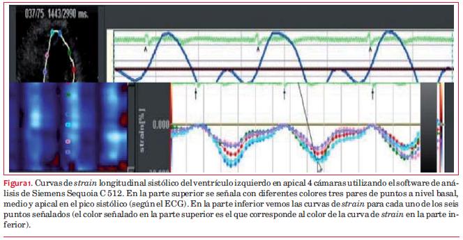
<body><![CDATA[<li><font color="#1f1a17" face="CentSchbook BT" size="2"> Embarazo.&nbsp;     </font></li>           <li><font color="#1f1a17" face="CentSchbook BT" size="2"> No consentir la utilizaci&oacute;n de datos para la difusi&oacute;n cient&iacute;fica de los  mismos.&nbsp;     </font></li>         </ul>         <p align="left"><font color="#1f1a17" face="CentSchbook BT" size="2"> Se realiz&oacute; cuestionario y examen f&iacute;sico registrando sexo, edad, PA, peso,  talla, y se calcul&oacute; el &aacute;rea de superficie corporal seg&uacute;n la f&oacute;rmula de  Dubois y Dubois<font color="#1f1a17" face="CentSchbook BT" size="1"><sup>(<a href="#28-">28</a>)</sup></font><font color="#1f1a17" face="CentSchbook BT" size="2">. Posteriormente se realiz&oacute; ecocardiograma Doppler color  y <i>strain</i> longitudinal regional y global del VI y VD.&nbsp;</font> </font></p>        <multicol gutter="18" cols="2"></multicol>     <p align="left"><font color="#1f1a17" face="CentSchbook BT" size="2"> Para el an&aacute;lisis de los datos se dividi&oacute; la poblaci&oacute;n por sexo y comparamos  los datos de SLGVI y SLGVD obtenidos de nuestra poblaci&oacute;n sana uruguaya  con otra poblaci&oacute;n de referencia internacional. El trabajo de referencia  para comparar el SLGVI es el &uacute;nico en la literatura internacional que describe  una cohorte normal utilizando el mismo ecocardi&oacute;grafo (Siemens Sequoia  C 512) y la misma metodolog&iacute;a de medici&oacute;n del <i>strain</i> (VVI)<font color="#1f1a17" face="CentSchbook BT" size="1"><sup>(<a href="#25-">25</a>)</sup></font><font color="#1f1a17" face="CentSchbook BT" size="2">. Para comparar  el SLGVD obtenido en nuestra poblaci&oacute;n con otra de referencia, no encontramos  ning&uacute;n trabajo publicado que utilice el mismo ec&oacute;grafo, por lo tanto lo  comparamos con el trabajo m&aacute;s grande publicado de valores normales en SLGVD</font><font color="#1f1a17" face="CentSchbook BT" size="1"><sup>(<a href="#27-">27</a>)</sup></font><font color="#1f1a17" face="CentSchbook BT" size="2">,  que usa similar metodolog&iacute;a (VVI).&nbsp;</font> </font></p>         <p align="left"><font color="#1f1a17" face="Candara" size="2"> <b>Estudio ecocardiogr&aacute;fico&nbsp;</b> </font></p>         <p align="left"><font color="#1f1a17" face="CentSchbook BT" size="2"> El estudio se realiz&oacute; con ecocardi&oacute;grafo Siemens Sequoia C-512 y transductor  de 2,5-4 MHz. Se realizaron enfoques convencionales para mediciones morfol&oacute;gicas  de cuatro cavidades. Como medidas funcionales del VI y VD se valor&oacute; la  media de la onda S del Doppler tisular promediando el valor pico obtenido  en el anillo lateral y septal de la v&aacute;lvula mitral y a nivel de la excursi&oacute;n  sist&oacute;lica del plano del anillo tricusp&iacute;deo. Se valor&oacute; mediante modo M la  ESAT como par&aacute;metro avalado de valoraci&oacute;n de funci&oacute;n sist&oacute;lica global del  VD. Posteriormente se obtuvieron im&aacute;genes en apical 4 y 2 c&aacute;maras con segunda  arm&oacute;nica, alta resoluci&oacute;n temporal (60-100 im&aacute;genes/s) y se&ntilde;al electrocardiogr&aacute;fica  de buena definici&oacute;n, realizando en forma <i>off-line</i> la cuantificaci&oacute;n de  la FEVI: se calcularon los vol&uacute;menes telediast&oacute;licos y telesist&oacute;licos del  VI mediante m&eacute;todo de Simpson modificado, y el <i>strain</i> longitudinal sist&oacute;lico  (valor pico) regional y global (tomando bordes endoc&aacute;rdicos), calculando  los valores sist&oacute;licos m&aacute;ximos de cada regi&oacute;n: basal, medio y apical de  los enfoques 2 y 4 c&aacute;maras. La media de cada grupo de los seis valores  de 2 y 4 c&aacute;maras corresponden al <i>strain</i> longitudinal global (SLG) de 2  y 4 c&aacute;maras, respectivamente, y la media de los valores de los 12 segmentos  se consider&oacute; el valor del SLG (ver <a name="f1."></a><a href="#f1">figura 1</a>). El SLG del VD se cuantific&oacute;  en apical 4 c&aacute;maras, se eligieron tres pares de puntos endoc&aacute;rdicos (basal,  medio y apical) y la media de los valores de los seis segmentos se consider&oacute;  el valor general del VD (SLG VD). Tanto para el an&aacute;lisis del SLG VI como  para SLG VD se excluyeron los puntos con mala ac&uacute;stica.&nbsp; </font></p>         <p align="left">    <br> </p>         <p><a name="f1"></a><img style="width: 664px; height: 345px;" alt="" src="/img/revistas/ruc/v31n2/2a08f1.jpg"><a href="#f1."></a></p>         ]]></body>
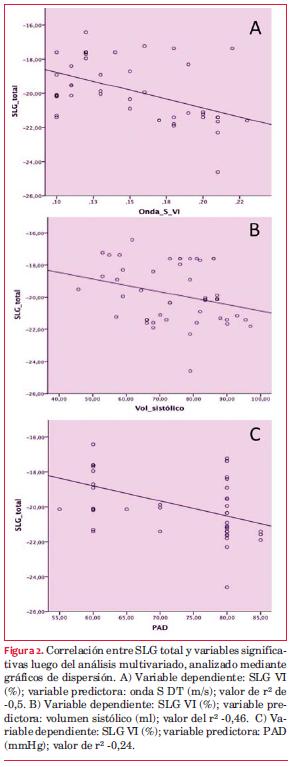
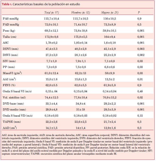
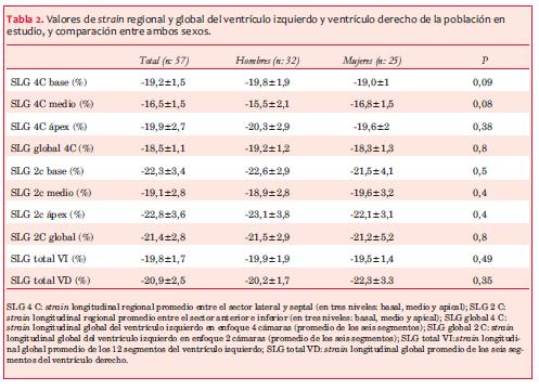
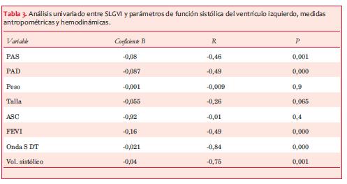
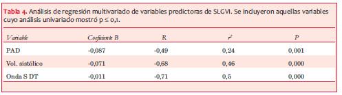
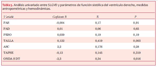
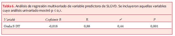
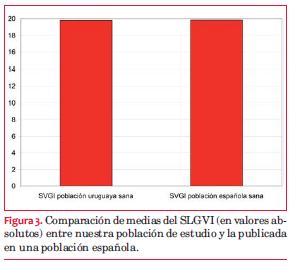
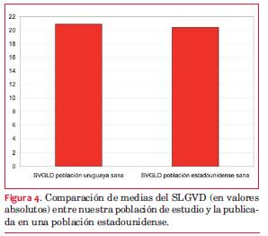
<body><![CDATA[<p></p>         <p align="left"><font color="#1f1a17" face="CentSchbook BT" size="2"> Previo a la realizaci&oacute;n de este estudio se realiz&oacute; un trabajo de<i> factibilidad  y</i><i> </i><i>reproducibilidad intra e interobservador </i>de<i> </i>SLGVI que avalara la utilizaci&oacute;n  de este m&eacute;todo en trabajos cient&iacute;ficos. De los resultados destacamos una  excelente factibilidad de los segmentos analizados, 148 de 150 totales  (98,7), y una excelente reproducibilidad intra e interobservador, valorado  por el coeficiente de correlaci&oacute;n intraclase, ambos con valores superiores  a 0,75<font color="#1f1a17" face="CentSchbook BT" size="1"><sup>(<a href="#29-">29</a>)</sup></font><font color="#1f1a17" face="CentSchbook BT" size="2">.</font><font color="#1f1a17" face="CentSchbook BT" size="1"><sup><i>&nbsp;</i></sup></font> </font></p>         <p align="left"><font color="#1f1a17" face="Candara" size="2"> <b>An&aacute;lisis estad&iacute;stico&nbsp;</b> </font></p>         <p align="left"><font color="#1f1a17" face="CentSchbook BT" size="2"> Las variables cualitativas se expresan como valor absoluto (%), las cuantitativas  en media &plusmn; DS. Se comprob&oacute; normalidad con test de Shapiro-Wilk, por lo  que el an&aacute;lisis comparativo entre subgrupos se realiz&oacute; con test de t de  Student para muestras independientes. Se realiz&oacute; correlaci&oacute;n de Pearson  entre SLG VI y SLG VD con par&aacute;metros antropom&eacute;tricos, hemodin&aacute;micos y funcionales  del VI y VD, y an&aacute;lisis uni y multivariado incluyendo medidas funcionales  del VI y VD. Los valores de SLG VI y VD se compararon con otras medidas  de referencia internacional mediante test de t para una muestra. Los datos  se expresan en diferencias de medias e IC95%. Se consider&oacute; significativa  una p &lt; 0,05. Los c&aacute;lculos se realizaron con el paquete estad&iacute;stico SPSS  v20 (SPSS Inc. Chicago, Illinois, Estados Unidos). Dados los antecedentes  del equipo respecto a la relaci&oacute;n entre la FEVI y el <i>strain </i>longitudinal  apical, <font color="#1f1a17" face="CentSchbook BT" size="2">como forma de an&aacute;lisis </font><font color="#1f1a17" face="CentSchbook BT" size="2"><i>post-hoc</i></font><font color="#1f1a17" face="CentSchbook BT" size="2"> analizamos la correlaci&oacute;n entre  la FEVI y la onda S del Doppler tisular, y la correlaci&oacute;n de esta &uacute;ltima  con el </font><font color="#1f1a17" face="CentSchbook BT" size="2"><i>strain</i></font><font color="#1f1a17" face="CentSchbook BT" size="2"> longitudinal regional apical de 4 c&aacute;maras.&nbsp;</font> </font></p>          <p>&nbsp;</p>     <multicol gutter="18" cols="2"></multicol>     <p align="left"><font color="#d62437" face="Candara" size="2"> <b>Resultados&nbsp;</b> </font></p>         <p align="left"><font color="#1f1a17" face="CentSchbook BT" size="2"> <font color="#1f1a17" face="CentSchbook BT" size="2">Se incluy&oacute; un total de 57 sujetos, de los cuales 25 (43,8%) eran mujeres.  La edad fue de 35,6&nbsp;&plusmn;&nbsp;9,9 a&ntilde;os. En la <a name="t1."></a><a href="#t1.">tabla 1</a> se indican las caracter&iacute;sticas  demogr&aacute;ficas, antropom&eacute;tricas, hemodin&aacute;micas y ecocardiogr&aacute;ficas (anat&oacute;micas  y funcionales) de esta poblaci&oacute;n. Los hombres presentaron medidas antropom&eacute;tricas  y anat&oacute;micas ecocardiogr&aacute;ficas estad&iacute;sticamente m&aacute;s altas que las mujeres;  por el contrario, no hubo diferencias estad&iacute;sticamente significativas en  la FEVI, onda S del Doppler tisular del VI y VD y la excursi&oacute;n sist&oacute;lica  del plano de anillo tricusp&iacute;deo (TAPSE). Los valores de SLG y regional  del VI y VD se exponen en la <a name="t2."></a><a href="#t2"><span style="text-decoration: underline;">tabla 2</span></a>. Se analizaron 645 de los 684 segmentos  pautados del </font><font color="#1f1a17" face="CentSchbook BT" size="2"><i>strain</i></font><font color="#1f1a17" face="CentSchbook BT" size="2"> longitudinal del VI y 314 de los 342 del VD. No hubo  diferencias estad&iacute;sticamente significativas entre la media de los valores  de </font><font color="#1f1a17" face="CentSchbook BT" size="2"><i>strain</i></font><font color="#1f1a17" face="CentSchbook BT" size="2"> regional y global del VI y VD entre sexos. En cuanto a la distribuci&oacute;n  de los valores a nivel regional del VI, tanto en 2 como en 4 c&aacute;maras se  registra un mismo patr&oacute;n de distribuci&oacute;n del </font><font color="#1f1a17" face="CentSchbook BT" size="2"><i>strain</i></font><font color="#1f1a17" face="CentSchbook BT" size="2"> longitudinal regional,  con el valor m&aacute;s negativo a nivel del &aacute;pex, luego la base y el valor menos  negativo en nivel medio, explicado por la orientaci&oacute;n helicoidal de las  fibras mioc&aacute;rdicas. Tambi&eacute;n se evidencia que el valor del SLG y regional  del VI de 2 c&aacute;maras es m&aacute;s negativo (mayor deformaci&oacute;n) que el encontrado  en el SLG y regional del VI de 4 c&aacute;maras. La correlaci&oacute;n entre el SLGVI  con otras medidas funcionales ventriculares y medidas antropom&eacute;tricas se  muestra en las <a name="t3."></a><a href="#t3">tablas 3</a> y <a name="t4."></a><a href="#t4">4</a>. En el an&aacute;lisis univariado se comprob&oacute; una  fuerte correlaci&oacute;n estad&iacute;stica entre el SLGVI y el pico de onda S del Doppler  tisular (R: -0,84; p&lt;0,001), el volumen sist&oacute;lico (R: 0,75; p: 0,001),</font><font color="#1f1a17" face="CentSchbook BT" size="2"><i>  </i></font><font color="#1f1a17" face="CentSchbook BT" size="2">y d&eacute;bil con la FEVI (R: -0,49; p: 0,001), PA diast&oacute;lica (PAD) (R: -0,46;  p:</font><font color="#1f1a17" face="CentSchbook BT" size="2"><i> </i></font><font color="#1f1a17" face="CentSchbook BT" size="2">0,001) y PA sist&oacute;lica (PAS) (R: -0,49; p:&lt;0,001). En an&aacute;lisis multivariado  los &uacute;nicos predictores del SLGVI fueron la onda S del Doppler tisular (B=-0,011;  p&lt;0,001), volumen sist&oacute;lico (B=-0,071; p&lt;0,001) y PAD (B=-0,087; p=0,001)  <a name="f2."></a>(<a href="#f2">figura 2</a>). La correlaci&oacute;n entre el SLGVD con medidas antropom&eacute;tricas y  funcionales del VD se muestra en las <a name="t5."></a><a href="#t5">tablas 5</a> y <a name="t6."></a><a href="#t6">6</a>. En el an&aacute;lisis multivariado  el &uacute;nico predictor result&oacute; ser la onda S del Doppler tisular (R: -0,88,  r</font><font color="#1f1a17" face="CentSchbook BT" size="1"><sup>2</sup></font><font color="#1f1a17" face="CentSchbook BT" size="2">: 0,44; p: 0,001). Al comparar estos valores con datos internacionales</font><font color="#1f1a17" face="CentSchbook BT" size="1"><sup>(25)  </sup></font><font color="#1f1a17" face="CentSchbook BT" size="2">vemos que nuestro valor de media de SLGVI fue de -19,8% &plusmn; 1,7%, mientras  que el de la poblaci&oacute;n espa&ntilde;ola fue de -19,84% &plusmn; 2,9%, con diferencia de  medias de 0,04 (IC95%: -0,51, &ndash; 0,52; p: 0,98), marcando una sorprendente  similitud en cuanto a valores de referencia <span style="text-decoration: underline;"><a name="f3."></a>(<a href="#f3">figura 3</a>)</span>. En referencia al  SLGVD, se compar&oacute; con una poblaci&oacute;n estadounidense de referencia</font><font color="#1f1a17" face="CentSchbook BT" size="1"><sup>(<a href="#27-">27</a>)</sup></font><font color="#1f1a17" face="CentSchbook BT" size="2"> y  encontramos que la media fue de -20,9% &plusmn; 2,5% versus -20,4 &plusmn; 3,2% en esta  &uacute;ltima. Las diferencias de medias fue para el VD (%): -0,5 (95%IC -1,2  &ndash; 0,17; p: 0,13) <a name="f4."></a>(<a href="#f4"><span style="text-decoration: underline;">figura 4</span></a>).&nbsp;&nbsp;&nbsp;&nbsp;&nbsp;</font> </font>   <basefont size="3"> </p>         <p align="left"><font color="#1f1a17" face="CentSchbook BT" size="1"> <font color="#1f1a17" face="Candara" size="2"><b><a name="t1"></a><a href="#t1."></a></b></font><img style="width: 663px; height: 682px;" alt="" src="/img/revistas/ruc/v31n2/2a08t1.jpg"></font></p>     <p align="left"><font color="#1f1a17" face="CentSchbook BT" size="1">&nbsp;</font><basefont size="3"> </p>         <p align="left"><font color="#1f1a17" face="CentSchbook BT" size="1"> <font color="#1f1a17" face="CentSchbook BT" size="1"><b><a name="t2"></a><a href="#t2."></a></b><img style="width: 497px; height: 360px;" alt="" src="/img/revistas/ruc/v31n2/2a08t2.jpg"></font></font></p>     ]]></body>
<body><![CDATA[<p align="left"><font color="#1f1a17" face="CentSchbook BT" size="1"><font color="#1f1a17" face="CentSchbook BT" size="1"></font>&nbsp;</font><basefont size="3"> </p>         <p align="left"><font color="#1f1a17" face="Candara" size="2"> <font color="#1f1a17" face="Candara" size="2"><b><a name="t3"></a><img style="width: 497px; height: 263px;" alt="" src="/img/revistas/ruc/v31n2/2a08t3.jpg"><a href="#t3."></a></b></font><span style="font-weight: bold;"></span></font></p>     <p align="left"><font color="#1f1a17" face="Candara" size="2"><span style="font-weight: bold;"></span> </font></p>    <basefont size="3">     <p align="left"><font color="#1f1a17" face="Candara" size="2"> <font color="#1f1a17" face="Candara" size="2"><b><a name="t4"></a><img style="width: 497px; height: 139px;" alt="" src="/img/revistas/ruc/v31n2/2a08t4.jpg"><a href="#t4."></a></b></font></font></p>     <p align="left"><font color="#1f1a17" face="Candara" size="2"> </font></p>    <basefont size="3">     <p align="left"><font color="#1f1a17" face="Candara" size="2"> <font color="#1f1a17" face="Candara" size="2"><b><a name="t5"></a><img style="width: 599px; height: 257px;" alt="" src="/img/revistas/ruc/v31n2/2a08t5.jpg"><a href="#t5."></a></b></font><span style="font-weight: bold;"></span></font></p>     <p align="left"><font color="#1f1a17" face="Candara" size="2"><span style="font-weight: bold;"></span> </font></p>    <basefont size="3">     <p align="left"><font color="#1f1a17" face="Candara" size="2"> <font color="#1f1a17" face="Candara" size="2"><b><a name="t6"></a><img style="width: 599px; height: 124px;" alt="" src="/img/revistas/ruc/v31n2/2a08t6.jpg"><a href="#t6."></a></b></font></font></p>     <p align="left"><font color="#1f1a17" face="Candara" size="2"> </font></p>        <br>        ]]></body>
<body><![CDATA[<p>    <br>   <a name="f2"></a>&nbsp;<a href="#t2."></a><img style="width: 297px; height: 767px;" alt="" src="/img/revistas/ruc/v31n2/2a08f2.jpg"></p>         <p>&nbsp;    <br>  <a name="f3"></a>&nbsp;<a href="#f3."></a><img style="width: 297px; height: 262px;" alt="" src="/img/revistas/ruc/v31n2/2a08f3.jpg"> </p>         <p></p>         <p>    <br>  <a name="f4"></a>&nbsp;<a href="#f4."></a><img style="width: 298px; height: 262px;" alt="" src="/img/revistas/ruc/v31n2/2a08f4.jpg"></p>         <p></p>     <multicol gutter="18" cols="2"></multicol>     <p align="left"><font color="#d62437" face="Candara" size="2"> <b>Discusi&oacute;n&nbsp;</b> </font></p>         <p align="left"><font color="#1f1a17" face="CentSchbook BT" size="2"> Los hallazgos del presente trabajo son el resultado de un trabajo met&oacute;dico  utilizando una t&eacute;cnica innovadora, sin experiencia en el medio y con escasa  bibliograf&iacute;a internacional. Consideramos que se trata de una propuesta  original para la regi&oacute;n que aporta valores de referencia nacionales para  el SLGVI y SLGVD. Discutiremos los resultados en base a los objetivos propuestos.&nbsp; </font></p>         ]]></body>
<body><![CDATA[<p align="left"><font color="#1f1a17" face="Candara" size="2"> <b>Valores de referencia de la deformaci&oacute;n mioc&aacute;rdica del ventr&iacute;culo izquierdo  y derecho&nbsp;</b> </font></p>         <p align="left"><font color="#1f1a17" face="CentSchbook BT" size="2"> Si bien existen varios tipos de deformaci&oacute;n mioc&aacute;rdica, longitudinal, radial  y circunferencial, hay consenso en cuanto a que el par&aacute;metro de mayor aplicabilidad  cl&iacute;nica y proyecci&oacute;n es el SLG<font color="#1f1a17" face="CentSchbook BT" size="1"><sup>(<a href="#19-">19</a>)</sup></font><font color="#1f1a17" face="CentSchbook BT" size="2">, por lo que centramos la investigaci&oacute;n  en este. Seg&uacute;n la gu&iacute;a de ecocardiograf&iacute;a actual es necesaria la b&uacute;squeda  de consenso sobre los valores de SLG y los laboratorios deber&iacute;an establecer  valores propios de normalidad. En el caso del VVI la evidencia es escasa,  y los m&eacute;todos heterog&eacute;neos midiendo SLG a nivel del subepicardio o del  subendocardio no contemplan la medici&oacute;n del <i>strain</i> en los 17 segmentos  del VI. Trabajos previos en poblaci&oacute;n normal utilizan cohortes de pacientes  remitidos por indicaci&oacute;n cl&iacute;nica a realizarse examen ecocardiogr&aacute;fico,  incluyendo aquellos que resultaran normales; sin embargo, las recomendaciones  de valores de referencia ecocardiogr&aacute;ficos deber&iacute;an obtenerse a partir  de una muestra aleatoria de voluntarios sanos. Existe solamente un trabajo  para el VI y el VD que tome en cuenta todos los segmentos ventriculares,  que analice solamente el subendocardio y que la cohorte sean voluntarios  sanos reclutados para este estudio</font><font color="#1f1a17" face="CentSchbook BT" size="1"><sup>(<a href="#27-">27)</a></sup></font><font color="#1f1a17" face="CentSchbook BT" size="2">. Nuestra muestra fue obtenida de  forma exigente en cuanto a criterios de inclusi&oacute;n con el objetivo de incluir  a personas sanas, evitando sesgos en los valores.&nbsp;</font> </font></p>          <p>&nbsp;</p>     <multicol gutter="18" cols="2"></multicol>     <p align="left"><font color="#1f1a17" face="CentSchbook BT" size="2"> <font color="#1f1a17" face="CentSchbook BT" size="2">El valor obtenido de SLGVI (-19,8% &plusmn; 1,7%) es comparable con el referido  en el metaan&aacute;lisis m&aacute;s importante publicado</font><font color="#1f1a17" face="CentSchbook BT" size="1"><sup>(<a href="#20-">20</a>)</sup></font><font color="#1f1a17" face="CentSchbook BT" size="2">, sobre un total de 24 estudios  con casi 2.600 pacientes, cuya media fue de -19,7% &plusmn; 2,1%. Debemos recordar  que este an&aacute;lisis incluye una poblaci&oacute;n heterog&eacute;nea y utiliza diferentes  modo y forma de medici&oacute;n del </font><font color="#1f1a17" face="CentSchbook BT" size="2"><i>strain</i></font><font color="#1f1a17" face="CentSchbook BT" size="2">. Sin embargo, cuando comparamos los  datos con un trabajo con igual metodolog&iacute;a al nuestro</font><font color="#1f1a17" face="CentSchbook BT" size="1"><sup>(35)</sup></font><font color="#1f1a17" face="CentSchbook BT" size="2">, encontramos  tambi&eacute;n similitudes sorprendentes. El valor de media de SLG en nuestra  poblaci&oacute;n, por VVI, es, en consecuencia, comparable al de la literatura,  generando confiabilidad en el resultado.&nbsp;</font> </font></p>      <multicol gutter="18" cols="2"></multicol>     <p align="left"><font color="#1f1a17" face="CentSchbook BT" size="2"> <font color="#1f1a17" face="CentSchbook BT" size="2">En cuanto a la distribuci&oacute;n de los valores a nivel regional del VI, tanto  en 2 como en 4 c&aacute;maras se destaca un mismo patr&oacute;n de distribuci&oacute;n del SLG  regional, donde el valor m&aacute;s negativo est&aacute; a nivel del &aacute;pex, luego la base  y el valor menos negativo en el medio. Tambi&eacute;n evidenciamos que el valor  del SLG y regional del VI de 2 c&aacute;maras es m&aacute;s negativo (mayor deformaci&oacute;n)  que el encontrado en el SLG y regional del VI de 4 c&aacute;maras.</font><font color="#1f1a17" face="CentSchbook BT" size="1"><sup> </sup></font><font color="#1f1a17" face="CentSchbook BT" size="2">Con el SLGVD  sucedi&oacute; lo mismo en cuanto a comparabilidad</font><font color="#1f1a17" face="CentSchbook BT" size="1"><sup>(<a href="#27-">27</a>)</sup></font><font color="#1f1a17" face="CentSchbook BT" size="2">.&nbsp;</font> </font></p>         <p align="left"><font color="#1f1a17" face="Candara" size="2"> <b>Efecto demogr&aacute;fico en la deformaci&oacute;n mioc&aacute;rdica&nbsp;</b> </font></p>         <p align="left"><font color="#1f1a17" face="CentSchbook BT" size="2"> Si bien hasta hace cinco a&ntilde;os el efecto de la edad en la deformaci&oacute;n era  un tema debatido, actualmente se acepta que no hay efecto significativo  de la edad sobre los valores normales del SLG<font color="#1f1a17" face="CentSchbook BT" size="1"><sup>(<a href="#20-">20</a>,<a href="#21-">21</a></sup></font><font color="#1f1a17" face="CentSchbook BT" size="1"><sup>)</sup></font><font color="#1f1a17" face="CentSchbook BT" size="2">. Nuestra cohorte no  nos permite valorar esta hip&oacute;tesis por el estrecho rango de edades.&nbsp;</font> </font></p>         <p align="left"><font color="#1f1a17" face="CentSchbook BT" size="2"> Lo que s&iacute; es un tema de debate actual, con evidencias contrapuestas, es  el efecto del sexo en la deformaci&oacute;n mioc&aacute;rdica longitudinal del VI. Hay  trabajos que lo niegan<font color="#1f1a17" face="CentSchbook BT" size="1"><sup>(<a href="#20-">20</a>,<a href="#22-">22</a>,<a name="32--"></a><a href="#32-">32</a>)</sup></font><font color="#1f1a17" face="CentSchbook BT" size="2">, y otros que se&ntilde;alan que las mujeres tienen  un SLG entre 1%-2% m&aacute;s negativo que los hombres</font><font color="#1f1a17" face="CentSchbook BT" size="1"><sup>(21,<a name="33--"></a>33)</sup></font><font color="#1f1a17" face="CentSchbook BT" size="2">,</font><font color="#1f1a17" face="CentSchbook BT" size="1"><sup> </sup></font><font color="#1f1a17" face="CentSchbook BT" size="2">encontrando esta  discrepancia en la escasa informaci&oacute;n que existe para el VD</font><font color="#1f1a17" face="CentSchbook BT" size="1"><sup>(<a href="#27-">27</a>,<a name="34--"></a><a href="#34-">3</a><a href="#34-">4</a>,<a name="35--"></a><a href="#35-">35</a>)</sup></font><font color="#1f1a17" face="CentSchbook BT" size="2">.  En nuestra serie tampoco encontramos diferencias por sexo en SLGVI y SLGVD.&nbsp;</font> </font></p>         <p align="left"><font color="#1f1a17" face="Candara" size="2"> <b>Efecto de las medidas antropom&eacute;tricas y factores hemodin&aacute;micos en la determinaci&oacute;n  del SLG&nbsp;</b> </font></p>         <p align="left"><font color="#1f1a17" face="CentSchbook BT" size="2"> Si bien en estudios importantes sobre valores de referencia para adultos  sanos del SLGVI estos factores no demostraron ser predictores determinantes<font color="#1f1a17" face="CentSchbook BT" size="1"><sup>(<a href="#21-">21</a>,<a href="#27-">27</a>,<a href="#33-">33</a>)</sup></font><font color="#1f1a17" face="CentSchbook BT" size="2">,  el metaan&aacute;lisis de Yingchoncharoen</font><font color="#1f1a17" face="CentSchbook BT" size="1"><sup>(<a href="#20-">20</a>)</sup></font><font color="#1f1a17" face="CentSchbook BT" size="1"><sup> </sup></font><font color="#1f1a17" face="CentSchbook BT" size="2">mostr&oacute; una asociaci&oacute;n de la PAS  con el SLGVI como &uacute;nico factor determinante del mismo, no encontr&aacute;ndose  que los valores antropom&eacute;tricos est&eacute;n asociados.&nbsp;</font> </font></p>         ]]></body>
<body><![CDATA[<p align="left"><font color="#1f1a17" face="CentSchbook BT" size="2"> A nivel del SLGVD ninguno de los factores hemodin&aacute;micos y antropom&eacute;tricos  ha demostrado tener capacidad de predicci&oacute;n de la deformaci&oacute;n mioc&aacute;rdica  del VD<font color="#1f1a17" face="CentSchbook BT" size="1"><sup>(<a href="#27-">27</a>-<a href="#35-">35</a>)</sup></font><font color="#1f1a17" face="CentSchbook BT" size="2">.&nbsp;</font> </font></p>         <p align="left"><font color="#1f1a17" face="CentSchbook BT" size="2"> Nuestros hallazgos tampoco muestran que los valores antropom&eacute;tricos sean  factores determinantes del SLG del VI y VD en una poblaci&oacute;n sana. En cuanto  a los factores hemodin&aacute;micos como determinantes del SLGVD, no hubo factores  de predicci&oacute;n, pero a nivel del SLGVI la PAD fue el &uacute;nico factor determinante,  con un r<font color="#1f1a17" face="CentSchbook BT" size="1"><sup>2</sup></font><font color="#1f1a17" face="CentSchbook BT" size="2"></font><font color="#1f1a17" face="CentSchbook BT" size="1"><sup> </sup></font><font color="#1f1a17" face="CentSchbook BT" size="2">de 0,24, significando que solamente un cuarto del SLGVI depende  de esta variable. Esta vinculaci&oacute;n de la PAD con el SLGVI no est&aacute; referida  en la literatura. &iquest;Por qu&eacute; este par&aacute;metro hemodin&aacute;mico de la di&aacute;stole podr&iacute;a  influir sobre un par&aacute;metro tan sensible de la funci&oacute;n sist&oacute;lica del VI  como el SLG? Analizando la fisiolog&iacute;a del ciclo card&iacute;aco desde el punto  de vista del bucle presi&oacute;n-volumen y su correlaci&oacute;n con las curvas de presi&oacute;n  a&oacute;rtica, vemos que la PAD corresponde al inicio del per&iacute;odo eyectivo sist&oacute;lico,  fase en la cual el ventr&iacute;culo se ve independizado de otras condiciones  de carga; por lo tanto, modificaciones de la PAD podr&iacute;an determinar cambios  en la funci&oacute;n sist&oacute;lica precoz.&nbsp;</font> </font></p>         <p align="left"><font color="#1f1a17" face="CentSchbook BT" size="2"> Con respecto al an&aacute;lisis de correlaci&oacute;n entre el <i>strain</i> y la PA, remarcamos  que a diferencia del <i>strain</i> longitudinal sist&oacute;lico, el <i>strain rate</i> es el  &iacute;ndice que es menos dependiente de las condiciones de carga, &iacute;ndice no  estimado en este trabajo y que deber&iacute;a ser incluido en futuras investigaciones.&nbsp; </font></p>         <p align="left"><font color="#1f1a17" face="Candara" size="2"> <b>Par&aacute;metros ecocardiogr&aacute;ficos de funci&oacute;n ventricular sist&oacute;lica como predictores  determinante del strain longitudinal global&nbsp;</b> </font></p>         <p align="left"><font color="#1f1a17" face="CentSchbook BT" size="2"> En la evaluaci&oacute;n de la funci&oacute;n sist&oacute;lica del VI el uso de nuevas t&eacute;cnicas  ecocardiogr&aacute;ficas ha demostrado que el SLG es un buen predictor de disfunci&oacute;n  sist&oacute;lica precoz, aunque su correlaci&oacute;n con la FEVI medida por ecocardiograma  bidimensional es d&eacute;bil<font color="#1f1a17" face="CentSchbook BT" size="1"><sup>(36,37)</sup></font><font color="#1f1a17" face="CentSchbook BT" size="2">, dado que enfocan aspectos diferentes, uno  es &iacute;ndice de funci&oacute;n de c&aacute;mara y el otro de fibra.&nbsp;</font> </font></p>         <p align="left"><font color="#1f1a17" face="CentSchbook BT" size="2"> <font color="#1f1a17" face="CentSchbook BT" size="2">Recientemente, publicamos</font><font color="#1f1a17" face="CentSchbook BT" size="1"><sup>(<a href="#30-">30</a>)</sup></font><font color="#1f1a17" face="CentSchbook BT" size="2"> que en una poblaci&oacute;n sana utilizando VVI</font><font color="#1f1a17" face="CentSchbook BT" size="2"><i>  </i></font><font color="#1f1a17" face="CentSchbook BT" size="2">hab&iacute;a una d&eacute;bil correlaci&oacute;n entre la FEVI y el SLGVI, y que el &uacute;nico predictor  de FEVI result&oacute; ser el SLG regional apical de 4 c&aacute;maras, proponi&eacute;ndolo,  en el escenario de FEVI normal, como &uacute;nica medida de </font><font color="#1f1a17" face="CentSchbook BT" size="2"><i>strain</i></font><font color="#1f1a17" face="CentSchbook BT" size="2"> por su capacidad  predictiva y f&aacute;cil acceso. Profundizando en el an&aacute;lisis del valor del </font><font color="#1f1a17" face="CentSchbook BT" size="2"><i>strain  </i></font><font color="#1f1a17" face="CentSchbook BT" size="2">con la funci&oacute;n sist&oacute;lica, corroboramos los hallazgos previos entre el SLGVI  y la FEVI</font><font color="#1f1a17" face="CentSchbook BT" size="1"><sup>(<a href="#30-">30</a>)</sup></font><font color="#1f1a17" face="CentSchbook BT" size="2">, destacando que del an&aacute;lisis de regresi&oacute;n multivariado, tanto  la onda S del Doppler tisular como el volumen sist&oacute;lico del VI presentaron  capacidad de predicci&oacute;n estad&iacute;sticamente significativa con el SLGVI, marc&aacute;ndose  que por cada disminuci&oacute;n de 0,01 m/s del valor del Doppler tisular el SLG  se reduce 0,011 (0,071%). Si analizamos el valor estad&iacute;stico de estos hallazgos  (en base al an&aacute;lisis del r</font><font color="#1f1a17" face="CentSchbook BT" size="1"><sup>2</sup></font><font color="#1f1a17" face="CentSchbook BT" size="2">) vemos que hay una dependencia entre el SLGVI  y estos valores en el entorno de 50%, marcando una asociaci&oacute;n estad&iacute;sticamente  fuerte. Dado que el DT y el </font><font color="#1f1a17" face="CentSchbook BT" size="2"><i>strain</i></font><font color="#1f1a17" face="CentSchbook BT" size="2"> tienen su fortaleza en el escenario  de etapas precoces de la disfunci&oacute;n sist&oacute;lica, no llama la atenci&oacute;n la  asociaci&oacute;n encontrada.&nbsp;</font> </font></p>         <p align="left"><font color="#1f1a17" face="CentSchbook BT" size="2"> <font color="#1f1a17" face="CentSchbook BT" size="2">En cuanto al VD no hay datos de la literatura respecto a predictores de  funci&oacute;n sist&oacute;lica por este m&eacute;todo. En tal sentido, los resultados fueron  innovadores dado que evidenciaron la capacidad predictora del Doppler tisular  del VD y del ESAT, con el SLGVD, demostrando el an&aacute;lisis de regresi&oacute;n multivariado  que el &uacute;nico factor estad&iacute;sticamente significativo fue la onda S del Doppler  tisular; cada disminuci&oacute;n de 0,01 m/s del valor del Doppler tisular, el  SLG se reduce 0,018% (o sea, se hace menos negativo), y dado el valor del  r</font><font color="#1f1a17" face="CentSchbook BT" size="2"></font><font color="#1f1a17" face="CentSchbook BT" size="1"><sup>2</sup></font><font color="#1f1a17" face="CentSchbook BT" size="2"> vemos una asociaci&oacute;n de dependencia estad&iacute;stica de aproximadamente 50.  Si analizamos los valores de SLGVI y VD con el Doppler tisular de cada  ventr&iacute;culo vemos que hubo paralelismo entre ambos, con una importante correlaci&oacute;n  entre el SLG y el Doppler tisular. Esta es una hip&oacute;tesis motivadora para  seguir profundizando en el tema, marcando su aplicabilidad en un grupo  importante de pacientes para la detecci&oacute;n precoz de la disfunci&oacute;n sist&oacute;lica.&nbsp;</font> </font></p>          <p>  <multicol gutter="18" cols="2"></multicol> </p>        <p align="left"><font color="#1f1a17" face="Candara" size="2"> <b>Limitaciones&nbsp;</b> </font></p>     <ul>           <li><font color="#1f1a17" face="CentSchbook BT" size="2"> La poblaci&oacute;n estudiada tiene un estrecho rango etario.&nbsp;     </font></li>           ]]></body>
<body><![CDATA[<li><font color="#1f1a17" face="CentSchbook BT" size="2"> La muestra es de conveniencia y no obtenida en forma aleatoria.&nbsp;     </font></li>           <li><font color="#1f1a17" face="CentSchbook BT" size="2"> Los resultados se limitan al m&eacute;todo VVI y de acuerdo al m&eacute;todo de obtenci&oacute;n  explicado. No es generalizable a otras modalidades de <i>speckle tracking</i>.&nbsp;     </font></li>           <li><font color="#1f1a17" face="CentSchbook BT" size="2"> El tama&ntilde;o muestral es aceptable para una precisi&oacute;n de 90% en el resultado  obtenido, aunque el <i>n</i> es peque&ntilde;o y de un &uacute;nico centro.&nbsp;     </font></li>          <br>         </ul>         <p align="left"><font color="#d62437" face="Candara" size="2"> <b>Conclusiones&nbsp;</b> </font></p>         <p align="left"><font color="#1f1a17" face="CentSchbook BT" size="2"> Se evalu&oacute; la deformaci&oacute;n normal del miocardio en una cohorte de voluntarios  sanos por m&eacute;todo de <i>strain</i> (VVI), valores de SLG y regional del VI y VD  como lo exigen las recomendaciones actuales.&nbsp; </font></p>         <p align="left"><font color="#1f1a17" face="CentSchbook BT" size="2"> Se presentan valores de normalidad de SLG de VI y VD para una poblaci&oacute;n  sana uruguaya y se compara con la evidencia internacional, no encontrando  diferencias significativas en cuanto a la media de SLG del VI y VD.&nbsp; </font></p>         <p align="left"><font color="#1f1a17" face="CentSchbook BT" size="2"> Se demuestra la capacidad de predicci&oacute;n sobre el SLGVI de la PAD, el pico  de onda S del Doppler tisular del VI y el volumen sist&oacute;lico y del Doppler  tisular del VD sobre el SLGVD. Estos hallazgos ayudan a complementar la  informaci&oacute;n que nos brinda el <i>strain</i> y permite abrir futuras l&iacute;neas de  investigaci&oacute;n.&nbsp; </font></p>        <br>         ]]></body>
<body><![CDATA[<p align="left"><font color="#d62437" face="Candara" size="2"> <b>Bibliograf&iacute;a&nbsp;</b> </font></p>         <!-- ref --><p align="left"><font color="#1f1a17" face="CentSchbook BT" size="1"><a name="1-"></a> <a href="#1-10--">1</a>.&nbsp;&nbsp;&nbsp;&nbsp;<b>Torrent-Guasp F. </b>La estructuraci&oacute;n macrosc&oacute;pica del miocardio ventricular.  Rev Esp Cardiol 1980;33:265-87.    <font color="#1f1a17" face="Courier New" size="1">&nbsp;</font> </font></p>         <!-- ref --><p align="left"><font color="#1f1a17" face="CentSchbook BT" size="1"><a name="2-"></a> <a href="#1-10--">2</a>.&nbsp;&nbsp;&nbsp;&nbsp;<b>Torrent-Guasp F.</b> Estructura y funci&oacute;n del coraz&oacute;n. Rev Esp Cardiol 1998;  51:91-102.    <font color="#1f1a17" face="Courier New" size="1">&nbsp;</font> </font></p>         <!-- ref --><p align="left"><font color="#1f1a17" face="CentSchbook BT" size="1"><a name="3-"></a> <a href="#1-10--">3</a>.&nbsp;&nbsp;&nbsp;&nbsp;<b>Torrent-Guasp F, Buckberg GD, Clemente CD, Ballester-Rod&eacute;s M, Carreras-Costa  F, Flotats A. </b>Las razones de la estructura y mec&aacute;nica del coraz&oacute;n. Rev  Lat Cardiol 2000; 21:159-67.    <font color="#1f1a17" face="Courier New" size="1">&nbsp;</font> </font></p>         <!-- ref --><p align="left"><font color="#1f1a17" face="CentSchbook BT" size="1"><a name="4-"></a> <a href="#1-10--">4</a>.&nbsp;&nbsp;&nbsp;&nbsp;<b>Torrent-Guasp F. </b>La mec&aacute;nica ventricular. Rev Lat Cardiol 2001;22:48-55.    <font color="#1f1a17" face="Courier New" size="1">&nbsp;</font> </font></p>         <!-- ref --><p align="left"><font color="#1f1a17" face="CentSchbook BT" size="1"><a name="5-"></a> <a href="#1-10--">5</a>.&nbsp;&nbsp;&nbsp;&nbsp;<b>Torrent-Guasp F, Kocica MJ, Corno AF, Komeda M, Carreras-Costa F, Flotats  A, et al. </b>Towards new understanding of the heart structure and function.  Eur J Cardiothorac Surg 2005;27:191-201.    <font color="#1f1a17" face="Courier New" size="1">&nbsp;</font> </font></p>         <!-- ref --><p align="left"><font color="#1f1a17" face="CentSchbook BT" size="1"><a name="6-"></a> <a href="#1-10--">6</a>.&nbsp;&nbsp;&nbsp;&nbsp;<b>Torrent-Guasp F, Ballester M, Buckberg GD, Carreras F, Flotats A, Carri&oacute;  I, et al.</b> Spatial orientation of the ventricular muscle band: physiologic  contribution and surgical implications. J Thorac Cardiovasc Surg 2001;122:389-92.    <font color="#1f1a17" face="Courier New" size="1">&nbsp;</font> </font></p>         <p align="left"><font color="#1f1a17" face="CentSchbook BT" size="1"><a name="7-"></a> <a href="#1-10--">7</a>.&nbsp;&nbsp;&nbsp;&nbsp;<b>Poveda F, Gil D,Mart&iacute;n E, Andaluz A, Ballester M, Carreras F.</b> Estudio  tractogr&aacute;fico de la anatom&iacute;a helicoidal del miocardio ventricular mediante  resonancia magn&eacute;tica por tensor de difusi&oacute;n. Rev Esp Cardiol 2013;66:782&ndash;90.<font color="#1f1a17" face="Courier New" size="1">&nbsp;</font> </font></p>         <!-- ref --><p align="left"><font color="#1f1a17" face="CentSchbook BT" size="1"><a name="8-"></a> <a href="#1-10--">8</a>.&nbsp;&nbsp;&nbsp;&nbsp;<b>Blessberger H, Binder T.</b> Two dimensional speckle tracking echocardiography:  basic principles. Herat 2010;96:716-22.    <font color="#1f1a17" face="Courier New" size="1">&nbsp;</font> </font></p>         <!-- ref --><p align="left"><font color="#1f1a17" face="CentSchbook BT" size="1"><a name="9-"></a> <a href="#1-10--">9</a>.&nbsp;&nbsp;&nbsp;&nbsp;<b>Sengupta PP, Krishnamoorthy VK, Korinek J,Narula J, Vannan MA, Lester  SJ, et al.</b> Left ventricular form and function revisited: applied translational  science to cardiovascular ultrasound imaging. J Am Soc Echocardiogr 2007;20:539-51.    <font color="#1f1a17" face="Courier New" size="1">&nbsp;</font> </font></p>         <!-- ref --><p align="left"><font color="#1f1a17" face="CentSchbook BT" size="1"><a name="10-"></a> <a href="#1-10--">10</a>.&nbsp;&nbsp;&nbsp;&nbsp;<b>Brown J, Jelkins C,Marwick TH. </b>Use of myocardial strain to assess global  left ventricular function: a comparison with cardiac magnetic resonance  and 3-dimensional echocardiography. Am Heart J 2009; 157:1:102e1-e5.    <font color="#1f1a17" face="Courier New" size="1">&nbsp;</font> </font></p>         ]]></body>
<body><![CDATA[<!-- ref --><p align="left"><font color="#1f1a17" face="CentSchbook BT" size="1"><a name="11-"></a> <a href="#11-15--">11</a>.&nbsp;&nbsp;&nbsp;&nbsp;<b>Pavlopoulos H, Nihoyannopoulos P. </b>Strain and strain rate deformation  parameters: from tissue Doppler to 2D speckle tracking. Int J Cardiovasc  Imaging 2008;24:479-91.    ?<font color="#1f1a17" face="Courier New" size="1">&nbsp;</font> </font></p>         <!-- ref --><p align="left"><font color="#1f1a17" face="CentSchbook BT" size="1"><a name="12-"></a> <a href="#11-15--">12</a>.&nbsp;&nbsp;&nbsp;&nbsp;<b>Marwick TH. </b>Measurement of strain and strain rate by echocardiography:  ready for prime time? J Am Coll Cardiol 2006;47:1313-27.    <font color="#1f1a17" face="Courier New" size="1">&nbsp;</font> </font></p>         <!-- ref --><p align="left"><font color="#1f1a17" face="CentSchbook BT" size="1"><a name="13-"></a> <a href="#11-15--">13</a>.&nbsp;&nbsp;&nbsp;&nbsp;<b>Perry R, De Pasquale CG, Chew DP, Joseph MX. </b>Assessment of early diastolic  left ventricular function by two-dimensional echocardiographic speckle  tracking. Eur J Echocardiogr 2008;9:791-5.    <font color="#1f1a17" face="Courier New" size="1">&nbsp;</font> </font></p>         <!-- ref --><p align="left"><font color="#1f1a17" face="CentSchbook BT" size="1"><a name="14-"></a> <a href="#11-15--">14</a>.&nbsp;&nbsp;&nbsp;&nbsp;<b>Cianciulli T, Prieto O(H), Desseno A, Stefano P. </b>Deformaci&oacute;n mioc&aacute;rdica.  En:Cianculli T, Prezioso H,Lax L. Nuevas t&eacute;cnicas en ecocardiograf&iacute;a. Bs  As: Journal; 2011. p 15-49.    <font color="#1f1a17" face="Courier New" size="1">&nbsp;</font> </font></p>         <!-- ref --><p align="left"><font color="#1f1a17" face="CentSchbook BT" size="1"><a name="15-"></a> <a href="#11-15--">15</a>.&nbsp;&nbsp;&nbsp;&nbsp;<b>Hurlburt HM, Aurigemma GP, Hill JC,Narayanan A, Gaasch WH, Vinch CS,  et al.</b> Direct ultrasound measurement of longitudinal, circumferential,  and radial strain using 2-dimensional strain imaging in normal adults.  Echocardiography. 2007; 24:723-31.    <font color="#1f1a17" face="Courier New" size="1">&nbsp;</font> </font></p>         ]]></body>
<body><![CDATA[<p align="left"><font color="#1f1a17" face="CentSchbook BT" size="1"><a name="16-"></a> <a href="#16-18--">16</a>.&nbsp;&nbsp;&nbsp;&nbsp;<b>Vasan RS, Larson MG, Benjamin EJ, Evans JC, Reiss CK, Levy D.</b> Congestive  heart failure in subjects with normal versus reduced left ventricular ejection  fraction: prevalence and mortality in a population-based cohort. J Am Coll  Cardiol 1999;33: 1948&ndash;55.<font color="#1f1a17" face="Courier New" size="1">&nbsp;</font> </font></p>      <multicol gutter="18" cols="2"></multicol>     <p align="left"><font color="#1f1a17" face="CentSchbook BT" size="1"><a name="17-"></a> <a href="#16-18--">17</a>.&nbsp;&nbsp;&nbsp;&nbsp;<b>Mondillo S, Galderisi M, Mele D, Cameli M, Lomoriello VS, Zac&agrave; V, et  al.</b> Speckle-tracking echocardiography: a new technique for assessing myocardial  function. J Ultrasound Med. 2011;30:71&ndash;83.<font color="#1f1a17" face="Courier New" size="1">&nbsp;</font> </font></p>         <p align="left"><font color="#1f1a17" face="CentSchbook BT" size="1"><a name="18-"></a> <a href="#16-18--">18</a>.&nbsp;&nbsp;&nbsp;&nbsp;<b>Mor-Avi V, Lang RM, Badano LP, Belohlavek M, Cardim NM, Derumeaux G,  et al.</b> Current and evolving echocardiographic techniques for the quantitative  evaluation of cardiac mechanics: ASE/EAE consensus statement on methodology  and indications endorsed by the Japanese Society of&nbsp;</font></p>       <p align="left"><font color="#1f1a17" face="CentSchbook BT" size="1">&nbsp; &nbsp; &nbsp; &nbsp; &nbsp; &nbsp; &nbsp; &nbsp; Echocardiography. Eur  J Echocardiogr 2011;12:167&ndash;205.<font color="#1f1a17" face="Courier New" size="1">&nbsp;</font> </font></p>         <p align="left"><font color="#1f1a17" face="CentSchbook BT" size="1"><a name="19-"></a> <a href="#19-30--">19</a>.&nbsp;&nbsp;&nbsp;&nbsp;<b>Lang RM, Badano LP, Mor-Avi V, Afilalo J, Armstrong A, Ernande L, et  al.</b> Recommendations for cardiac chamber quantification by echocardiography  in adults: an update from the American Society of Echocardiography and  the European Association of Cardiovascular Imaging. J Am Soc&nbsp;</font></p>       <p align="left"><font color="#1f1a17" face="CentSchbook BT" size="1">&nbsp; &nbsp; &nbsp; &nbsp; &nbsp; &nbsp; &nbsp; &nbsp;Echocardiogr  2015;28(1):1-39e14.<font color="#1f1a17" face="Courier New" size="1">&nbsp;</font> </font></p>         <!-- ref --><p align="left"><font color="#1f1a17" face="CentSchbook BT" size="1"><a name="20-"></a> <a href="#19-30--">20</a>.&nbsp;&nbsp;&nbsp;&nbsp;<b>Yingchoncharoen T, Agarwal S, Popovic ZB, Marwick TH. </b>Normal ranges  of left ventricular strain: a meta-analysis. J Am Soc Echocardiogr 2013;26:185-91.    <font color="#1f1a17" face="Courier New" size="1">&nbsp;</font> </font></p>         <!-- ref --><p align="left"><font color="#1f1a17" face="CentSchbook BT" size="1"><a name="21-"></a> <a href="#19-30--">21</a>.&nbsp;&nbsp;&nbsp;&nbsp;<b>Kocabay G, Muraru D, Peluso D, Cucchini U, Mihaila S, Padayattil-Jose  S, et al.</b> Mec&aacute;nica ventricular izquierda normal mediante ecocardiograf&iacute;a  speckle tracking bidimensional. Valores de referencia para adultos sanos.  Rev Esp Cardiol 2014;67:651-8.    <font color="#1f1a17" face="Courier New" size="1">&nbsp;</font> </font></p>         ]]></body>
<body><![CDATA[<p align="left"><font color="#1f1a17" face="CentSchbook BT" size="1"><a name="22-"></a> <a href="#19-30--">22</a>.&nbsp;&nbsp;&nbsp;&nbsp;<b>Reckefuss N, Butz T, Horstkotte D, Faber L. </b>Evaluation of longitudinal  and radial left ventricular function by two-dimensional speckle-tracking  echocardiography in a large cohort of normal probands. Int J Cardiovasc  Imaging 2011;27:515&ndash;26.<font color="#1f1a17" face="Courier New" size="1">&nbsp;</font> </font></p>         <p align="left"><font color="#1f1a17" face="CentSchbook BT" size="1"><a name="23-"></a> <a href="#19-30--">23</a>.&nbsp;&nbsp;&nbsp;&nbsp;<b>Marwick TH, Leano RL, Brown J, Sun JP, Hoffmann R, Lysyansky P, et al.  </b>Myocardial strain measurement with 2-dimensional speckle-tracking echocardiography:  definition of normal range. JACC Cardiovasc Imaging 2009;2:80&ndash;4.<font color="#1f1a17" face="Courier New" size="1">&nbsp;</font> </font></p>         <p align="left"><font color="#1f1a17" face="CentSchbook BT" size="1"><a name="24-"></a> <a href="#19-30--">24</a>.&nbsp;&nbsp;&nbsp;&nbsp;<b>Kuznetsova T, Herbots L, Richart T, D&rsquo;Hooge J, Thijs L, Fagard RH, et  al. </b>Left ventricular strain and strain rate in a general population. EurHeart  J. 2008;29:2014-23. <font color="#1f1a17" face="Courier New" size="1">&nbsp;</font> </font></p>         <!-- ref --><p align="left"><font color="#1f1a17" face="CentSchbook BT" size="1"><a name="25-"></a> <a href="#19-30--">25</a>.&nbsp;&nbsp;&nbsp;&nbsp;<b>Rodr&iacute;guez-Bail&oacute;n I, Jim&eacute;nez-Navarro MF, P&eacute;rez-Gonz&aacute;lez R, Garc&iacute;a-Orta  R, Morillo-Velarde R, Teresa-Galv&aacute;n E.</b> Deformaci&oacute;n ventricular izquierda  en ecocardiograf&iacute;a bidimensional: valores y tiempos en sujetos normales.  Rev Esp Cardiol 2010;63:1195-9.    <font color="#1f1a17" face="Courier New" size="1">&nbsp;</font> </font></p>         <!-- ref --><p align="left"><font color="#1f1a17" face="CentSchbook BT" size="1"><a name="26-"></a> <a href="#19-30--">26</a>.&nbsp;&nbsp;&nbsp;&nbsp;<b>Carasso S, Biaggi P, Rakowski H,Mutlak D, Lessick J, Aronson D, et al.</b>  Velocity Vector Imaging: standard tissue-tracking results acquired in normals.  The VVI-STRAIN Study. J Am Soc Echocardiogr 2012;25:543-52.    <font color="#1f1a17" face="Courier New" size="1">&nbsp;</font> </font></p>         <p align="left"><font color="#1f1a17" face="CentSchbook BT" size="1"><a name="27-"></a> <a href="#19-30--">27</a>.&nbsp;&nbsp;&nbsp;&nbsp;<b>Fine NM, Shah AA, Han I-Y, Yu Y, Hsiao J-f, Koshino Y, et al.</b> Left and  right ventricular strain and strain rate measurement in normal adults using  velocity vector imaging: an assessment of reference values and intersystem  agreement. Int J Cardiovasc Imaging 2013;29:571&ndash;80.<font color="#1f1a17" face="Courier New" size="1">&nbsp;</font> </font></p>         <p align="left"><font color="#1f1a17" face="CentSchbook BT" size="1"><a name="28-"></a> <a href="#19-30--">28</a>.&nbsp;&nbsp;&nbsp;&nbsp;<b>Du Bois D,Du Bois EF.</b>Clinicalcalorimetry.A formula to estimate the approximate  surface area if height and weight be known. Arch Intern Med (Chic) 1916;17:863&ndash;71.<font color="#1f1a17" face="Courier New" size="1">&nbsp;</font> </font></p>         <!-- ref --><p align="left"><font color="#1f1a17" face="CentSchbook BT" size="1"><a name="29-"></a> <a href="#19-30--">29</a>.&nbsp;&nbsp;&nbsp;&nbsp;<b>Parma G, Florio L.</b> Factibilidad y reproducibilidad intra e interobservador  de strainlongitudinal global por m&eacute;todo Vector VelocityImaging en una poblaci&oacute;n  adulta. Rev Urug Cardiol 2013; 28Suppl 1: 2-73.    <font color="#1f1a17" face="Courier New" size="1">&nbsp;</font> </font></p>         <p align="left"><font color="#1f1a17" face="CentSchbook BT" size="1"><a name="30-"></a> <a href="#19-30--">30</a>.&nbsp;&nbsp;&nbsp;&nbsp;<b>Parma G, Florio L, Dayan V, Martinez F, Lluberas N, Lluberas R.</b> Strain  longitudinal apical 4 c&aacute;maras por vector velocityimaging: prometedor predictor  de fracci&oacute;n de eyecci&oacute;n de ventr&iacute;culo izquierdo en sujetos sanos. Rev Esp  Cardiol 2015;68:351&ndash;2.<font color="#1f1a17" face="Courier New" size="1">&nbsp;</font> </font></p>         <p align="left"><font color="#1f1a17" face="CentSchbook BT" size="1"><a name="31-"></a> 31.&nbsp;&nbsp;&nbsp;&nbsp;<b>Sunderman FWJr. </b>Current concepts of &ldquo;normal values&rdquo;, &ldquo;reference values&rdquo;,  and &ldquo;discrimination values&rdquo;, in clinical chemistry. Clin Chem 1975;21:  1873&ndash;7.<font color="#1f1a17" face="Courier New" size="1">&nbsp;</font> </font></p>         <p align="left"><font color="#1f1a17" face="CentSchbook BT" size="1"><a name="32-"></a> <a href="#32-">32</a>.&nbsp;&nbsp;&nbsp;&nbsp;<b>Marcus KA, Mavinkurve-Groothuis AM, Barends M, vanDijk A, Feuth T, De  Korte C, et al. </b>Reference values for myocardial two-dimensional strain  echocardiography in a healthy pediatric and young adult cohort. J Am Soc  Echocardiogr 2011; 24:625&ndash;36.<font color="#1f1a17" face="Courier New" size="1">&nbsp;</font> </font></p>         <p align="left"><font color="#1f1a17" face="CentSchbook BT" size="1"><a name="33-"></a> <a href="#33--">33</a>.&nbsp;&nbsp;&nbsp;&nbsp;<b>Dalen H, Thorstensen A, Aase SA, Ingul CB, Torp H, Vatten LJ, et al.</b>  Segmental and global longitudinal strain and strain rate based on echocardiography  of 1266 healthy individuals: the HUNT study in Norway. Eur J Echocardiogr  2010;11:176&ndash;83.<font color="#1f1a17" face="Courier New" size="1">&nbsp;</font> </font></p>         <p align="left"><font color="#1f1a17" face="CentSchbook BT" size="1"><a name="34-"></a> <a href="#34--">34</a>.&nbsp;&nbsp;&nbsp;&nbsp;<b>Weidemann F, Eyskens B, Jamal F, Mertens L, Kowalski M, D&rsquo;Hooge J, et  al.</b> Quantification of regional left and right ventricular radial and longitudinal  function in healthy children using ultrasound-based strain rate and strain  imaging. J Am Soc Echocardiogr 2002;15:20&ndash;8.<font color="#1f1a17" face="Courier New" size="1">&nbsp;</font> </font></p>         <p align="left"><font color="#1f1a17" face="CentSchbook BT" size="1"><a name="35-"></a>&nbsp;<a href="#35--">35</a>.&nbsp;&nbsp;&nbsp;&nbsp;<b>Kutty S, Deatsman SL, Nugent ML, Russell D, Frommelt PC.</b> Assessment  of regional right ventricular velocities, strain, and displacement in normal  children using velocity vector imaging. Echocardiography 2008; 25:294&ndash;307.<font color="#1f1a17" face="Courier New" size="1">&nbsp;</font> </font></p>         <p align="left"><font color="#1f1a17" face="CentSchbook BT" size="1">&nbsp;36.&nbsp;&nbsp;&nbsp;&nbsp;<b>Takigiku K, Takeuchi M, Izumi C, Yuda S, Sakata K, Ohte N, et al.</b> Normal  range of left ventricular 2-dimensional strain: Japanese Ultrasound Speckle  Tracking of the Left Ventricle (JUSTICE) study. Circ J 2012;76:2623&ndash;32.<font color="#1f1a17" face="Courier New" size="1">&nbsp;</font> </font></p>         <p align="left"><font color="#1f1a17" face="CentSchbook BT" size="1">&nbsp;37.&nbsp;&nbsp;&nbsp;&nbsp;<b>Feigenbaum H, Mastouri R, Sawada S.</b> A practical approach to using strain  echocardiography to evaluate the left ventricle. Circ J 2012;76:1550&ndash;5.<font color="#1f1a17" face="Courier New" size="1">&nbsp;</font> </font></p>          <p>  </p>        ]]></body>
<body><![CDATA[ ]]></body><back>
<ref-list>
<ref id="B1">
<label>1</label><nlm-citation citation-type="journal">
<person-group person-group-type="author">
<name>
<surname><![CDATA[Torrent-Guasp]]></surname>
<given-names><![CDATA[F]]></given-names>
</name>
</person-group>
<article-title xml:lang="es"><![CDATA[La estructuración macroscópica del miocardio ventricular]]></article-title>
<source><![CDATA[Rev Esp Cardiol]]></source>
<year>1980</year>
<volume>33</volume>
<page-range>265-87</page-range></nlm-citation>
</ref>
<ref id="B2">
<label>2</label><nlm-citation citation-type="journal">
<person-group person-group-type="author">
<name>
<surname><![CDATA[Torrent-Guasp]]></surname>
<given-names><![CDATA[F]]></given-names>
</name>
</person-group>
<article-title xml:lang="es"><![CDATA[Estructura y función del corazón]]></article-title>
<source><![CDATA[Rev Esp Cardiol]]></source>
<year>1998</year>
<volume>51</volume>
<page-range>91-102</page-range></nlm-citation>
</ref>
<ref id="B3">
<label>3</label><nlm-citation citation-type="journal">
<person-group person-group-type="author">
<name>
<surname><![CDATA[Torrent-Guasp]]></surname>
<given-names><![CDATA[F]]></given-names>
</name>
<name>
<surname><![CDATA[Buckberg]]></surname>
<given-names><![CDATA[GD]]></given-names>
</name>
<name>
<surname><![CDATA[Clemente]]></surname>
<given-names><![CDATA[CD]]></given-names>
</name>
<name>
<surname><![CDATA[Ballester-Rodés]]></surname>
<given-names><![CDATA[M]]></given-names>
</name>
<name>
<surname><![CDATA[Carreras-Costa]]></surname>
<given-names><![CDATA[F]]></given-names>
</name>
<name>
<surname><![CDATA[Flotats]]></surname>
<given-names><![CDATA[A]]></given-names>
</name>
</person-group>
<article-title xml:lang="es"><![CDATA[Las razones de la estructura y mecánica del corazón]]></article-title>
<source><![CDATA[Rev Lat Cardiol]]></source>
<year>2000</year>
<volume>21</volume>
<page-range>159-67</page-range></nlm-citation>
</ref>
<ref id="B4">
<label>4</label><nlm-citation citation-type="journal">
<person-group person-group-type="author">
<name>
<surname><![CDATA[Torrent-Guasp]]></surname>
<given-names><![CDATA[F]]></given-names>
</name>
</person-group>
<article-title xml:lang="es"><![CDATA[La mecánica ventricular]]></article-title>
<source><![CDATA[Rev Lat Cardiol]]></source>
<year>2001</year>
<volume>22</volume>
<page-range>48-55</page-range></nlm-citation>
</ref>
<ref id="B5">
<label>5</label><nlm-citation citation-type="journal">
<person-group person-group-type="author">
<name>
<surname><![CDATA[Torrent-Guasp]]></surname>
<given-names><![CDATA[F]]></given-names>
</name>
<name>
<surname><![CDATA[Kocica]]></surname>
<given-names><![CDATA[MJ]]></given-names>
</name>
<name>
<surname><![CDATA[Corno]]></surname>
<given-names><![CDATA[AF]]></given-names>
</name>
<name>
<surname><![CDATA[Komeda]]></surname>
<given-names><![CDATA[M]]></given-names>
</name>
<name>
<surname><![CDATA[Carreras-Costa]]></surname>
<given-names><![CDATA[F]]></given-names>
</name>
<name>
<surname><![CDATA[Flotats]]></surname>
<given-names><![CDATA[A]]></given-names>
</name>
</person-group>
<article-title xml:lang="en"><![CDATA[Towards new understanding of the heart structure and function]]></article-title>
<source><![CDATA[Eur J Cardiothorac Surg]]></source>
<year>2005</year>
<volume>27</volume>
<page-range>191-201</page-range></nlm-citation>
</ref>
<ref id="B6">
<label>6</label><nlm-citation citation-type="">
<person-group person-group-type="author">
<name>
<surname><![CDATA[Torrent-Guasp]]></surname>
<given-names><![CDATA[F]]></given-names>
</name>
<name>
<surname><![CDATA[Buckberg]]></surname>
<given-names><![CDATA[GD]]></given-names>
</name>
<name>
<surname><![CDATA[Flotats]]></surname>
<given-names><![CDATA[A]]></given-names>
</name>
<name>
<surname><![CDATA[Carrió]]></surname>
<given-names><![CDATA[I]]></given-names>
</name>
</person-group>
<article-title xml:lang="en"><![CDATA[Spatial orientation of the ventricular muscle band: physiologic contribution and surgical implications]]></article-title>
<source><![CDATA[]]></source>
<year></year>
</nlm-citation>
</ref>
<ref id="B7">
<label>7</label><nlm-citation citation-type="journal">
<person-group person-group-type="author">
<name>
<surname><![CDATA[Poveda]]></surname>
<given-names><![CDATA[F]]></given-names>
</name>
<name>
<surname><![CDATA[Gil D,Martín]]></surname>
<given-names><![CDATA[E]]></given-names>
</name>
<name>
<surname><![CDATA[Andaluz]]></surname>
<given-names><![CDATA[A]]></given-names>
</name>
<name>
<surname><![CDATA[Ballester]]></surname>
<given-names><![CDATA[M]]></given-names>
</name>
<name>
<surname><![CDATA[Carreras]]></surname>
<given-names><![CDATA[F]]></given-names>
</name>
</person-group>
<article-title xml:lang="es"><![CDATA[Estudio tractográfico de la anatomía helicoidal del miocardio ventricular mediante resonancia magnética por tensor de difusión]]></article-title>
<source><![CDATA[Rev Esp Cardiol]]></source>
<year>2013</year>
<volume>66</volume>
<page-range>782-90</page-range></nlm-citation>
</ref>
<ref id="B8">
<label>8</label><nlm-citation citation-type="journal">
<person-group person-group-type="author">
<name>
<surname><![CDATA[Blessberger]]></surname>
<given-names><![CDATA[H]]></given-names>
</name>
<name>
<surname><![CDATA[Binder]]></surname>
<given-names><![CDATA[T]]></given-names>
</name>
</person-group>
<article-title xml:lang="en"><![CDATA[Two dimensional speckle tracking echocardiography: basic principles]]></article-title>
<source><![CDATA[Herat]]></source>
<year>2010</year>
<volume>96</volume>
<page-range>716-22</page-range></nlm-citation>
</ref>
<ref id="B9">
<label>9</label><nlm-citation citation-type="journal">
<person-group person-group-type="author">
<name>
<surname><![CDATA[Sengupta]]></surname>
<given-names><![CDATA[PP]]></given-names>
</name>
<name>
<surname><![CDATA[Krishnamoorthy]]></surname>
<given-names><![CDATA[VK]]></given-names>
</name>
<name>
<surname><![CDATA[Korinek]]></surname>
<given-names><![CDATA[J]]></given-names>
</name>
<name>
<surname><![CDATA[Narula]]></surname>
<given-names><![CDATA[J]]></given-names>
</name>
<name>
<surname><![CDATA[Vannan]]></surname>
<given-names><![CDATA[MA]]></given-names>
</name>
<name>
<surname><![CDATA[Lester]]></surname>
<given-names><![CDATA[SJ]]></given-names>
</name>
</person-group>
<article-title xml:lang="en"><![CDATA[Left ventricular form and function revisited: applied translational science to cardiovascular ultrasound imaging]]></article-title>
<source><![CDATA[J Am Soc Echocardiogr]]></source>
<year>2007</year>
<volume>20</volume>
<page-range>539-51</page-range></nlm-citation>
</ref>
<ref id="B10">
<label>10</label><nlm-citation citation-type="journal">
<person-group person-group-type="author">
<name>
<surname><![CDATA[Brown]]></surname>
<given-names><![CDATA[J]]></given-names>
</name>
<name>
<surname><![CDATA[Jelkins]]></surname>
<given-names><![CDATA[C]]></given-names>
</name>
<name>
<surname><![CDATA[Marwick]]></surname>
<given-names><![CDATA[TH]]></given-names>
</name>
</person-group>
<article-title xml:lang="en"><![CDATA[Use of myocardial strain to assess global left ventricular function: a comparison with cardiac magnetic resonance and 3-dimensional echocardiography]]></article-title>
<source><![CDATA[Am Heart J]]></source>
<year>2009</year>
<volume>157</volume>
<numero>1</numero>
<issue>1</issue>
</nlm-citation>
</ref>
<ref id="B11">
<label>11</label><nlm-citation citation-type="journal">
<person-group person-group-type="author">
<name>
<surname><![CDATA[Pavlopoulos]]></surname>
<given-names><![CDATA[H]]></given-names>
</name>
<name>
<surname><![CDATA[Nihoyannopoulos]]></surname>
<given-names><![CDATA[P]]></given-names>
</name>
</person-group>
<article-title xml:lang="en"><![CDATA[Strain and strain rate deformation parameters: from tissue Doppler to 2D speckle tracking]]></article-title>
<source><![CDATA[Int J Cardiovasc Imaging]]></source>
<year>2008</year>
<volume>24</volume>
<page-range>479-91</page-range></nlm-citation>
</ref>
<ref id="B12">
<label>12</label><nlm-citation citation-type="journal">
<person-group person-group-type="author">
<name>
<surname><![CDATA[Marwick]]></surname>
<given-names><![CDATA[TH]]></given-names>
</name>
</person-group>
<article-title xml:lang="en"><![CDATA[Measurement of strain and strain rate by echocardiography: ready for prime time?]]></article-title>
<source><![CDATA[J Am Coll Cardiol]]></source>
<year>2006</year>
<volume>47</volume>
<page-range>1313-27</page-range></nlm-citation>
</ref>
<ref id="B13">
<label>13</label><nlm-citation citation-type="journal">
<person-group person-group-type="author">
<name>
<surname><![CDATA[Perry]]></surname>
<given-names><![CDATA[R]]></given-names>
</name>
<name>
<surname><![CDATA[De Pasquale]]></surname>
<given-names><![CDATA[CG]]></given-names>
</name>
<name>
<surname><![CDATA[Chew]]></surname>
<given-names><![CDATA[DP]]></given-names>
</name>
<name>
<surname><![CDATA[Joseph]]></surname>
<given-names><![CDATA[MX]]></given-names>
</name>
</person-group>
<article-title xml:lang="en"><![CDATA[Assessment of early diastolic left ventricular function by two-dimensional echocardiographic speckle tracking]]></article-title>
<source><![CDATA[Eur J Echocardiogr]]></source>
<year>2008</year>
<volume>9</volume>
<page-range>791-5</page-range></nlm-citation>
</ref>
<ref id="B14">
<label>14</label><nlm-citation citation-type="book">
<person-group person-group-type="author">
<name>
<surname><![CDATA[Cianciulli]]></surname>
<given-names><![CDATA[T]]></given-names>
</name>
<name>
<surname><![CDATA[Prieto]]></surname>
<given-names><![CDATA[O(H)]]></given-names>
</name>
<name>
<surname><![CDATA[Desseno]]></surname>
<given-names><![CDATA[A]]></given-names>
</name>
<name>
<surname><![CDATA[Stefano]]></surname>
<given-names><![CDATA[P]]></given-names>
</name>
</person-group>
<article-title xml:lang="es"><![CDATA[Deformación miocárdica]]></article-title>
<person-group person-group-type="editor">
<name>
<surname><![CDATA[Cianculli]]></surname>
<given-names><![CDATA[T]]></given-names>
</name>
<name>
<surname><![CDATA[Prezioso]]></surname>
<given-names><![CDATA[H]]></given-names>
</name>
<name>
<surname><![CDATA[Lax]]></surname>
<given-names><![CDATA[L]]></given-names>
</name>
</person-group>
<source><![CDATA[Nuevas técnicas en ecocardiografía]]></source>
<year>2011</year>
<page-range>15-49</page-range><publisher-loc><![CDATA[Bs As ]]></publisher-loc>
<publisher-name><![CDATA[Journal]]></publisher-name>
</nlm-citation>
</ref>
<ref id="B15">
<label>15</label><nlm-citation citation-type="journal">
<person-group person-group-type="author">
<name>
<surname><![CDATA[Hurlburt]]></surname>
<given-names><![CDATA[HM]]></given-names>
</name>
<name>
<surname><![CDATA[Aurigemma]]></surname>
<given-names><![CDATA[GP]]></given-names>
</name>
<name>
<surname><![CDATA[Hill]]></surname>
<given-names><![CDATA[JC]]></given-names>
</name>
<name>
<surname><![CDATA[Narayanan]]></surname>
<given-names><![CDATA[A]]></given-names>
</name>
<name>
<surname><![CDATA[Gaasch]]></surname>
<given-names><![CDATA[WH]]></given-names>
</name>
<name>
<surname><![CDATA[Vinch]]></surname>
<given-names><![CDATA[CS]]></given-names>
</name>
</person-group>
<article-title xml:lang="en"><![CDATA[Direct ultrasound measurement of longitudinal, circumferential, and radial strain using 2-dimensional strain imaging in normal adults]]></article-title>
<source><![CDATA[Echocardiography]]></source>
<year>2007</year>
<volume>24</volume>
<page-range>723-31</page-range></nlm-citation>
</ref>
<ref id="B16">
<label>16</label><nlm-citation citation-type="journal">
<person-group person-group-type="author">
<name>
<surname><![CDATA[Vasan]]></surname>
<given-names><![CDATA[RS]]></given-names>
</name>
<name>
<surname><![CDATA[Larson]]></surname>
<given-names><![CDATA[MG]]></given-names>
</name>
<name>
<surname><![CDATA[Benjamin]]></surname>
<given-names><![CDATA[EJ]]></given-names>
</name>
<name>
<surname><![CDATA[Evans]]></surname>
<given-names><![CDATA[JC]]></given-names>
</name>
<name>
<surname><![CDATA[Reiss]]></surname>
<given-names><![CDATA[CK]]></given-names>
</name>
<name>
<surname><![CDATA[Levy]]></surname>
<given-names><![CDATA[D]]></given-names>
</name>
</person-group>
<article-title xml:lang="en"><![CDATA[Congestive heart failure in subjects with normal versus reduced left ventricular ejection fraction: prevalence and mortality in a population-based cohort]]></article-title>
<source><![CDATA[J Am Coll Cardiol]]></source>
<year>1999</year>
<numero>33</numero>
<issue>33</issue>
<page-range>1948-55</page-range></nlm-citation>
</ref>
<ref id="B17">
<label>17</label><nlm-citation citation-type="journal">
<person-group person-group-type="author">
<name>
<surname><![CDATA[Mondillo]]></surname>
<given-names><![CDATA[S]]></given-names>
</name>
<name>
<surname><![CDATA[Galderisi]]></surname>
<given-names><![CDATA[M]]></given-names>
</name>
<name>
<surname><![CDATA[Mele]]></surname>
<given-names><![CDATA[D]]></given-names>
</name>
<name>
<surname><![CDATA[Cameli]]></surname>
<given-names><![CDATA[M]]></given-names>
</name>
<name>
<surname><![CDATA[Lomoriello]]></surname>
<given-names><![CDATA[VS]]></given-names>
</name>
<name>
<surname><![CDATA[Zacà]]></surname>
<given-names><![CDATA[V]]></given-names>
</name>
</person-group>
<article-title xml:lang="en"><![CDATA[Speckle-tracking echocardiography: a new technique for assessing myocardial function]]></article-title>
<source><![CDATA[J Ultrasound Med]]></source>
<year>2011</year>
<volume>30</volume>
<page-range>71-83</page-range></nlm-citation>
</ref>
<ref id="B18">
<label>18</label><nlm-citation citation-type="journal">
<person-group person-group-type="author">
<name>
<surname><![CDATA[Mor-Avi]]></surname>
<given-names><![CDATA[V]]></given-names>
</name>
<name>
<surname><![CDATA[Lang]]></surname>
<given-names><![CDATA[RM]]></given-names>
</name>
<name>
<surname><![CDATA[Badano]]></surname>
<given-names><![CDATA[LP]]></given-names>
</name>
<name>
<surname><![CDATA[Belohlavek]]></surname>
<given-names><![CDATA[M]]></given-names>
</name>
<name>
<surname><![CDATA[Cardim]]></surname>
<given-names><![CDATA[NM]]></given-names>
</name>
<name>
<surname><![CDATA[Derumeaux]]></surname>
<given-names><![CDATA[G]]></given-names>
</name>
</person-group>
<article-title xml:lang="en"><![CDATA[Current and evolving echocardiographic techniques for the quantitative evaluation of cardiac mechanics: ASE/EAE consensus statement on methodology and indications endorsed by the Japanese Society of Echocardiography]]></article-title>
<source><![CDATA[Eur J Echocardiogr]]></source>
<year>2011</year>
<volume>12</volume>
<page-range>167-205</page-range></nlm-citation>
</ref>
<ref id="B19">
<label>19</label><nlm-citation citation-type="journal">
<person-group person-group-type="author">
<name>
<surname><![CDATA[Lang]]></surname>
<given-names><![CDATA[RM]]></given-names>
</name>
<name>
<surname><![CDATA[Badano]]></surname>
<given-names><![CDATA[LP]]></given-names>
</name>
<name>
<surname><![CDATA[Mor-Avi]]></surname>
<given-names><![CDATA[V]]></given-names>
</name>
<name>
<surname><![CDATA[Afilalo]]></surname>
<given-names><![CDATA[J]]></given-names>
</name>
<name>
<surname><![CDATA[Armstrong]]></surname>
<given-names><![CDATA[A]]></given-names>
</name>
<name>
<surname><![CDATA[Ernande]]></surname>
<given-names><![CDATA[L]]></given-names>
</name>
</person-group>
<article-title xml:lang="en"><![CDATA[Recommendations for cardiac chamber quantification by echocardiography in adults: an update from the American Society of Echocardiography and the European Association of Cardiovascular Imaging]]></article-title>
<source><![CDATA[J Am Soc Echocardiogr]]></source>
<year>2015</year>
<volume>28</volume>
<numero>1</numero>
<issue>1</issue>
<page-range>1-39e14</page-range></nlm-citation>
</ref>
<ref id="B20">
<label>20</label><nlm-citation citation-type="journal">
<person-group person-group-type="author">
<name>
<surname><![CDATA[Yingchoncharoen]]></surname>
<given-names><![CDATA[T]]></given-names>
</name>
<name>
<surname><![CDATA[Agarwal]]></surname>
<given-names><![CDATA[S]]></given-names>
</name>
<name>
<surname><![CDATA[Popovic]]></surname>
<given-names><![CDATA[ZB]]></given-names>
</name>
<name>
<surname><![CDATA[Marwick]]></surname>
<given-names><![CDATA[TH]]></given-names>
</name>
</person-group>
<article-title xml:lang="en"><![CDATA[Normal ranges of left ventricular strain: a meta-analysis]]></article-title>
<source><![CDATA[J Am Soc Echocardiogr]]></source>
<year>2013</year>
<volume>26</volume>
<page-range>185-91</page-range></nlm-citation>
</ref>
<ref id="B21">
<label>21</label><nlm-citation citation-type="journal">
<person-group person-group-type="author">
<name>
<surname><![CDATA[Kocabay]]></surname>
<given-names><![CDATA[G]]></given-names>
</name>
<name>
<surname><![CDATA[Muraru]]></surname>
<given-names><![CDATA[D]]></given-names>
</name>
<name>
<surname><![CDATA[Peluso]]></surname>
<given-names><![CDATA[D]]></given-names>
</name>
<name>
<surname><![CDATA[Cucchini]]></surname>
<given-names><![CDATA[U]]></given-names>
</name>
<name>
<surname><![CDATA[Mihaila]]></surname>
<given-names><![CDATA[S]]></given-names>
</name>
<name>
<surname><![CDATA[Padayattil-Jose]]></surname>
<given-names><![CDATA[S]]></given-names>
</name>
</person-group>
<article-title xml:lang="en"><![CDATA[Mecánica ventricular izquierda normal mediante ecocardiografía speckle tracking bidimensional: Valores de referencia para adultos sanos]]></article-title>
<source><![CDATA[Rev Esp Cardiol]]></source>
<year>2014</year>
<volume>67</volume>
<page-range>651-8</page-range></nlm-citation>
</ref>
<ref id="B22">
<label>22</label><nlm-citation citation-type="journal">
<person-group person-group-type="author">
<name>
<surname><![CDATA[Reckefuss]]></surname>
<given-names><![CDATA[N]]></given-names>
</name>
<name>
<surname><![CDATA[Butz]]></surname>
<given-names><![CDATA[T]]></given-names>
</name>
<name>
<surname><![CDATA[Horstkotte]]></surname>
<given-names><![CDATA[D]]></given-names>
</name>
<name>
<surname><![CDATA[Faber]]></surname>
<given-names><![CDATA[L]]></given-names>
</name>
</person-group>
<article-title xml:lang="en"><![CDATA[Evaluation of longitudinal and radial left ventricular function by two-dimensional speckle-tracking echocardiography in a large cohort of normal probands]]></article-title>
<source><![CDATA[Int J Cardiovasc Imaging]]></source>
<year>2011</year>
<volume>27</volume>
<page-range>515-26</page-range></nlm-citation>
</ref>
<ref id="B23">
<label>23</label><nlm-citation citation-type="journal">
<person-group person-group-type="author">
<name>
<surname><![CDATA[Marwick]]></surname>
<given-names><![CDATA[TH]]></given-names>
</name>
<name>
<surname><![CDATA[Brown]]></surname>
<given-names><![CDATA[J]]></given-names>
</name>
<name>
<surname><![CDATA[Sun]]></surname>
<given-names><![CDATA[JP]]></given-names>
</name>
<name>
<surname><![CDATA[Hoffmann]]></surname>
<given-names><![CDATA[R]]></given-names>
</name>
<name>
<surname><![CDATA[Lysyansky]]></surname>
<given-names><![CDATA[P]]></given-names>
</name>
</person-group>
<article-title xml:lang="en"><![CDATA[Myocardial strain measurement with 2-dimensional speckle-tracking echocardiography: definition of normal range]]></article-title>
<source><![CDATA[JACC Cardiovasc Imaging]]></source>
<year>2009</year>
<volume>2</volume>
<page-range>80-4</page-range></nlm-citation>
</ref>
<ref id="B24">
<label>24</label><nlm-citation citation-type="journal">
<person-group person-group-type="author">
<name>
<surname><![CDATA[Kuznetsova]]></surname>
<given-names><![CDATA[T]]></given-names>
</name>
<name>
<surname><![CDATA[Herbots]]></surname>
<given-names><![CDATA[L]]></given-names>
</name>
<name>
<surname><![CDATA[Richart]]></surname>
<given-names><![CDATA[T]]></given-names>
</name>
<name>
<surname><![CDATA[D&#8217;Hooge]]></surname>
<given-names><![CDATA[J]]></given-names>
</name>
<name>
<surname><![CDATA[Thijs]]></surname>
<given-names><![CDATA[L]]></given-names>
</name>
<name>
<surname><![CDATA[Fagard]]></surname>
<given-names><![CDATA[RH]]></given-names>
</name>
</person-group>
<article-title xml:lang="en"><![CDATA[Left ventricular strain and strain rate in a general population]]></article-title>
<source><![CDATA[EurHeart J.]]></source>
<year>2008</year>
<volume>29</volume>
<page-range>2014-23</page-range></nlm-citation>
</ref>
<ref id="B25">
<label>25</label><nlm-citation citation-type="journal">
<person-group person-group-type="author">
<name>
<surname><![CDATA[Rodríguez-Bailón]]></surname>
<given-names><![CDATA[I]]></given-names>
</name>
<name>
<surname><![CDATA[Jiménez-Navarro]]></surname>
<given-names><![CDATA[MF]]></given-names>
</name>
<name>
<surname><![CDATA[Pérez-González]]></surname>
<given-names><![CDATA[R]]></given-names>
</name>
<name>
<surname><![CDATA[García-Orta]]></surname>
<given-names><![CDATA[R]]></given-names>
</name>
<name>
<surname><![CDATA[Morillo-Velarde]]></surname>
<given-names><![CDATA[R]]></given-names>
</name>
<name>
<surname><![CDATA[Teresa-Galván]]></surname>
<given-names><![CDATA[E]]></given-names>
</name>
</person-group>
<article-title xml:lang="es"><![CDATA[Deformación ventricular izquierda en ecocardiografía bidimensional: valores y tiempos en sujetos normales]]></article-title>
<source><![CDATA[Rev Esp Cardiol]]></source>
<year>2010</year>
<volume>63</volume>
<page-range>1195-9</page-range></nlm-citation>
</ref>
<ref id="B26">
<label>26</label><nlm-citation citation-type="journal">
<person-group person-group-type="author">
<name>
<surname><![CDATA[Carasso]]></surname>
<given-names><![CDATA[S]]></given-names>
</name>
<name>
<surname><![CDATA[Biaggi]]></surname>
<given-names><![CDATA[P]]></given-names>
</name>
<name>
<surname><![CDATA[Rakowski]]></surname>
<given-names><![CDATA[H]]></given-names>
</name>
<name>
<surname><![CDATA[Mutlak]]></surname>
<given-names><![CDATA[D]]></given-names>
</name>
<name>
<surname><![CDATA[Lessick]]></surname>
<given-names><![CDATA[J]]></given-names>
</name>
<name>
<surname><![CDATA[Aronson]]></surname>
<given-names><![CDATA[D]]></given-names>
</name>
</person-group>
<article-title xml:lang="en"><![CDATA[Velocity Vector Imaging: standard tissue-tracking results acquired in normals]]></article-title>
<source><![CDATA[J Am Soc Echocardiogr]]></source>
<year>2012</year>
<volume>25</volume>
<page-range>543-52</page-range></nlm-citation>
</ref>
<ref id="B27">
<label>27</label><nlm-citation citation-type="journal">
<person-group person-group-type="author">
<name>
<surname><![CDATA[Fine]]></surname>
<given-names><![CDATA[NM]]></given-names>
</name>
<name>
<surname><![CDATA[Shah]]></surname>
<given-names><![CDATA[AA]]></given-names>
</name>
<name>
<surname><![CDATA[Han]]></surname>
<given-names><![CDATA[I-Y]]></given-names>
</name>
<name>
<surname><![CDATA[Yu]]></surname>
<given-names><![CDATA[Y]]></given-names>
</name>
<name>
<surname><![CDATA[J-f]]></surname>
<given-names><![CDATA[Hsiao]]></given-names>
</name>
<name>
<surname><![CDATA[Koshino]]></surname>
<given-names><![CDATA[Y]]></given-names>
</name>
</person-group>
<article-title xml:lang="en"><![CDATA[Left and right ventricular strain and strain rate measurement in normal adults using velocity vector imaging: an assessment of reference values and intersystem agreement]]></article-title>
<source><![CDATA[Int J Cardiovasc Imaging]]></source>
<year>2013</year>
<volume>29</volume>
<page-range>571-80</page-range></nlm-citation>
</ref>
<ref id="B28">
<label>28</label><nlm-citation citation-type="journal">
<person-group person-group-type="author">
<name>
<surname><![CDATA[Du Bois]]></surname>
<given-names><![CDATA[D]]></given-names>
</name>
<name>
<surname><![CDATA[Du Bois]]></surname>
<given-names><![CDATA[EF]]></given-names>
</name>
</person-group>
<article-title xml:lang="en"><![CDATA[Clinicalcalorimetry: A formula to estimate the approximate surface area if height and weight be known]]></article-title>
<source><![CDATA[Arch Intern Med (Chic)]]></source>
<year>1916</year>
<volume>17</volume>
<page-range>863-71</page-range></nlm-citation>
</ref>
<ref id="B29">
<label>29</label><nlm-citation citation-type="journal">
<person-group person-group-type="author">
<name>
<surname><![CDATA[Parma]]></surname>
<given-names><![CDATA[G]]></given-names>
</name>
<name>
<surname><![CDATA[Florio]]></surname>
<given-names><![CDATA[L]]></given-names>
</name>
</person-group>
<article-title xml:lang="en"><![CDATA[Factibilidad y reproducibilidad intra e interobservador de strainlongitudinal global por método Vector VelocityImaging en una población adulta]]></article-title>
<source><![CDATA[Rev Urug Cardiol]]></source>
<year>2013</year>
<volume>28</volume>
<numero>^s1</numero>
<issue>^s1</issue>
<supplement>1</supplement>
<page-range>2-73</page-range></nlm-citation>
</ref>
<ref id="B30">
<label>30</label><nlm-citation citation-type="journal">
<person-group person-group-type="author">
<name>
<surname><![CDATA[Parma]]></surname>
<given-names><![CDATA[G]]></given-names>
</name>
<name>
<surname><![CDATA[Florio]]></surname>
<given-names><![CDATA[L]]></given-names>
</name>
<name>
<surname><![CDATA[Dayan]]></surname>
<given-names><![CDATA[V]]></given-names>
</name>
<name>
<surname><![CDATA[Martinez]]></surname>
<given-names><![CDATA[F]]></given-names>
</name>
<name>
<surname><![CDATA[Lluberas]]></surname>
<given-names><![CDATA[N]]></given-names>
</name>
<name>
<surname><![CDATA[Lluberas]]></surname>
<given-names><![CDATA[R]]></given-names>
</name>
</person-group>
<article-title xml:lang="es"><![CDATA[Strain longitudinal apical 4 cámaras por vector velocityimaging: prometedor predictor de fracción de eyección de ventrículo izquierdo en sujetos sanos]]></article-title>
<source><![CDATA[Rev Esp Cardiol]]></source>
<year></year>
<volume>2015</volume>
<numero>68</numero>
<issue>68</issue>
<page-range>351-2</page-range></nlm-citation>
</ref>
<ref id="B31">
<label>31</label><nlm-citation citation-type="journal">
<person-group person-group-type="author">
<name>
<surname><![CDATA[FWJr]]></surname>
<given-names><![CDATA[Sunderman]]></given-names>
</name>
</person-group>
<article-title xml:lang="en"><![CDATA[Current concepts of &#8220;normal values&#8221;, &#8220;reference values&#8221;, and &#8220;discrimination values&#8221;, in clinical chemistry]]></article-title>
<source><![CDATA[Clin Chem]]></source>
<year>1975</year>
<volume>21</volume>
<page-range>1873-7</page-range></nlm-citation>
</ref>
<ref id="B32">
<label>32</label><nlm-citation citation-type="journal">
<person-group person-group-type="author">
<name>
<surname><![CDATA[Marcus]]></surname>
<given-names><![CDATA[KA]]></given-names>
</name>
<name>
<surname><![CDATA[Mavinkurve-Groothuis]]></surname>
<given-names><![CDATA[AM]]></given-names>
</name>
<name>
<surname><![CDATA[Barends]]></surname>
<given-names><![CDATA[M]]></given-names>
</name>
<name>
<surname><![CDATA[vanDijk]]></surname>
<given-names><![CDATA[A]]></given-names>
</name>
<name>
<surname><![CDATA[Feuth]]></surname>
<given-names><![CDATA[T]]></given-names>
</name>
<name>
<surname><![CDATA[De Korte]]></surname>
<given-names><![CDATA[C]]></given-names>
</name>
</person-group>
<article-title xml:lang="en"><![CDATA[Reference values for myocardial two-dimensional strain echocardiography in a healthy pediatric and young adult cohort]]></article-title>
<source><![CDATA[J Am Soc Echocardiogr]]></source>
<year>2011</year>
<volume>24</volume>
<page-range>625-36</page-range></nlm-citation>
</ref>
<ref id="B33">
<label>33</label><nlm-citation citation-type="journal">
<person-group person-group-type="author">
<name>
<surname><![CDATA[Dalen]]></surname>
<given-names><![CDATA[H]]></given-names>
</name>
<name>
<surname><![CDATA[Thorstensen]]></surname>
<given-names><![CDATA[A]]></given-names>
</name>
<name>
<surname><![CDATA[Aase]]></surname>
<given-names><![CDATA[SA]]></given-names>
</name>
<name>
<surname><![CDATA[Ingul]]></surname>
<given-names><![CDATA[CB]]></given-names>
</name>
<name>
<surname><![CDATA[Torp]]></surname>
<given-names><![CDATA[H]]></given-names>
</name>
<name>
<surname><![CDATA[Vatten]]></surname>
<given-names><![CDATA[LJ]]></given-names>
</name>
</person-group>
<article-title xml:lang="en"><![CDATA[Segmental and global longitudinal strain and strain rate based on echocardiography of 1266 healthy individuals: the HUNT study in Norway]]></article-title>
<source><![CDATA[Eur J Echocardiogr]]></source>
<year>2010</year>
<volume>11</volume>
<page-range>176-83</page-range></nlm-citation>
</ref>
<ref id="B34">
<label>34</label><nlm-citation citation-type="journal">
<person-group person-group-type="author">
<name>
<surname><![CDATA[Weidemann]]></surname>
<given-names><![CDATA[F]]></given-names>
</name>
<name>
<surname><![CDATA[Eyskens]]></surname>
<given-names><![CDATA[B]]></given-names>
</name>
<name>
<surname><![CDATA[Jamal]]></surname>
<given-names><![CDATA[F]]></given-names>
</name>
<name>
<surname><![CDATA[Mertens]]></surname>
<given-names><![CDATA[L]]></given-names>
</name>
<name>
<surname><![CDATA[Kowalski]]></surname>
<given-names><![CDATA[M]]></given-names>
</name>
<name>
<surname><![CDATA[D&#8217;Hooge]]></surname>
<given-names><![CDATA[J]]></given-names>
</name>
</person-group>
<article-title xml:lang="en"><![CDATA[Quantification of regional left and right ventricular radial and longitudinal function in healthy children using ultrasound-based strain rate and strain imaging]]></article-title>
<source><![CDATA[J Am Soc Echocardiogr]]></source>
<year></year>
<volume>2002</volume>
<numero>15</numero>
<issue>15</issue>
<page-range>20-8</page-range></nlm-citation>
</ref>
<ref id="B35">
<label>35</label><nlm-citation citation-type="journal">
<person-group person-group-type="author">
<name>
<surname><![CDATA[Kutty]]></surname>
<given-names><![CDATA[S]]></given-names>
</name>
<name>
<surname><![CDATA[Deatsman]]></surname>
<given-names><![CDATA[SL]]></given-names>
</name>
<name>
<surname><![CDATA[Nugent]]></surname>
<given-names><![CDATA[ML]]></given-names>
</name>
<name>
<surname><![CDATA[Russell]]></surname>
<given-names><![CDATA[D]]></given-names>
</name>
<name>
<surname><![CDATA[Frommelt]]></surname>
<given-names><![CDATA[PC]]></given-names>
</name>
</person-group>
<article-title xml:lang="en"><![CDATA[Assessment of regional right ventricular velocities, strain, and displacement in normal children using velocity vector imaging]]></article-title>
<source><![CDATA[Echocardiography]]></source>
<year>2008</year>
<volume>25</volume>
<page-range>294-307</page-range></nlm-citation>
</ref>
<ref id="B36">
<label>36</label><nlm-citation citation-type="journal">
<person-group person-group-type="author">
<name>
<surname><![CDATA[Takigiku]]></surname>
<given-names><![CDATA[K]]></given-names>
</name>
<name>
<surname><![CDATA[Takeuchi]]></surname>
<given-names><![CDATA[M]]></given-names>
</name>
<name>
<surname><![CDATA[Izumi]]></surname>
<given-names><![CDATA[C]]></given-names>
</name>
<name>
<surname><![CDATA[Yuda]]></surname>
<given-names><![CDATA[S]]></given-names>
</name>
<name>
<surname><![CDATA[Sakata]]></surname>
<given-names><![CDATA[K]]></given-names>
</name>
<name>
<surname><![CDATA[Ohte]]></surname>
<given-names><![CDATA[N]]></given-names>
</name>
</person-group>
<article-title xml:lang="en"><![CDATA[Normal range of left ventricular 2-dimensional strain: Japanese Ultrasound Speckle Tracking of the Left Ventricle (JUSTICE) study]]></article-title>
<source><![CDATA[Circ J]]></source>
<year>2012</year>
<volume>76</volume>
<page-range>2623-32</page-range></nlm-citation>
</ref>
<ref id="B37">
<label>37</label><nlm-citation citation-type="journal">
<person-group person-group-type="author">
<name>
<surname><![CDATA[Feigenbaum]]></surname>
<given-names><![CDATA[H]]></given-names>
</name>
<name>
<surname><![CDATA[Mastouri]]></surname>
<given-names><![CDATA[R]]></given-names>
</name>
<name>
<surname><![CDATA[Sawada]]></surname>
<given-names><![CDATA[S]]></given-names>
</name>
</person-group>
<article-title xml:lang="en"><![CDATA[A practical approach to using strain echocardiography to evaluate the left ventricle]]></article-title>
<source><![CDATA[Circ J]]></source>
<year>2012</year>
<volume>76</volume>
<page-range>1550-5</page-range></nlm-citation>
</ref>
</ref-list>
</back>
</article>