<?xml version="1.0" encoding="ISO-8859-1"?><article xmlns:mml="http://www.w3.org/1998/Math/MathML" xmlns:xlink="http://www.w3.org/1999/xlink" xmlns:xsi="http://www.w3.org/2001/XMLSchema-instance">
<front>
<journal-meta>
<journal-id>1688-0420</journal-id>
<journal-title><![CDATA[Revista Uruguaya de Cardiología]]></journal-title>
<abbrev-journal-title><![CDATA[Rev.Urug.Cardiol.]]></abbrev-journal-title>
<issn>1688-0420</issn>
<publisher>
<publisher-name><![CDATA[Sociedad Uruguaya de Cardiología]]></publisher-name>
</publisher>
</journal-meta>
<article-meta>
<article-id>S1688-04202015000100008</article-id>
<title-group>
<article-title xml:lang="es"><![CDATA[Relevancia clínica del ecocardiograma transesofágico en pacientes con ataque cerebrovascular isquémico: Estudio observacional, analítico]]></article-title>
<article-title xml:lang="en"><![CDATA[Clinical relevance of transesophageal echocardiography in patients with ischemic stroke: Observational, analytical study]]></article-title>
</title-group>
<contrib-group>
<contrib contrib-type="author">
<name>
<surname><![CDATA[Florio]]></surname>
<given-names><![CDATA[Lucía]]></given-names>
</name>
<xref ref-type="aff" rid="A01"/>
</contrib>
<contrib contrib-type="author">
<name>
<surname><![CDATA[Martínez]]></surname>
<given-names><![CDATA[Fabián]]></given-names>
</name>
<xref ref-type="aff" rid="A01"/>
</contrib>
<contrib contrib-type="author">
<name>
<surname><![CDATA[Parma]]></surname>
<given-names><![CDATA[Gabriel]]></given-names>
</name>
<xref ref-type="aff" rid="A01"/>
</contrib>
<contrib contrib-type="author">
<name>
<surname><![CDATA[Lluberas]]></surname>
<given-names><![CDATA[Natalia]]></given-names>
</name>
<xref ref-type="aff" rid="A01"/>
</contrib>
<contrib contrib-type="author">
<name>
<surname><![CDATA[Pazos]]></surname>
<given-names><![CDATA[Arturo]]></given-names>
</name>
<xref ref-type="aff" rid="A01"/>
</contrib>
<contrib contrib-type="author">
<name>
<surname><![CDATA[Fajardo]]></surname>
<given-names><![CDATA[Ana]]></given-names>
</name>
<xref ref-type="aff" rid="A01"/>
</contrib>
<contrib contrib-type="author">
<name>
<surname><![CDATA[Américo]]></surname>
<given-names><![CDATA[Carlos]]></given-names>
</name>
<xref ref-type="aff" rid="A01"/>
</contrib>
<contrib contrib-type="author">
<name>
<surname><![CDATA[Camejo]]></surname>
<given-names><![CDATA[Claudia]]></given-names>
</name>
<xref ref-type="aff" rid="A02"/>
</contrib>
<contrib contrib-type="author">
<name>
<surname><![CDATA[Legnani]]></surname>
<given-names><![CDATA[Cecilia]]></given-names>
</name>
<xref ref-type="aff" rid="A02"/>
</contrib>
</contrib-group>
<aff id="A01">
<institution><![CDATA[,Universidad de la República Hospital de Clínicas Centro Cardiovascular Universitario]]></institution>
<addr-line><![CDATA[ ]]></addr-line>
</aff>
<aff id="A02">
<institution><![CDATA[,Universidad de la República Hospital de Clínicas Centro Cardiovascular Universitario]]></institution>
<addr-line><![CDATA[ ]]></addr-line>
</aff>
<pub-date pub-type="pub">
<day>00</day>
<month>04</month>
<year>2015</year>
</pub-date>
<pub-date pub-type="epub">
<day>00</day>
<month>04</month>
<year>2015</year>
</pub-date>
<volume>30</volume>
<numero>1</numero>
<fpage>39</fpage>
<lpage>47</lpage>
<copyright-statement/>
<copyright-year/>
<self-uri xlink:href="http://www.scielo.edu.uy/scielo.php?script=sci_arttext&amp;pid=S1688-04202015000100008&amp;lng=en&amp;nrm=iso"></self-uri><self-uri xlink:href="http://www.scielo.edu.uy/scielo.php?script=sci_abstract&amp;pid=S1688-04202015000100008&amp;lng=en&amp;nrm=iso"></self-uri><self-uri xlink:href="http://www.scielo.edu.uy/scielo.php?script=sci_pdf&amp;pid=S1688-04202015000100008&amp;lng=en&amp;nrm=iso"></self-uri><abstract abstract-type="short" xml:lang="es"><p><![CDATA[Antecedentes: la búsqueda de cardioaortoembolia es prioritaria en pacientes con ataque cerebrovascular isquémico (ACVi). El ecocardiograma transesofágico (ETE) tiene mayor sensibilidad que el transtorácico (ETT) para detectar fuentes de cardioaortoembolia. Sin embargo, su indicación y relevancia no están totalmente definidas en la valoración inicial de estos pacientes. Objetivo: explorar la relevancia clínica de la realización sistemática de ETE en pacientes con ACVi. Método: estudio de corte transversal en el que 100 pacientes consecutivos con diagnóstico de ACVi, que otorgaron consentimiento, fueron incluidos prospectivamente entre febrero de 2012 y marzo de 2014. Dos cardiólogos independientes, ciegos entre sí, realizaron ETT y ETE durante el ingreso hospitalario de cada paciente. Otro cardiólogo, según la historia clínica, electrocardiograma (ECG) y ETT definió riesgo de embolia y tratamiento siguiendo pautas habituales. Según el resultado del ETE, se respondió: ¿Cambió la estimación de riesgo embólico? ¿Cambió la indicación terapéutica? Se consideró clínicamente relevante un número de ETE necesario (NEN) £ 10 y £ 15 por cada respuesta afirmativa, respectivamente. Secundariamente se estudió la capacidad diagnóstica del ETT versus ETE para cardioaortoembolia. Resultado: cincuenta hombres (50%); edad 64 ± 12 años, 18 con fibrilación auricular y 94 con infarto cerebral. NEN para cambiar una estimación de riesgo 2,85, IC 95% (2,3-3,9) y para cambiar un tratamiento 10, IC 95% (6,3-24,3). El ETT mostró una sensibilidad para fuente de cardioaortoembolia de 43,9% (IC 95% 31,0-56,7) y una especificidad de 81,4% (IC 95% 69,8-93,0) comparado con ETE tomado como gold standard. Conclusión: el ETE aporta información relevante para estimación de riesgo embólico y definición terapéutica en pacientes con ACVi, esto último sin significación estadística.]]></p></abstract>
<abstract abstract-type="short" xml:lang="en"><p><![CDATA[Background: search of cardiac and aortic source of emboli is crucial in managing acute cerebrovascular diseases (ACVD). Systematic use of transesophageal echocardiography (TEE) has not yet been defined, even though TEE is much more sensitive than transthoracic echocardiography (TTE). Purpose: to explore clinical relevance of systematic use of TEE in ACVD. Method: consecutive patients diagnosed with a new ischemic stroke or transient ischemic attack (TIA), have been assessed in-hospital between 2/2012 and 3/2014. Two independent cardiologists blinded to each other performed TTE and TEE. Another cardiologist, considering clinical history and TTE, determined embolic risk and established treatment according to clinical guidelines. Considering TEE result, these questions were answered: ¿Did TEE result change risk assessment? ¿Did TEE result change treatment indication (new drug indication, new goal for an old one or new intervention)?. It was considered as clinically relevant a needed number of TEE (NNT) £10 for risk assessment and £ 15 for treatment under each affirmative answer. Results: one hundred patients (50 men, age 64±12 years) were recruited; 18 patients with atrial fibrillation; 94 patients with stroke. NNT: 2,85 IC 95%(2,3- 3,9), to change one assessment and 10 IC 95%(6,3-24,3) to change one treatment. Diagnostic value of TTE vs TEE (as gold standard) was: TTE sensibility 43,9% (31,0-56,7) and specificity 81,4% (69,8-93,0). Conclusions: TEE gives clinically relevant information to change risk embolic assessment in TIA/stroke patients, but this information was not statistical significant in order to define treatment changes.]]></p></abstract>
<kwd-group>
<kwd lng="es"><![CDATA[ACCIDENTE CEREBROVASCULAR]]></kwd>
<kwd lng="es"><![CDATA[ECOCARDIOGRAFÍA TRANSESOFÁGICA]]></kwd>
<kwd lng="en"><![CDATA[STROKE]]></kwd>
<kwd lng="en"><![CDATA[TRANSESOPHAGEAL ECHOCARDIOGRAPHY]]></kwd>
</kwd-group>
</article-meta>
</front><body><![CDATA[ <div class="Section1">      <p style="margin: 0cm 0cm 0.0001pt;">  <multicol gutter="18" cols="2"></multicol><font face="Verdana"> <span style="font-size: 10pt; ">  Art&iacute;culo original&nbsp;</span><span style="font-size: 10pt; font-family: 10; "> </span> <span style="font-size: 10pt; ">    <br>       </span></font>       <span style="color: rgb(31, 26, 23);"><o:p></o:p></span></p>            <p style="margin: 0cm 0cm 0.0001pt;"><font face="Verdana"> <span style="font-size: 10pt; font-family: 10; ">&nbsp;</span></font><span style="font-size: 10pt; font-family: Verdana;"><o:p></o:p></span></p>            <p style="margin: 0cm 0cm 0.0001pt;"><b style=""><font face="Verdana" size="4"> Relevancia cl&iacute;nica del ecocardiograma transesof&aacute;gico en pacientes con ataque cerebrovascular isqu&eacute;mico.&nbsp; Estudio observacional, anal&iacute;tico</font><o:p></o:p></b></p>            <p style="margin: 0cm 0cm 0.0001pt;"><font face="Verdana"><b style=""> <span style="font-size: 10pt; font-family: 10;"><o:p>&nbsp;</o:p></span></b></font></p>            <p style="margin: 0cm 0cm 0.0001pt;"><b style=""><font face="Verdana" size="3"> <span style="font-family: 10; ">Clinical relevance of transesophageal echocardiography in patients with ischemic stroke. Observational, analytical study</span></font><span style="font-family: Verdana; color: rgb(31, 26, 23);"><o:p></o:p></span></b></p>            <p style="margin: 0cm 0cm 0.0001pt;"><font face="Verdana"> <span style="font-size: 10pt; font-family: 10; ">&nbsp; </span> </font><span style="font-size: 10pt; font-family: Verdana;"><o:p></o:p></span></p>            <p style="margin: 0cm 0cm 0.0001pt;"><span style="font-size: 10pt; "> <font face="Verdana">Dres</font></span><font face="Verdana"><span style="font-size: 10pt; ">. Luc&iacute;a Florio<a name="1aa"></a></span><span style="font-size: 10pt; color: rgb(218, 37, 29)"><a href="#1a"><sup>1</sup></a></span><span style="font-size: 10pt; ">, Fabi&aacute;n Mart&iacute;nez</span><span style="font-size: 10pt; color: rgb(218, 37, 29)"><a href="#1a"><sup>1</sup></a></span><span style="font-size: 10pt; ">,  </span> <span style="font-size: 10pt; ">Gabriel Parma</span><span style="font-size: 10pt; color: rgb(218, 37, 29)"><a href="#1a"><sup>1</sup></a></span><span style="font-size: 10pt; ">,  </span> <span style="font-size: 10pt; ">Natalia Lluberas</span><span style="font-size: 10pt; color: rgb(218, 37, 29)"><a href="#1a"><sup>1</sup></a></span><span style="font-size: 10pt; ">,  </span> <span style="font-size: 10pt; ">Arturo Pazos</span><span style="font-size: 10pt; color: rgb(218, 37, 29)"><a href="#1a"><sup>1</sup></a></span><span style="font-size: 10pt; ">,</span></font><o:p></o:p></p>            <p style="margin: 0cm 0cm 0.0001pt;"><font face="Verdana"> <span style="font-size: 10pt; ">Ana Fajardo</span><span style="font-size: 10pt; color: rgb(218, 37, 29)"><a href="#1a"><sup>1</sup></a></span><span style="font-size: 10pt; ">, Carlos Am&eacute;rico</span><span style="font-size: 10pt; color: rgb(218, 37, 29)"><a href="#1a"><sup>1</sup></a></span><span style="font-size: 10pt; ">,  </span> <span style="font-size: 10pt; ">Claudia Camejo</span><sup><span style="font-size: 10pt; "><a name="2aa"></a></span><span style="font-size: 10pt; color: rgb(218, 37, 29)"><a href="#2a">2</a></span></sup><span style="font-size: 10pt; ">,  </span> <span style="font-size: 10pt; ">Cecilia Legnani</span><span style="font-size: 10pt; color: rgb(218, 37, 29)"><a href="#2a"><sup>2</sup></a></span><span style="font-size: 10pt; ">&nbsp; </span> </font> <o:p></o:p></p>            ]]></body>
<body><![CDATA[<p style="margin: 0cm 0cm 0.0001pt;"><font face="Verdana"> <span style="font-size: 10pt; font-family: 10;"><o:p>&nbsp;</o:p></span></font></p>            <p style="margin: 0cm 0cm 0.0001pt;"> <font face="Verdana"> <span style="font-size: 10pt; color: rgb(31, 26, 23)"><a href="#1aa">1</a></span><span style="font-size: 10pt; ">. Centro Cardiovascular Universitario. Unidad de Imagen Card&iacute;aca. Hospital de Cl&iacute;nicas. UdelaR    <br>      <a name="2a"></a></span> <span style="font-size: 10pt; color: rgb(31, 26, 23)"><a href="#2aa">2</a></span><span style="font-size: 10pt; ">. Centro Cardiovascular Universitario. Unidad de Ataque CerebroVascular. Hospital de Cl&iacute;nicas UdelaR</span></font><o:p></o:p></p>            <p style="margin: 0cm 0cm 0.0001pt;"><font face="Verdana"> <span style="font-size: 10pt; ">    <br>       <span style="">Correspondencia: </span>Dra. Luc&iacute;a Florio. Correo electr&oacute;nico: </span> <span style="font-size: 10pt; color: rgb(31, 26, 23)"> <a href="mailto:lfloriol@adinet.com.uy">lfloriol@adinet.com.uy</a></span></font><o:p></o:p></p>            <p style="margin: 0cm 0cm 0.0001pt;"><font face="Verdana"> <span style="font-size: 10pt; font-family: 10; ">    <br>       Recibido febrero 26, 2015; aceptado marzo 25, 2015.&nbsp; </span></font><span style="font-size: 10pt; font-family: Verdana;"><o:p></o:p></span></p>            <p style="margin: 0cm 0cm 0.0001pt;"><font face="Verdana"> <span style="font-size: 10pt; ">&nbsp;</span></font><o:p></o:p></p>            <p style="margin: 0cm 0cm 0.0001pt;"><font face="Verdana"> <span style="font-size: 10pt; font-family: 10;"><o:p>&nbsp;</o:p></span></font></p>            <p style="margin: 0cm 0cm 0.0001pt;"><font face="Verdana"> <span style="font-size: 10pt; font-family: 10; ">Resumen</span><span style="font-size: 10pt; font-family: 10; ">&nbsp; </span> </font><span style="font-size: 10pt; font-family: Verdana;"><o:p></o:p></span></p>            ]]></body>
<body><![CDATA[<p style="margin: 0cm 0cm 0.0001pt;"><font face="Verdana"> <span style="font-size: 10pt; font-family: 10; ">Antecedentes:</span><span style="font-size: 10pt; "> la b&uacute;squeda de cardioaortoembolia es prioritaria en pacientes con ataque cerebrovascular isqu&eacute;mico (ACVi). El ecocardiograma transesof&aacute;gico (ETE) tiene mayor sensibilidad que el transtor&aacute;cico (ETT) para detectar fuentes de cardioaortoembolia. Sin embargo, su indicaci&oacute;n y relevancia no est&aacute;n totalmente definidas en la valoraci&oacute;n inicial de estos pacientes.    <br>       <span style="">Objetivo:</span> explorar la relevancia cl&iacute;nica de la realizaci&oacute;n sistem&aacute;tica de ETE en pacientes con ACVi.    <br>       <span style="">M&eacute;todo:</span> estudio de corte transversal en el que 100 pacientes consecutivos con diagn&oacute;stico de ACVi, que otorgaron consentimiento, fueron incluidos prospectivamente entre febrero de 2012 y marzo de 2014. Dos cardi&oacute;logos independientes, ciegos entre s&iacute;, realizaron ETT y ETE durante el ingreso hospitalario de cada paciente. Otro cardi&oacute;logo, seg&uacute;n la historia cl&iacute;nica, electrocardiograma (ECG) y ETT defini&oacute; riesgo de embolia y tratamiento siguiendo pautas habituales. Seg&uacute;n el resultado del ETE, se respondi&oacute;: &iquest;Cambi&oacute; la estimaci&oacute;n de riesgo emb&oacute;lico? &iquest;Cambi&oacute; la indicaci&oacute;n terap&eacute;utica? Se consider&oacute; cl&iacute;nicamente relevante un n&uacute;mero de ETE necesario (NEN) &pound; 10 y &pound; 15 por cada respuesta afirmativa, respectivamente. Secundariamente se estudi&oacute; la capacidad diagn&oacute;stica del ETT versus ETE para cardioaortoembolia.    <br>       <span style="">Resultado:</span> cincuenta hombres (50%); edad 64 &plusmn; 12 a&ntilde;os, 18 con fibrilaci&oacute;n auricular y 94 con infarto cerebral. NEN para cambiar una estimaci&oacute;n de riesgo 2,85, IC 95% (2,3-3,9) y para cambiar un tratamiento 10, IC 95% (6,3-24,3). El ETT mostr&oacute; una sensibilidad para fuente de cardioaortoembolia de 43,9% (IC 95% 31,0-56,7) y una especificidad de 81,4% (IC 95% 69,8-93,0) comparado con ETE tomado como <span style="">gold standard</span>.    <br>       <span style="">Conclusi&oacute;n:</span> el ETE aporta informaci&oacute;n relevante para estimaci&oacute;n de riesgo emb&oacute;lico y definici&oacute;n terap&eacute;utica en pacientes con ACVi, esto &uacute;ltimo sin significaci&oacute;n estad&iacute;stica.</span></font><o:p></o:p></p>            <p style="margin: 0cm 0cm 0.0001pt;"><font face="Verdana"> <span style="font-size: 10pt; font-family: 10; ">&nbsp; </span> </font><span style="font-size: 10pt; font-family: Verdana;"><o:p></o:p></span></p>            <p style="margin: 0cm 0cm 0.0001pt;"><font face="Verdana"> <span style="font-size: 10pt; font-family: 10; ">Palabras clave:    <br>       &nbsp;&nbsp;&nbsp;&nbsp;ACCIDENTE CEREBROVASCULAR    <br>       </span></font></p>  <font face="Verdana">  <span style="font-size: 10pt; font-family: 10; ">     &nbsp;&nbsp;&nbsp;&nbsp;ECOCARDIOGRAF&Iacute;A TRANSESOF&Aacute;GICA</span><font size="2">    <br>     ]]></body>
<body><![CDATA[<br>   </font></font><span style="font-size: 10pt; font-family: Verdana;"><o:p></o:p></span> <font face="Verdana"> <span style="font-size: 10pt; font-family: 10; ">Summary</span><span style="font-size: 10pt; font-family: 10; ">&nbsp; </span> </font><span style="font-size: 10pt; font-family: Verdana;"><o:p></o:p></span>          <p style="margin: 0cm 0cm 0.0001pt;"><font face="Verdana"> <span style="font-size: 10pt; font-family: 10; ">Background:</span><span style="font-size: 10pt; "> search of cardiac and aortic source of emboli is crucial in managing acute cerebrovascular diseases (ACVD). Systematic use of transesophageal echocardiography (TEE) has not yet been defined, even though TEE is much more sensitive than transthoracic echocardiography (TTE).    <br>       <span style="">Purpose:</span> to explore clinical relevance of systematic use of TEE in ACVD.    <br>       <span style="">Method:</span> consecutive patients diagnosed with a new ischemic stroke or transient ischemic attack (TIA), have been assessed in-hospital between 2/2012 and 3/2014. Two independent cardiologists blinded to each other performed TTE and TEE. Another cardiologist, considering clinical history and TTE, determined embolic risk and established treatment according to clinical guidelines. Considering TEE result, these questions were answered: &iquest;Did TEE result change risk assessment? &iquest;Did TEE result change treatment indication (new drug indication, new goal for an old one or new intervention)<span class="GramE">?.</span> It was considered as clinically relevant a needed number of TEE (NNT) &pound;10 for risk assessment and &nbsp;&pound; 15 for treatment under each affirmative answer.    <br>       <span style="">Results:</span> one hundred patients (50 men, age 64&plusmn;12 years)&nbsp;were recruited;&nbsp;18 patients with atrial fibrillation; 94 patients with stroke. NNT: 2,85 IC 95%(2,3- 3,9), to change one assessment and 10 IC 95%(6,3-24,3) to change one treatment. Diagnostic value of TTE vs TEE (as gold standard) was: TTE sensibility 43,9% (31,0-56,7) and specificity 81,4% (69,8-93,0).     <br>       <span style="">Conclusions: </span>TEE gives clinically relevant information to change risk embolic assessment in TIA/stroke patients, but this information was not statistical significant in order to define treatment changes.</span></font><o:p></o:p></p>            <p style="margin: 0cm 0cm 0.0001pt;"><font face="Verdana"> <span style="font-size: 10pt; font-family: 10; ">&nbsp; </span> </font><span style="font-size: 10pt; font-family: Verdana;"><o:p></o:p></span></p>            <p style="margin: 0cm 0cm 0.0001pt;"><font face="Verdana"> <span style="font-size: 10pt; font-family: 10; ">Key words:    <br>       &nbsp;&nbsp;&nbsp;&nbsp;STROKE    <br>       </span></font></p>  <font face="Verdana">  <span style="font-size: 10pt; font-family: 10; ">     &nbsp;&nbsp;&nbsp;&nbsp;TRANSESOPHAGEAL ECHOCARDIOGRAPHY&nbsp; </span></font><span style="font-size: 10pt; font-family: Verdana;"><o:p></o:p></span>      <font face="Verdana" size="2">          ]]></body>
<body><![CDATA[<br>      <br>  </font>  <multicol gutter="18" cols="2"></multicol><font face="Verdana">  <span style="font-size: 10pt; ">  Introducción</span><span style="font-size: 10pt; ">&nbsp;</span></font><o:p></o:p><font face="Verdana"><font size="2">          <br>  </font><span style="font-size: 10pt; font-family: 10; ">&nbsp;</span></font><span style="font-size: 10pt; font-family: Verdana;"><o:p></o:p></span><font face="Verdana"><font size="2">          <br>  </font>  <span style="font-size: 10pt; font-family: 10; ">El ataque cerebrovascular (ACV) es una de las primeras causas de muerte y la primera causa de discapacidad en adultos en Uruguay y a nivel <span class="GramE">internacional<sup>(</sup></span></span><sup><span style="font-size: 10pt; font-family: 10; "><a name="1."></a></span><span style="font-size: 10pt; font-family: 10; color: rgb(31, 26, 23)"><a href="#1">1</a></span><span style="font-size: 10pt; font-family: 10; ">-<a name="2."></a><a name="3."></a><a name="4."></a></span><span style="font-size: 10pt; font-family: 10; color: rgb(31, 26, 23)"><a href="#4">4</a></span><span style="font-size: 10pt; font-family: 10; ">)</span></sup><span style="font-size: 10pt; font-family: 10; ">; su tratamiento implica un alto costo para la sociedad en t&eacute;rminos de diagn&oacute;stico, tratamiento y rehabilitaci&oacute;n</span><sup><span style="font-size: 10pt; font-family: 10; ">(<a name="5."></a></span><span style="font-size: 10pt; font-family: 10; color: rgb(31, 26, 23)"><a href="#5">5</a></span><span style="font-size: 10pt; font-family: 10; ">-<a name="6."></a><a name="7."></a></span><span style="font-size: 10pt; font-family: 10; color: rgb(31, 26, 23)"><a href="#7">7</a></span><span style="font-size: 10pt; font-family: 10; ">)</span></sup><span style="font-size: 10pt; font-family: 10; ">. La etiolog&iacute;a isqu&eacute;mica var&iacute;a entre 60% y 90%</span><sup><span style="font-size: 10pt; font-family: 10; ">(<a name="8."></a></span><span style="font-size: 10pt; font-family: 10; color: rgb(31, 26, 23)"><a href="#8">8</a></span><span style="font-size: 10pt; font-family: 10; ">,<a name="9."></a></span><span style="font-size: 10pt; font-family: 10; color: rgb(31, 26, 23)"><a href="#9">9</a></span><span style="font-size: 10pt; font-family: 10; ">)</span></sup><span style="font-size: 10pt; font-family: 10; "> seg&uacute;n la regi&oacute;n geogr&aacute;fica considerada. Por etiolog&iacute;a isqu&eacute;mica se entiende que hay una disminuci&oacute;n cr&iacute;tica del flujo sangu&iacute;neo cerebral regional con la posibilidad de determinar lesiones irreversibles (infarto cerebral). Los infartos cerebrales se clasifican en diferentes subtipos seg&uacute;n los mecanismos etiopatog&eacute;nicos involucrados en base a criterios cl&iacute;nicos, imagenol&oacute;gicos y paracl&iacute;nicos: ateromatosis de grandes arterias, cardioembolia, lacunar e indeterminado (sin causa aparente, con m&aacute;s de una causa, con estudios insuficientes) o de otras etiolog&iacute;as <span class="GramE">inhabituales<sup>(</sup></span></span><sup><span style="font-size: 10pt; font-family: 10; "><a name="10."></a></span><span style="font-size: 10pt; font-family: 10; color: rgb(31, 26, 23)"><a href="#10">10</a></span><span style="font-size: 10pt; font-family: 10; ">)</span></sup><span style="font-size: 10pt; font-family: 10; ">. El accidente isqu&eacute;mico transitorio (AIT) (amenaza de infarto) comparte los mecanismos etiol&oacute;gicos del infarto cerebral. La cardioembolia causa entre 20% y 50% de los ACVisqu&eacute;micos (ACVi), estos son t&iacute;picamente m&aacute;s grandes, m&uacute;ltiples y <span class="GramE">recurrentes<sup>(</sup></span></span><sup><span style="font-size: 10pt; font-family: 10; color: rgb(31, 26, 23)"><a href="#10">10</a></span><span style="font-size: 10pt; font-family: 10; ">-<a name="11."></a><a name="12."></a></span><span style="font-size: 10pt; font-family: 10; color: rgb(31, 26, 23)"><a href="#12">12</a></span><span style="font-size: 10pt; font-family: 10; ">)</span></sup><span style="font-size: 10pt; font-family: 10; ">, siendo la fibrilaci&oacute;n auricular la etiolog&iacute;a m&aacute;s frecuente. En algunos casos los resultados de la b&uacute;squeda etiol&oacute;gica son negativos y en otros hay m&aacute;s de un mecanismo posible de ACVi en un mismo paciente, lo que dificulta su definici&oacute;n (ACVi de etiolog&iacute;a incierta). Algunos hallazgos frecuentes como la aortopat&iacute;a <span class="GramE">ateroscler&oacute;tica<sup>(</sup></span></span><sup><span style="font-size: 10pt; font-family: 10; color: rgb(31, 26, 23)"><a href="#11">11</a></span><span style="font-size: 10pt; font-family: 10; ">-<a name="13."></a><a name="14."></a><a name="15."></a><a name="16."></a><a name="17."></a></span><span style="font-size: 10pt; font-family: 10; color: rgb(31, 26, 23)"><a href="#17">17</a></span><span style="font-size: 10pt; font-family: 10; ">)</span></sup><span style="font-size: 10pt; font-family: 10; "> no est&aacute;n contemplados en las clasificaciones etiopatog&eacute;nicas cl&aacute;sicas y el tratamiento espec&iacute;fico no est&aacute; absolutamente definido m&aacute;s all&aacute; de la prevenci&oacute;n secundaria. El ecocardiograma transesof&aacute;gico (ETE) es el instrumento m&aacute;s importante para el diagn&oacute;stico de cardioembolia con una reconocida superioridad diagn&oacute;stica en t&eacute;rminos de sensibilidad y especificidad respecto al ecocardiograma transtor&aacute;cico (ETT<span class="GramE">)<sup>(</sup></span></span><sup><span style="font-size: 10pt; font-family: 10; color: rgb(31, 26, 23)"><a href="#18">18</a></span><span style="font-size: 10pt; font-family: 10; ">)</span></sup><span style="font-size: 10pt; font-family: 10; ">. La b&uacute;squeda etiol&oacute;gica importa principalmente por las definiciones terap&eacute;uticas que emerjan de su aporte diagn&oacute;stico y que <span class="GramE">pueden</span> variar desde la prevenci&oacute;n cardiovascular secundaria cl&aacute;sica, la anticoagulaci&oacute;n, el tratamiento antibi&oacute;tico o el eventual intervencionismo card&iacute;aco (por ejemplo, tumor card&iacute;aco). La estratificaci&oacute;n del riesgo emb&oacute;lico tambi&eacute;n es relevante al definir potenciales cambios terap&eacute;uticos (por ejemplo, antiagregaci&oacute;n versus anticoagulaci&oacute;n) e independientemente de ellos permite individualizar conceptos de pron&oacute;stico y controles.&nbsp; </span></font><span style="font-size: 10pt; font-family: Verdana;"><o:p></o:p></span>          <p style="margin: 0cm 0cm 0.0001pt;"><font face="Verdana"> <span style="font-size: 10pt; font-family: 10; ">Sin embargo, la evidencia acerca del rol del ETE en el estudio inicial y rutinario de los pacientes con ACVi es limitada y contradictoria, incluso en menores de 45 a&ntilde;os, en quienes aumenta la probabilidad de encontrar una fuente cardioemb&oacute;lica no sospechada por la cl&iacute;nica, como en casos de prolapso de v&aacute;lvula mitral, foramen oval permeable, aneurisma del septum interauricular, comunicaci&oacute;n interauricular, endocarditis tromb&oacute;tica no bacteriana y <span class="GramE">mixomas<sup>(</sup></span></span><sup><span style="font-size: 10pt; font-family: 10; "><a name="18."></a></span><span style="font-size: 10pt; font-family: 10; color: rgb(31, 26, 23)"><a href="#18">18</a></span><span style="font-size: 10pt; font-family: 10; ">,<a name="19."></a></span><span style="font-size: 10pt; font-family: 10; color: rgb(31, 26, 23)"><a href="#19">19</a></span><span style="font-size: 10pt; font-family: 10; ">)</span></sup><span style="font-size: 10pt; font-family: 10; ">.&nbsp; </span> </font><span style="font-size: 10pt; font-family: Verdana;"><o:p></o:p></span></p>        <font face="Verdana">        <span style="font-size: 10pt; font-family: 10; ">Nos proponemos como objetivo explorar la relevancia cl&iacute;nica de la realizaci&oacute;n sistem&aacute;tica de ETE en pacientes con ACVi y su importancia en la definici&oacute;n diagn&oacute;stica y terap&eacute;utica, y en forma secundaria valorar la capacidad diagn&oacute;stica del ETT en comparaci&oacute;n con ETE para la identificaci&oacute;n de fuentes de cardioaortoembolia. Con este prop&oacute;sito comenzamos por realizar un estudio observacional, anal&iacute;tico, multidisciplinario, que involucra a unidades especializadas en el estudio del ACV y de imagenolog&iacute;a card&iacute;aca.&nbsp; </span> </font><span style="font-size: 10pt; font-family: Verdana;"><o:p></o:p></span>      <font face="Verdana"><font size="2">          <br>  </font>  <span style="font-size: 10pt; font-family: 10;"><o:p>&nbsp;</o:p></span><font size="2">          <br>  </font>  <span style="font-size: 10pt; ">M&eacute;todo</span></font><o:p></o:p><font face="Verdana"><font size="2">     <br>      <br>  </font>  <span style="font-size: 10pt; font-family: 10; ">Se realiz&oacute; un estudio de corte transversal en el que se incluyeron en forma prospectiva pacientes consecutivos, valorados por neur&oacute;logos de la Unidad de Ataque Cerebrovascular del Centro Cardio Vascular Universitario (CCVU), con diagn&oacute;stico de ACVi, que otorgaron consentimiento informado entre febrero de 2012 y marzo de 2014. El proyecto fue aprobado por el comit&eacute; de &eacute;tica institucional.&nbsp; </span></font><span style="font-size: 10pt; font-family: Verdana;"><o:p></o:p></span>          <p style="margin: 0cm 0cm 0.0001pt;"><font face="Verdana"> <span style="font-size: 10pt; font-family: 10; ">Los neur&oacute;logos constituyeron el equipo asistencial responsable directo del paciente, diagnosticaron el ACVi y definieron la naturaleza y etiopatogenia del mismo. Los cardi&oacute;logos especializados en imagen realizaron el ETT y el ETE y un cardi&oacute;logo cl&iacute;nico defini&oacute; los puntos diagn&oacute;sticos y de tratamiento que se explican m&aacute;s adelante. Ambos equipos analizaron los resultados en conjunto.&nbsp; </span></font><span style="font-size: 10pt; font-family: Verdana;"><o:p></o:p></span></p>            ]]></body>
<body><![CDATA[<p style="margin: 0cm 0cm 0.0001pt;"><font face="Verdana"> <span style="font-size: 10pt; font-family: 10; ">Un total de 116 pacientes consecutivos fueron considerados, de los cuales 12 no otorgaron consentimiento y tres no se realizaron ETE por tener contraindicaciones para el mismo; en un paciente la intubaci&oacute;n esof&aacute;gica no fue posible. Finalmente, 100 pacientes fueron incluidos en el an&aacute;lisis.&nbsp; </span></font><span style="font-size: 10pt; font-family: Verdana;"><o:p></o:p></span></p>            <p style="margin: 0cm 0cm 0.0001pt;"><font face="Verdana"> <span style="font-size: 10pt; font-family: 10; ">Dos cardi&oacute;logos, con al menos nivel II de entrenamiento para ecocardiograf&iacute;a transtor&aacute;cica y transesof&aacute;gica seg&uacute;n <span class="GramE">ECOSIAC<sup>(</sup></span></span><sup><span style="font-size: 10pt; font-family: 10; "><a name="20."></a></span><span style="font-size: 10pt; font-family: 10; color: rgb(31, 26, 23)"><a href="#20">20</a></span><span style="font-size: 10pt; font-family: 10; ">)</span></sup><span style="font-size: 10pt; font-family: 10; ">, independientes y ciegos entre s&iacute;, realizaron ETT y ETE siguiendo criterios diagn&oacute;sticos preestablecidos para hallazgos principales (<a name="-ape1"></a></span><span style="font-size: 10pt; font-family: 10; color: rgb(31, 26, 23)"><a href="#ape1">ap&eacute;ndice 1</a></span><span style="font-size: 10pt; font-family: 10; ">) durante el ingreso hospitalario de cada paciente registrando los resultados. Los estudios se realizaron con ec&oacute;grafo Siemens Sequoia C512, traductor transtor&aacute;cico de 4 Mhz y sonda transesof&aacute;gica de 7 Mhz.&nbsp; </span></font><span style="font-size: 10pt; font-family: Verdana;"><o:p></o:p></span></p>            <p style="margin: 0cm 0cm 0.0001pt;"><font face="Verdana"> <span style="font-size: 10pt; font-family: 10; ">Un cardi&oacute;logo cl&iacute;nico diferente a los que realizaron la ecograf&iacute;a, seg&uacute;n la historia, el ECG y el ETT, defini&oacute; el riesgo de <span class="GramE">embolia<sup>(</sup></span></span><sup><span style="font-size: 10pt; font-family: 10; color: rgb(31, 26, 23)"><a href="#10">10</a></span><span style="font-size: 10pt; font-family: 10; ">,<a name="21."></a></span><span style="font-size: 10pt; font-family: 10; color: rgb(31, 26, 23)"><a href="#21">21</a></span><span style="font-size: 10pt; font-family: 10; ">)</span></sup><span style="font-size: 10pt; font-family: 10; "> (ap&eacute;ndices <a name="-ape2"></a> </span> <span style="font-size: 10pt; font-family: 10; color: rgb(31, 26, 23)"><a href="#ape2">2</a></span><span style="font-size: 10pt; font-family: 10; "> y <a name="-ape3"></a> </span> <span style="font-size: 10pt; font-family: 10; color: rgb(31, 26, 23)"><a href="#ape3">3</a></span><span style="font-size: 10pt; font-family: 10; ">) y el tratamiento. Para definir tratamiento se tomaron en cuenta las gu&iacute;as de pr&aacute;ctica cl&iacute;nica vigentes de fibrilaci&oacute;n auricular, hipertensi&oacute;n arterial, prevenci&oacute;n secundaria, etc&eacute;tera. Adem&aacute;s, puntualiz&oacute; las indicaciones de antiagregaci&oacute;n, anticoagulaci&oacute;n, tratamiento con estatinas, tratamiento con inhibidores de la enzima de conversi&oacute;n de angiotensina I a II, otro tratamiento farmacol&oacute;gico y tratamiento no farmacol&oacute;gico intervencionista. Luego, seg&uacute;n el resultado del ETE, respondi&oacute; las siguientes preguntas:&nbsp; </span></font><span style="font-size: 10pt; font-family: Verdana;"><o:p></o:p></span></p>            <p style="margin: 0cm 0cm 0.0001pt;"><font face="Verdana"> <span style="font-size: 10pt; font-family: 10; ">  1.&nbsp;&nbsp;&nbsp;&nbsp;&iquest;Cambi&oacute; la estimaci&oacute;n de riesgo emb&oacute;lico?&nbsp; </span></font><span style="font-size: 10pt; font-family: Verdana;"><o:p></o:p></span></p>            <p style="margin: 0cm 0cm 0.0001pt;"><font face="Verdana"> <span style="font-size: 10pt; font-family: 10; ">2.&nbsp;&nbsp;&nbsp;&nbsp;&iquest;Cambi&oacute; la indicaci&oacute;n terap&eacute;utica?&nbsp; </span></font><span style="font-size: 10pt; font-family: Verdana;"><o:p></o:p></span></p>        <font face="Verdana">        <span style="font-size: 10pt; font-family: 10; ">Se consider&oacute; un cambio de indicaci&oacute;n terap&eacute;utica la indicaci&oacute;n de nueva droga, una nueva meta terap&eacute;utica para una droga ya indicada o una nueva intervenci&oacute;n no farmacol&oacute;gica.&nbsp;</span><font size="2">        <br>  </font></font><span style="font-size: 10pt; font-family: Verdana;"><o:p></o:p></span>      <font face="Verdana">      <span style="font-size: 10pt; font-family: 10; ">El equipo investigador para dar un marco objetivo y cuantificable de la relevancia cl&iacute;nica de los diagn&oacute;sticos aportados por el ETE defini&oacute;, previo al inicio del trabajo y en forma num&eacute;rica, este concepto: un n&uacute;mero de ETE necesario (NEN) &pound; 10 para el cambio de la estimaci&oacute;n del riesgo emb&oacute;lico y &pound; 15 para el cambio de la indicaci&oacute;n terap&eacute;utica<span class="GramE">..</span> En forma secundaria se estudi&oacute; la capacidad diagn&oacute;stica del ETT versus ETE para el diagn&oacute;stico de fuente de cardioaortoembolia definiendo sensibilidad y especificidad del ETT tomando el ETE como referencia.&nbsp; </span></font><span style="font-size: 10pt; font-family: Verdana;"><o:p></o:p></span>      <font face="Verdana"><font size="2">          <br>  </font>  <span style="font-size: 10pt; font-family: 10; ">Las variables cuantitativas son expresadas en media(s) y desv&iacute;o est&aacute;ndar (DE), las categ&oacute;ricas en porcentajes y los resultados principales con su valor absoluto e IC95%.&nbsp; </span> </font><span style="font-size: 10pt; font-family: Verdana;"><o:p></o:p></span>      <font face="Verdana"><font size="2">          <br>  </font>  <span style="font-size: 10pt; font-family: 10;"><o:p>    <br>  </o:p></span><span style="font-size: 10pt; ">Resultados</span></font><o:p></o:p><font face="Verdana"><font size="2">          <br>  </font>  <span style="font-size: 10pt; font-family: 10; ">    ]]></body>
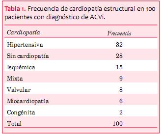
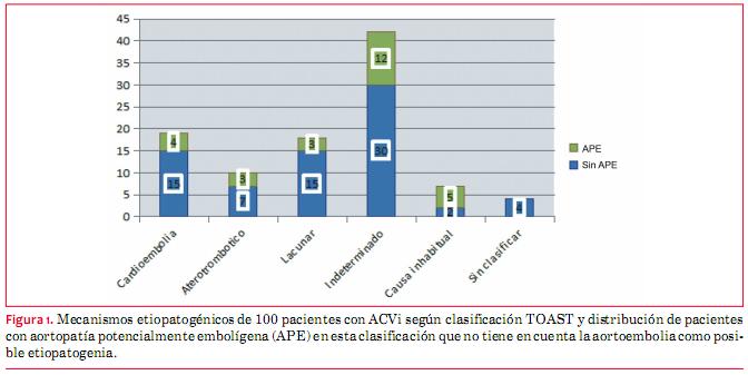
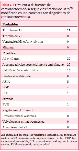
<body><![CDATA[<br>  Se reclutaron 100 pacientes, 50 hombres (50%); media de edad 64 &plusmn; 12 a&ntilde;os, 90% con al menos un factor de riesgo cardiovascular mayor, 18% con historia de fibrilaci&oacute;n auricular, 94% con diagn&oacute;stico de infarto cerebral y el 6% restante de AIT.&nbsp;  </span> </font><span style="font-size: 10pt; font-family: Verdana;"><o:p></o:p></span>          <p style="margin: 0cm 0cm 0.0001pt;"><font face="Verdana"> <span style="font-size: 10pt; font-family: 10; ">Los mecanismos etiopatog&eacute;nicos identificados de los ACVi, seg&uacute;n neur&oacute;logo especializado en el diagn&oacute;stico y tratamiento del ACVi, est&aacute;n representados en la </span> <span style="font-size: 10pt; font-family: 10; color: rgb(31, 26, 23)"> <a href="/img/revistas/ruc/v30n1/1a08g1.JPG">figura 1</a></span><span style="font-size: 10pt; font-family: 10; ">. Destacamos el alto porcentaje de pacientes con mecanismo indeterminado (42%), ya sea por ausencia de un mecanismo demostrado o por presencia de m&aacute;s de un mecanismo posible. La aortopat&iacute;a ateroscler&oacute;tica tor&aacute;cica est&aacute; presente en el 62% de los pacientes. De ellos, la aortopat&iacute;a potencialmente embol&iacute;gena (APE) se presenta en 27 pacientes, la cual no tiene representaci&oacute;n en la clasificaci&oacute;n cl&aacute;sica de TOAST</span><sup><span style="font-size: 10pt; font-family: 10; ">(</span><span style="font-size: 10pt; font-family: 10; color: rgb(31, 26, 23)"><a href="#10">10</a></span><span style="font-size: 10pt; font-family: 10; ">)</span></sup><span style="font-size: 10pt; font-family: 10; ">; de hecho, 17 pacientes de estos 27 se encuentran en el grupo indeterminados o sin clasificar (</span><span style="font-size: 10pt; font-family: 10; color: rgb(31, 26, 23)"><a href="/img/revistas/ruc/v30n1/1a08g1.JPG">figura 1</a></span><span style="font-size: 10pt; font-family: 10; ">). Se considera APE a aquellas placas ateromatosas localizadas en aorta ascendente o cayado (proximales) y que adem&aacute;s se presentan como complejas: ulceradas, protruyentes (midiendo m&aacute;s de 4 mm de altura) o con componente m&oacute;vil asociado<span style=""> (debris)</span>.</span></font></p>          <p style="margin: 0cm 0cm 0.0001pt;"><font face="Verdana"> <span style="font-size: 10pt; font-family: 10; ">En referencia al ecocardiograma, la fracci&oacute;n de eyecci&oacute;n del ventr&iacute;culo izquierdo (FEVI) fue de 57% &plusmn; 10% y predomin&oacute; la presencia de cardiopat&iacute;a estructural (</span><span style="font-size: 10pt; font-family: 10; color: rgb(31, 26, 23)"><a href="#t1">tabla 1</a></span><span style="font-size: 10pt; font-family: 10; ">).&nbsp; </span></font><span style="font-size: 10pt; font-family: Verdana;"><o:p></o:p></span></p>                <p style="margin: 0cm 0cm 0.0001pt;">&nbsp;</p>      <font face="Verdana">      <font size="2"><img style="width: 328px; height: 277px;" alt="" src="/img/revistas/ruc/v30n1/1a08t1.JPG">    <br>      </font>      <span style="font-size: 10pt; ">&nbsp;</span></font><o:p></o:p><font face="Verdana" size="2"> </font>      <p style="margin: 0cm 0cm 0.0001pt;"><font face="Verdana"> <span style="font-size: 10pt; font-family: 10; ">Las fuentes probables de cardioaortoembolia por ETE est&aacute;n ilustradas en la </span> <span style="font-size: 10pt; font-family: 10; color: rgb(31, 26, 23)"> <a href="#t2">tabla 2</a></span><span style="font-size: 10pt; font-family: 10; ">&nbsp;<span class="GramE"><sup>(</sup></span></span><sup><span style="font-size: 10pt; font-family: 10; "><a name="23."></a></span><span style="font-size: 10pt; font-family: 10; color: rgb(31, 26, 23)"><a href="#23">23</a></span><span style="font-size: 10pt; font-family: 10; ">)</span></sup><span style="font-size: 10pt; font-family: 10; ">. Destacamos que 10 de 11 pacientes con fuente probable asociaban tambi&eacute;n una fuente posible; a su vez, 57 pacientes del total ten&iacute;an al menos una fuente posible y 30 de ellos ten&iacute;an al menos dos fuentes posibles. Finalmente, 42 pacientes no ten&iacute;an fuente posible/probable.&nbsp; </span></font><span style="font-size: 10pt; font-family: Verdana;"><o:p></o:p></span></p>            <p style="margin: 0cm 0cm 0.0001pt;">&nbsp;</p>          <p style="margin: 0cm 0cm 0.0001pt;"><font face="Verdana"> <span style="font-size: 10pt; font-family: 10;"><a name="t2"></a><img style="width: 323px; height: 617px;" alt="" src="/img/revistas/ruc/v30n1/1a08t2.JPG"></span></font></p>          <p style="margin: 0cm 0cm 0.0001pt;"><font face="Verdana"> <span style="font-size: 10pt; ">&nbsp;</span></font><o:p></o:p></p>        <font face="Verdana">        <span style="font-size: 10pt; font-family: 10; ">No se registraron complicaciones mayores vinculadas al ETE; solamente como complicaci&oacute;n menor un hematoma de am&iacute;gdala de resoluci&oacute;n espont&aacute;nea (1%).&nbsp; </span> </font><span style="font-size: 10pt; font-family: Verdana;"><o:p></o:p></span>      <font face="Verdana"><font size="2">          <br>  </font><b> <span style="font-size: 10pt; font-family: 10; ">Objetivo primario&nbsp;</span></b></font><b style=""><font face="Verdana"><span style="font-size: 10pt; font-family: 10; "> </span> </font><span style="font-size: 10pt; font-family: Verdana;"><o:p></o:p></span></b> <font face="Verdana"><font size="2">          ]]></body>
<body><![CDATA[<br>  </font>  <span style="font-size: 10pt; font-family: 10; ">El NEN para cambiar una estimaci&oacute;n de riesgo fue de 2,85, IC 95% (2,3- 3,9) y para cambiar un tratamiento fue de 10,0, IC 95% (6,3-24,3). Los cambios de tratamientos realizados se muestran en la </span>  <span style="font-size: 10pt; font-family: 10; color: rgb(31, 26, 23)"> <a href="/img/revistas/ruc/v30n1/1a08t3.JPG">tabla 3</a></span><span style="font-size: 10pt; font-family: 10; ">. Los c&aacute;lculos del NEN fueron hechos usando el paquete estad&iacute;stico del libro Evidencias en Cardiolog&iacute;a III, de Doval y Tajer, editorial Gedic, 2014.&nbsp; </span></font><span style="font-size: 10pt; font-family: Verdana;"><o:p></o:p></span>          <font face="Verdana" size="2">              <br>  </font><span style="font-size: 10pt; font-family: Verdana;"><o:p></o:p></span> <font face="Verdana"><font size="2">    <br>      </font>      <b><span style="font-size: 10pt; font-family: 10; ">Objetivo secundario&nbsp;</span></b></font><b style=""><font face="Verdana"><span style="font-size: 10pt; font-family: 10; "> </span> </font><span style="font-size: 10pt; font-family: Verdana;"><o:p></o:p></span></b> <font face="Verdana"><font size="2">          <br>  </font>  <span style="font-size: 10pt; font-family: 10; ">El ETT mostr&oacute; una sensibilidad para el diagn&oacute;stico de cardioaortoembolia de 43,9% (IC 95% 31,0-56,7) y una especificidad de 81,4% (IC 95% 69,8-93,0) comparado con el ETE (</span><span style="font-size: 10pt; font-family: 10; color: rgb(31, 26, 23)"><a href="/img/revistas/ruc/v30n1/1a08t4.JPG">tabla 4</a></span><span style="font-size: 10pt; font-family: 10; ">); cuando se trata de diagnosticar trombos en orejuela izquierda, la sensibilidad del ETT fue nula.&nbsp; </span></font><span style="font-size: 10pt; font-family: Verdana;"><o:p></o:p></span>       <font face="Verdana" size="2">           <br>      <br>  </font><span style="font-size: 10pt; font-family: Verdana;"><o:p></o:p></span> <font face="Verdana"><span style="font-size: 10pt; ">Discusi&oacute;n</span></font><o:p></o:p><font face="Verdana"><font size="2">          <br>  </font>  <span style="font-size: 10pt; font-family: 10; ">    <br>  Se trata de un trabajo realizado en un servicio universitario con el concepto de integraci&oacute;n de equipos especializados de trabajo (unidad de ACV y unidad de imagen card&iacute;aca) con el objetivo de profundizar el conocimiento del rendimiento de las herramientas diagn&oacute;sticas disponibles. Encontramos que el ETE utilizado en forma rutinaria en la valoraci&oacute;n diagn&oacute;stica inicial de pacientes con ACVi aporta informaci&oacute;n cl&iacute;nicamente significativa, definiendo un cambio en la estimaci&oacute;n del riesgo emb&oacute;lico en aproximadamente uno de cada tres pacientes y un cambio terap&eacute;utico efectivo en uno de cada diez, si bien este &uacute;ltimo punto no tuvo significaci&oacute;n estad&iacute;stica.&nbsp;  </span></font><span style="font-size: 10pt; font-family: Verdana;"><o:p></o:p></span>          <p style="margin: 0cm 0cm 0.0001pt;"><font face="Verdana"> <span style="font-size: 10pt; font-family: 10; ">Los resultados encontrados respecto a la capacidad diagn&oacute;stica del ETE versus ETT para fuentes de cardioembolia son acordes a los reportados por otros autores. Destacamos un trabajo de De Bruijin y colaboradores que reportan una sensibilidad del ETE mayor al doble que el ETT y concluyen que el ETE es superior al ETT en el estudio de pacientes de todas las edades con <span class="GramE">ACVi<sup>(</sup></span></span><sup><span style="font-size: 10pt; font-family: 10; color: rgb(31, 26, 23)"><a href="#18">18</a></span><span style="font-size: 10pt; font-family: 10; ">)</span></sup><span style="font-size: 10pt; font-family: 10; ">; mientras que Namara concluye que el uso de ETE en este escenario cl&iacute;nico no solo es superior cl&iacute;nicamente, sino que adem&aacute;s es econ&oacute;micamente m&aacute;s rentable</span><sup><span style="font-size: 10pt; font-family: 10; ">(<a name="22."></a></span><span style="font-size: 10pt; font-family: 10; color: rgb(31, 26, 23)"><a href="#22">22</a></span><span style="font-size: 10pt; font-family: 10; ">)</span></sup><span style="font-size: 10pt; font-family: 10; ">. Es importante destacar de todas maneras que si bien la sensibilidad del ETT es mala para fuentes de cardioembolia en su globalidad y muy mala para trombos intrauriculares, la especificidad de los hallazgos es cl&iacute;nicamente aceptable.&nbsp; </span></font><span style="font-size: 10pt; font-family: Verdana;"><o:p></o:p></span></p>            <p style="margin: 0cm 0cm 0.0001pt;"><font face="Verdana"> <span style="font-size: 10pt; font-family: 10; ">A su vez, si bien no encontramos significaci&oacute;n estad&iacute;stica con los criterios de NEN definidos arbitrariamente, no llama la atenci&oacute;n que al hacer nuevos diagn&oacute;sticos y reclasificar el riesgo emb&oacute;lico de cada paciente, se ajuste o cambie la terap&eacute;utica, lo que sucedi&oacute; tambi&eacute;n en uno de cada ocho pacientes de la serie de de Bruijin</span><sup><span style="font-size: 10pt; font-family: 10; ">(</span><span style="font-size: 10pt; font-family: 10; color: rgb(31, 26, 23)"><a href="#18">18</a></span><span style="font-size: 10pt; font-family: 10; ">)</span></sup><span style="font-size: 10pt; font-family: 10; ">.&nbsp;</span></font></p>              ]]></body>
<body><![CDATA[<p style="margin: 0cm 0cm 0.0001pt;"><span style="font-size: 10pt; font-family: Verdana;"><o:p></o:p></span> <font face="Verdana"> <span style="font-size: 10pt; font-family: 10; ">Los trombos en la orejuela izquierda encontrados fueron todos en pacientes con historia de fibrilaci&oacute;n auricular (causa m&aacute;s frecuente de cardioembolia). El diagn&oacute;stico de trombo en orejuela izquierda implica un desaf&iacute;o pues debe considerar en el diagn&oacute;stico diferencial los m&uacute;sculos pect&iacute;neos. Debe tenerse presente, adem&aacute;s, que la orejuela izquierda puede estar constituida por m&aacute;s de un l&oacute;bulo que tambi&eacute;n pueden estar colonizados con trombos. Otro elemento importante es la medici&oacute;n de la velocidad de vaciado de la orejuela izquierda pues el hallazgo de un trombo es excepcional cuando las velocidades de vaciado son superiores a 30 cm/<span class="GramE">seg<sup>(</sup></span></span><sup><span style="font-size: 10pt; font-family: 10; color: rgb(31, 26, 23)"><a href="#23">23</a></span><span style="font-size: 10pt; font-family: 10; ">)</span></sup><span style="font-size: 10pt; font-family: 10; ">. La variabilidad interobservador para el diagn&oacute;stico de trombo en la orejuela izquierda resulta baja y la correlaci&oacute;n es excelente cuando se predefinen los criterios para su <span class="GramE">diagn&oacute;stico<sup>(<a name="24."></a></sup></span></span><sup><span style="font-size: 10pt; font-family: 10; color: rgb(31, 26, 23)"><a href="#24">24</a></span><span style="font-size: 10pt; font-family: 10; ">) </span> </sup> <span style="font-size: 10pt; font-family: 10; ">y el estudio es realizado por cardi&oacute;logos imagen&oacute;logos con experiencia</span><sup><span style="font-size: 10pt; font-family: 10; ">(<a name="25."></a></span><span style="font-size: 10pt; font-family: 10; color: rgb(31, 26, 23)"><a href="#25">25</a></span><span style="font-size: 10pt; font-family: 10; ">)</span></sup><span style="font-size: 10pt; font-family: 10; ">. Nuestro equipo, pese a no haber estimado su propia variabilidad para el diagn&oacute;stico de fuentes de cardioembolia, se apoya en estos dos preconceptos para validar sus resultados.&nbsp; </span></font><span style="font-size: 10pt; font-family: Verdana;"><o:p></o:p></span></p>            <p style="margin: 0cm 0cm 0.0001pt;"><font face="Verdana"> <span style="font-size: 10pt; font-family: 10; ">  &nbsp;En el escenario aislado del paciente con fibrilaci&oacute;n o flutter auricular en el cual no se plantea cardioversi&oacute;n y est&aacute; definida la anticoagulaci&oacute;n, la indicaci&oacute;n de ETE se considera inapropiada seg&uacute;n la gu&iacute;a de criterios para el uso de ecocardiograf&iacute;a transtor&aacute;cica y transesof&aacute;gica del 2007</span><sup><span style="font-size: 10pt; font-family: 10; ">(</span><span style="font-size: 10pt; font-family: 10; color: rgb(31, 26, 23)"><a href="#19">19</a></span><span style="font-size: 10pt; font-family: 10; ">)</span></sup><span style="font-size: 10pt; font-family: 10; ">. El equipo investigador se pregunt&oacute; &iquest;por qu&eacute; deber&iacute;amos hacer ETE a estos pacientes con ACVi que tienen indicaci&oacute;n previa de anticoagulaci&oacute;n? No obstante, en nuestra serie encontramos otras causas posibles de embolia concomitantes en pacientes con historia de fibrilaci&oacute;n auricular con y sin trombos en la orejuela, particularmente aortopat&iacute;a. Esto ya ha sido descrito por otros autores y explicado por la epidemiolog&iacute;a similar de ambas patolog&iacute;as cuya prevalencia aumenta en los m&aacute;s <span class="GramE">a&ntilde;osos<sup>(</sup></span></span><sup><span style="font-size: 10pt; font-family: 10; "><a name="26."></a></span><span style="font-size: 10pt; font-family: 10; color: rgb(31, 26, 23)"><a href="#26">26</a></span><span style="font-size: 10pt; font-family: 10; ">,<a name="27."></a></span><span style="font-size: 10pt; font-family: 10; color: rgb(31, 26, 23)"><a href="#27">27</a></span><span style="font-size: 10pt; font-family: 10; ">)</span></sup><span style="font-size: 10pt; font-family: 10; ">.&nbsp; </span></font><span style="font-size: 10pt; font-family: Verdana;"><o:p></o:p></span></p>            <p style="margin: 0cm 0cm 0.0001pt;"><font face="Verdana"> <span style="font-size: 10pt; font-family: 10; ">La presencia de aortopat&iacute;a incrementa el riesgo emb&oacute;lico si es proximal y compleja, pero tambi&eacute;n es conocido que la mera presencia de ateromatosis a&oacute;rtica amerita el inicio de prevenci&oacute;n vascular secundaria completa aun en ausencia de factores de riesgo cardiovascular o de otra evidencia de enfermedad vascular ateroscler&oacute;tica (por ejemplo: joven sin factores de riesgo cardiovascular con ACVi y fibrilaci&oacute;n auricular).&nbsp; </span></font><span style="font-size: 10pt; font-family: Verdana;"><o:p></o:p></span></p>            <p style="margin: 0cm 0cm 0.0001pt;"><font face="Verdana"> <span style="font-size: 10pt; font-family: 10; ">De acuerdo con los resultados de la alta prevalencia de aortopat&iacute;a y aortopat&iacute;a potencialmente embol&iacute;gena, y su exclusi&oacute;n en la clasificaci&oacute;n de TOAST para mecanismos etiopatog&eacute;nicos del ACVi, nos planteamos si no ser&iacute;a necesario considerar que se incluya a la aortopat&iacute;a potencialmente embol&iacute;gena como un mecanismo independiente</span><sup><span style="font-size: 10pt; font-family: 10; ">(</span><span style="font-size: 10pt; font-family: 10; color: rgb(31, 26, 23)"><a href="#11">11</a></span><span style="font-size: 10pt; font-family: 10; ">-</span><span style="font-size: 10pt; font-family: 10; color: rgb(31, 26, 23)"><a href="#18">18</a></span><span style="font-size: 10pt; font-family: 10; ">)</span></sup><span style="font-size: 10pt; font-family: 10; ">.&nbsp; </span></font><span style="font-size: 10pt; font-family: Verdana;"><o:p></o:p></span></p>            <p style="margin: 0cm 0cm 0.0001pt;"><font face="Verdana"> <span style="font-size: 10pt; font-family: 10; ">La concomitancia de potenciales fuentes de cardioaortoembolia en un mismo paciente y de otras causas posibles de ACVi no cardiog&eacute;nicas plantea la duda de cu&aacute;l es el mecanismo que efectivamente explic&oacute; el evento &iacute;ndice. Esta situaci&oacute;n ya es conocida; dos trabajos de Howirtz en 1992 y Wessels en 2005 estudiaron pacientes con ACVi catalogados como infartos lacunares (cuyo mecanismo postulado es la oclusi&oacute;n in situ de peque&ntilde;os vasos intracraneanos con microateromatosis o lipohialinosis) y encontraron que en un tercio era posible encontrar otro mecanismo, ya sea por patolog&iacute;a card&iacute;aca o de grandes <span class="GramE">arterias<sup>(</sup></span></span><sup><span style="font-size: 10pt; font-family: 10; "><a name="28."></a></span><span style="font-size: 10pt; font-family: 10; color: rgb(31, 26, 23)"><a href="#28">28</a></span><span style="font-size: 10pt; font-family: 10; ">,<a name="29."></a></span><span style="font-size: 10pt; font-family: 10; color: rgb(31, 26, 23)"><a href="#29">29</a></span><span style="font-size: 10pt; font-family: 10; ">)</span></sup><span style="font-size: 10pt; font-family: 10; ">.&nbsp; </span> </font><span style="font-size: 10pt; font-family: Verdana;"><o:p></o:p></span></p>            <p style="margin: 0cm 0cm 0.0001pt;"><font face="Verdana"> <span style="font-size: 10pt; font-family: 10; ">  Otro elemento importante a considerar de nuestros resultados es que la presencia de trombos en la orejuela izquierda en pacientes aparentemente anticoagulados adecuadamente con antivitaminas K plante&oacute; la necesidad de aumentar el rango de anticoagulaci&oacute;n de INR entre 2 y 3 a entre 2,5 y 3,5, o considerar el cambio a anticoagulantes orales no antivitamina K.&nbsp; </span> </font><span style="font-size: 10pt; font-family: Verdana;"><o:p></o:p></span></p>        <font face="Verdana">        <span style="font-size: 10pt; font-family: 10; ">Son limitaciones del presente trabajo el ser unic&eacute;ntrico e incluir un relativamente bajo n&uacute;mero de pacientes. A su vez la definici&oacute;n de relevancia cl&iacute;nica est&aacute; basada en el juicio cl&iacute;nico convenido por el equipo investigador. Todo esto limita la generalizaci&oacute;n de los resultados. Se trata de un estudio de car&aacute;cter exploratorio que permite crear nuevas hip&oacute;tesis de trabajo.&nbsp;</span><font size="2">        <br>  </font></font><span style="font-size: 10pt; font-family: Verdana;"><o:p></o:p></span>      <font face="Verdana">      <span style="font-size: 10pt; font-family: 10; ">Aportamos informaci&oacute;n a favor del uso del ETE en el estudio de pacientes con ACVi reciente. No dudamos de la importancia de optimizar el diagn&oacute;stico y la estimaci&oacute;n de riesgo emb&oacute;lico en este grupo de pacientes. De acuerdo a nuestros resultados, pese a que la indicaci&oacute;n de ETE es segura, rentable y aporta informaci&oacute;n relevante, no creemos que exista a&uacute;n evidencia suficiente para indicar el estudio en forma rutinaria. La gu&iacute;a cl&iacute;nica de manejo del paciente con enfermedad cerebrovascular aguda 2013, no menciona el rol del ETE en la evaluaci&oacute;n inmediata de estos pacientes</span><sup><span style="font-size: 10pt; font-family: 10; ">(<a name="30."></a></span><span style="font-size: 10pt; font-family: 10; color: rgb(31, 26, 23)"><a href="#30">30</a></span><span style="font-size: 10pt; font-family: 10; ">)</span></sup><span style="font-size: 10pt; font-family: 10; ">; por otra parte, en un art&iacute;culo de revisi&oacute;n del a&ntilde;o 2003 de Garc&iacute;a Fern&aacute;ndez, se destaca el rol del ETE para el diagn&oacute;stico de fuentes de cardioaortoembolia y aconseja el uso rutinario de este en pacientes con ACV menores de 50 a&ntilde;os, en pacientes con embolia y estenosis mitral reum&aacute;tica, frente a la sospecha de embolia paradojal, en el ACV a repetici&oacute;n y cuando el ETT es dudoso; por otro lado, se plantean como otras indicaciones posibles la fibrilaci&oacute;n auricular embol&iacute;gena, la sospecha de aortoembolia y la patolog&iacute;a valvular mitral<sup>(31)</sup>. En definitiva, estas indicaciones como globalidad dejan limitadas situaciones en las cuales no ser&iacute;a aconsejable realizar un ETE en el contexto del ACVi. Por supuesto que la individualizaci&oacute;n de la indicaci&oacute;n debe hacerse siempre valorando la oportunidad, evitando realizar maniobras semiinvasivas en pacientes inestables sabiendo que las mismas no van a definir cambios inmediatos de conducta. Por otro lado, en pacientes sin contraindicaciones para realizar ETE y con pron&oacute;stico vital y funcional aceptables, deber&iacute;a sopesarse siempre la importancia de optimizar su valoraci&oacute;n diagn&oacute;stica que conduzca a un cambio terap&eacute;utico que disminuya recurrencias y sus graves consecuencias.&nbsp; </span></font><span style="font-size: 10pt; font-family: Verdana;"><o:p></o:p></span>      <font face="Verdana"><font size="2">          <br>  </font>  <span style="font-size: 10pt; font-family: 10; ">Finalmente, es necesario avanzar en la definici&oacute;n de causalidad entre los hallazgos encontrados y el mecanismo de ACVi invocado. Para eso, intentaremos definir la relevancia de los hallazgos del ETE en el diagn&oacute;stico etiopat&oacute;genico del ACVi y planificamos un estudio de dise&ntilde;o caso-control que pueda orientar a causalidad, seg&uacute;n los hallazgos nos proponemos avanzar en un estudio prospectivo a largo plazo.&nbsp; </span></font><span style="font-size: 10pt; font-family: Verdana;"><o:p></o:p></span>      <font face="Verdana"><font size="2">          <br>  </font>  <span style="font-size: 10pt; font-family: 10;"><o:p>&nbsp;</o:p></span><font size="2">          <br>  </font>  <span style="font-size: 10pt; font-family: 10;"><o:p>    ]]></body>
<body><![CDATA[<br>  </o:p></span><span style="font-size: 10pt; ">Bibliograf&iacute;a</span></font><o:p></o:p><font face="Verdana" size="2"> </font>          <p style="margin: 0cm 0cm 0.0001pt;"><font face="Verdana"> <span style="font-size: 10pt; font-family: 10; ">&nbsp; </span> </font><span style="font-size: 10pt; font-family: Verdana;"><o:p></o:p></span></p>            <!-- ref --><p style="margin: 0cm 0cm 0.0001pt;"><font face="Verdana"> <span style="font-size: 10pt; font-family: 10; "><a name="1"></a> </span> <span style="font-size: 10pt; font-family: 10; color: rgb(31, 26, 23)"><a href="#1.">1</a></span><span style="font-size: 10pt; font-family: 10; ">.&nbsp;&nbsp;&nbsp;&nbsp;<span style="">Uruguay. Ministerio de Salud P&uacute;blica.</span> Mortalidad en el Uruguay: defunciones registradas seg&uacute;n causa de muerte,  Uruguay 2008. Montevideo: MSP, 2008 Disponible en: </span> <span style="font-size: 10pt; font-family: 10; color: rgb(31, 26, 23)"> <a href="http://www2.msp.gub.uy/ucepidemiologia_6490_1.html">http://www2.msp.gub.uy/ucepidemiologia_6490_1.html</a></span><span style="font-size: 10pt; font-family: 10; ">. [Consulta:].    &nbsp; </span> </font><span style="font-size: 10pt; font-family: Verdana;"><o:p></o:p></span></p>            <!-- ref --><p style="margin: 0cm 0cm 0.0001pt;"><font face="Verdana"> <span style="font-size: 10pt; font-family: 10; "><a name="2"></a> </span> <span style="font-size: 10pt; font-family: 10; color: rgb(31, 26, 23)"><a href="#2.">2</a></span><span style="font-size: 10pt; font-family: 10; ">.&nbsp;&nbsp;&nbsp;&nbsp;<span style="">Bonita R.</span> Epidemiology of stroke. Lancet 1992; 339(8789):342-4.    &nbsp; </span></font><span style="font-size: 10pt; font-family: Verdana;"><o:p></o:p></span></p>            <!-- ref --><p style="margin: 0cm 0cm 0.0001pt;"><font face="Verdana"> <span style="font-size: 10pt; font-family: 10; "><a name="3"></a> </span> <span style="font-size: 10pt; font-family: 10; color: rgb(31, 26, 23)"><a href="#3.">3</a></span><span style="font-size: 10pt; font-family: 10; ">.&nbsp;&nbsp;&nbsp;&nbsp;<span style="">Wolfe CD, Giroud M, Kolominsky-Rabas P, Dundas R, Lemesle M, Heuschmann P, et al.</span> Variations in stroke incidence and survival in 3 areas of Europe: European Registries of Stroke (EROS) Collaboration. Stroke 2000; 31(9):2074-9.    &nbsp; </span></font><span style="font-size: 10pt; font-family: Verdana;"><o:p></o:p></span></p>            <!-- ref --><p style="margin: 0cm 0cm 0.0001pt;"><font face="Verdana"> <span style="font-size: 10pt; font-family: 10; "><a name="4"></a> </span> <span style="font-size: 10pt; font-family: 10; color: rgb(31, 26, 23)"><a href="#4.">4</a></span><span style="font-size: 10pt; font-family: 10; ">.&nbsp;&nbsp;&nbsp;&nbsp;<span style="">Bamford J, Sandercock P, Dennis M, Warlow C, Jones L, McPherson K, et al.</span> A prospective study of acute cerebrovascular disease in the community: the Oxfordshire Community Stroke Project 1981-86. 1. Methodology, demography and incident cases of first-ever stroke. J Neurol Neurosurg Psychiatry 1988; 51(11):1373-80.    &nbsp; </span></font><span style="font-size: 10pt; font-family: Verdana;"><o:p></o:p></span></p>            ]]></body>
<body><![CDATA[<!-- ref --><p style="margin: 0cm 0cm 0.0001pt;"><font face="Verdana"> <span style="font-size: 10pt; font-family: 10; "><a name="5"></a> </span> <span style="font-size: 10pt; font-family: 10; color: rgb(31, 26, 23)"><a href="#5.">5</a></span><span style="font-size: 10pt; font-family: 10; ">.&nbsp;&nbsp;&nbsp;&nbsp;<span style="">Saka O, Serra V, Samyshkin Y, McGuire A, Wolfe CC. </span>Cost-effectiveness of a stroke unit care followed by early supported discharge. Stroke 2009; 40(1):24-9.    &nbsp; </span></font><span style="font-size: 10pt; font-family: Verdana;"><o:p></o:p></span></p>            <!-- ref --><p style="margin: 0cm 0cm 0.0001pt;"><font face="Verdana"> <span style="font-size: 10pt; font-family: 10; "><a name="6"></a> </span> <span style="font-size: 10pt; font-family: 10; color: rgb(31, 26, 23)"><a href="#6.">6</a></span><span style="font-size: 10pt; font-family: 10; ">.&nbsp;&nbsp;&nbsp;&nbsp;<span style="">Launois R, Giroud M, M&eacute;gnigb&ecirc;to AC, Le Lay K, Pr&eacute;sent&eacute; G, Mahagne MH, et al.</span> Estimating the cost-effectiveness of stroke units in France compared with conventional care. Stroke 2004; 35(3): 770-5.    &nbsp; </span></font><span style="font-size: 10pt; font-family: Verdana;"><o:p></o:p></span></p>            <!-- ref --><p style="margin: 0cm 0cm 0.0001pt;"><font face="Verdana"> <span style="font-size: 10pt; font-family: 10; "><a name="7"></a> </span> <span style="font-size: 10pt; font-family: 10; color: rgb(31, 26, 23)"><a href="#7.">7</a></span><span style="font-size: 10pt; font-family: 10; ">.&nbsp;&nbsp;&nbsp;&nbsp;<span style="">Zhu HF, Newcommon NN, Cooper ME, Green TL, Seal B, Klein G, et al; Calgary Stroke Program.</span> Impact of a stroke unit on length of hospital stay and in-hospital case fatality. Stroke 2009; 40(1):18-23.    &nbsp; </span></font><span style="font-size: 10pt; font-family: Verdana;"><o:p></o:p></span></p>            <!-- ref --><p style="margin: 0cm 0cm 0.0001pt;"><font face="Verdana"> <span style="font-size: 10pt; font-family: 10; "><a name="8"></a> </span> <span style="font-size: 10pt; font-family: 10; color: rgb(31, 26, 23)"><a href="#8.">8</a></span><span style="font-size: 10pt; font-family: 10; ">.&nbsp;&nbsp;&nbsp;&nbsp;<span style="">Sacco RL, Adams R, Albers G, Alberts MJ, Benavente O, Furie K, et al; American Heart Association/American Stroke Association Council on Stroke; Council on Cardiovascular Radiology and Intervention; American Academy of Neurology.</span> Guidelines for prevention of stroke in patients with ischemic stroke or transient ischemic attack: a statement for health care professionals from the American Heart Association/American Stroke Association Council on Stroke: co-sponsored by the Council on Cardiovascular Radiology and Intervention: the American Academy of Neurology affirms the value of this guideline. Circulation 2006; 113(10):e409-49.    &nbsp; </span></font><span style="font-size: 10pt; font-family: Verdana;"><o:p></o:p></span></p>            <!-- ref --><p style="margin: 0cm 0cm 0.0001pt;"><font face="Verdana"> <span style="font-size: 10pt; font-family: 10; ">  <a name="9"></a> </span> <span style="font-size: 10pt; font-family: 10; color: rgb(31, 26, 23)">  <a href="#9.">9</a></span><span style="font-size: 10pt; font-family: 10; ">.&nbsp;&nbsp;&nbsp;&nbsp;<span style="">Saposnik G, Del Brutto OH; Iberoamerican Society of Cerebrovascular Diseases.</span> Stroke in South America: a systematic review of incidence, prevalence, and stroke subtypes. Stroke 2003; 34(9): 2103-7.    &nbsp; </span></font><span style="font-size: 10pt; font-family: Verdana;"><o:p></o:p></span></p>            ]]></body>
<body><![CDATA[<!-- ref --><p style="margin: 0cm 0cm 0.0001pt;"><font face="Verdana"> <span style="font-size: 10pt; font-family: 10; "><a name="10"></a> </span> <span style="font-size: 10pt; font-family: 10; color: rgb(31, 26, 23)"><a href="#10.">10</a></span><span style="font-size: 10pt; font-family: 10; ">.&nbsp;&nbsp;&nbsp;&nbsp;<span style="">Adams HP Jr, Bendixen BH, Kappelle LJ, Biller J, Love BB, Gordon DL, et al. </span>Classification of subtype of acute ischemic stroke: definitions for use in a multicenter clinical trial. TOAST. Trial of Org 10172 in Acute Stroke Treatment. Stroke 1993; 24(1):35-41.    &nbsp; </span> </font><span style="font-size: 10pt; font-family: Verdana;"><o:p></o:p></span></p>            <!-- ref --><p style="margin: 0cm 0cm 0.0001pt;"><font face="Verdana"> <span style="font-size: 10pt; font-family: 10; "><a name="11"></a> </span> <span style="font-size: 10pt; font-family: 10; color: rgb(31, 26, 23)"><a href="#11.">11</a></span><span style="font-size: 10pt; font-family: 10; ">.&nbsp;&nbsp;&nbsp;&nbsp;<span style="">Furie KL, Kasner SE, Adams RJ, Albers GW, Bush RL, Fagan SC, et al; American Heart Association Stroke Council, Council on Cardiovascular Nursing, Council on Clinical Cardiology, and Interdisciplinary Council on Quality of Care and Outcomes Research.</span> Guidelines for the prevention of stroke in patients with stroke or transient ischemic attack: a guideline for health care professionals from the American Heart Association/American Stroke Association. Stroke 2011; 42(1):227-76.    &nbsp; </span> </font><span style="font-size: 10pt; font-family: Verdana;"><o:p></o:p></span></p>            <!-- ref --><p style="margin: 0cm 0cm 0.0001pt;"><font face="Verdana"> <span style="font-size: 10pt; font-family: 10; "><a name="12"></a> </span> <span style="font-size: 10pt; font-family: 10; color: rgb(31, 26, 23)"><a href="#12.">12</a></span><span style="font-size: 10pt; font-family: 10; ">.&nbsp;&nbsp;&nbsp;&nbsp;<span style="">Amarenco P, Duyckaerts C, Tzourio C, H&eacute;nin D, Bousser MG, Hauw JJ. </span>The prevalence of ulcerated plaques in the aortic arch in patients with stroke. N Engl J Med 1992; 326(4):221-5.    &nbsp; </span></font><span style="font-size: 10pt; font-family: Verdana;"><o:p></o:p></span></p>            <!-- ref --><p style="margin: 0cm 0cm 0.0001pt;"><font face="Verdana"> <span style="font-size: 10pt; font-family: 10; "><a name="13"></a> </span> <span style="font-size: 10pt; font-family: 10; color: rgb(31, 26, 23)"><a href="#13.">13</a></span><span style="font-size: 10pt; font-family: 10; ">.&nbsp;&nbsp;&nbsp;&nbsp;<span style="">Amarenco P, Cohen A, Tzourio C, Bertrand B, Hommel M, Besson G, et al.</span> Atherosclerotic disease of the aortic arch and the riskof ischemic stroke. N Engl J Med 1994; 331(22):1474-9.    &nbsp; </span> </font><span style="font-size: 10pt; font-family: Verdana;"><o:p></o:p></span></p>            <!-- ref --><p style="margin: 0cm 0cm 0.0001pt;"><font face="Verdana"> <span style="font-size: 10pt; font-family: 10; "><a name="14"></a> </span> <span style="font-size: 10pt; font-family: 10; color: rgb(31, 26, 23)"><a href="#14.">14</a></span><span style="font-size: 10pt; font-family: 10; ">.&nbsp;&nbsp;&nbsp;&nbsp;<span style="">Mitusch R, Doherty C, Wucherpfennig H, Memmesheimer C, Tepe C, Stierle U, et al. </span>Vascular events during follow-up in patients with aortic arch atherosclerosis. Stroke 1997; 28(1):36-9.    &nbsp; </span></font><span style="font-size: 10pt; font-family: Verdana;"><o:p></o:p></span></p>            ]]></body>
<body><![CDATA[<!-- ref --><p style="margin: 0cm 0cm 0.0001pt;"><font face="Verdana"> <span style="font-size: 10pt; font-family: 10; "><a name="15"></a> </span> <span style="font-size: 10pt; font-family: 10; color: rgb(31, 26, 23)"><a href="#15.">15</a></span><span style="font-size: 10pt; font-family: 10; ">.&nbsp;&nbsp;&nbsp;&nbsp;<span style="">Toyoda K, Yasaka M, Nagata S, Yamaguchi T.</span> Aortogenic embolic stroke: a transesophageal echocardiographic approach. Stroke 1992; 23(8):1056-61.    &nbsp; </span></font><span style="font-size: 10pt; font-family: Verdana;"><o:p></o:p></span></p>            <!-- ref --><p style="margin: 0cm 0cm 0.0001pt;"><font face="Verdana"> <span style="font-size: 10pt; font-family: 10; "><a name="16"></a> </span> <span style="font-size: 10pt; font-family: 10; color: rgb(31, 26, 23)"><a href="#16.">16</a></span><span style="font-size: 10pt; font-family: 10; ">.&nbsp;&nbsp;&nbsp;&nbsp;<span style="">Beltr&aacute;n A, Gonz&aacute;lez Moreno J<span class="GramE">,Pizzano</span> N,PousoJ, Besada E.</span> Ateromatosis protruyente de la aorta tor&aacute;cica proximal: uno de los factores de riesgo m&aacute;s marcados de los accidentes &nbsp;cerebrovasculares isqu&eacute;micos. Rev Urug Cardiol 1995; 10:62-72.    &nbsp; </span></font><span style="font-size: 10pt; font-family: Verdana;"><o:p></o:p></span></p>            <!-- ref --><p style="margin: 0cm 0cm 0.0001pt;"><font face="Verdana"> <span style="font-size: 10pt; font-family: 10; "><a name="17"></a> </span> <span style="font-size: 10pt; font-family: 10; color: rgb(31, 26, 23)"><a href="#17.">17</a></span><span style="font-size: 10pt; font-family: 10; ">.&nbsp;&nbsp;&nbsp;&nbsp;<span style="">deBruijn SF, Agema WR, Lammers GJ, van der Wall EE, Wolterbeek R, et al. </span>Transesophageal echocardiography is superior to transthoracic echocardiography in management of patients of any age with transient ischemic attack or stroke. Stroke 2006; 37(10):2531-4.    &nbsp; </span> </font><span style="font-size: 10pt; font-family: Verdana;"><o:p></o:p></span></p>            <!-- ref --><p style="margin: 0cm 0cm 0.0001pt;"><font face="Verdana"> <span style="font-size: 10pt; font-family: 10; "><a name="18"></a> </span> <span style="font-size: 10pt; font-family: 10; color: rgb(31, 26, 23)"><a href="#18.">18</a></span><span style="font-size: 10pt; font-family: 10; ">.&nbsp;&nbsp;&nbsp;&nbsp;<span style="">Douglas PS, Khandheria B, Stainback RF, Weissman NJ, Brindis RG, Patel MR, et al; American College of Cardiology Foundation Quality Strategic Directions Committee Appropriateness Criteria Working Group; American Society of Echocardiography; American College of Emergency Physicians; American Society of Nuclear Cardiology; Society for Cardiovascular Angiography and Interventions; Society of Cardiovascular Computed Tomography; Society for Cardiovascular Magnetic Resonance; American College of Chest Physicians; Society of Critical Care Medicine. </span>ACCF/ASE/ACEP/ASNC/SCAI/SCCT/SCMR 2007 appropriateness criteria for transthoracic and transesophageal echocardiography: a report of the American College of Cardiology Foundation Quality Strategic Directions Committee Appropriateness Criteria Working Group, American Society of Echocardiography, American College of Emergency Physicians, American Society of Nuclear Cardiology, Society for Cardiovascular Angiography and Interventions, Society of Cardiovascular Computed Tomography, and the Society for Cardiovascular Magnetic Resonance endorsed by the American College of Chest Physicians and the Society of Critical Care Medicine. J Am Coll Cardiol 2007; 50(2):187-204.    &nbsp; </span></font><span style="font-size: 10pt; font-family: Verdana;"><o:p></o:p></span></p>            <!-- ref --><p style="margin: 0cm 0cm 0.0001pt;"><font face="Verdana"> <span style="font-size: 10pt; font-family: 10; "><a name="19"></a> </span> <span style="font-size: 10pt; font-family: 10; color: rgb(31, 26, 23)"><a href="#19.">19</a></span><span style="font-size: 10pt; font-family: 10; ">.&nbsp;&nbsp;&nbsp;&nbsp;<span style="">Karalis DG, Chandrasekaran K, Victor MF, Ross JJ Jr, Mintz GS.</span> Recognition and embolic potential of intraaortic atherosclerotic debris. J Am Coll Cardiol 1991; 17(1):73-8.    &nbsp; </span></font><span style="font-size: 10pt; font-family: Verdana;"><o:p></o:p></span></p>            ]]></body>
<body><![CDATA[<!-- ref --><p style="margin: 0cm 0cm 0.0001pt;"><font face="Verdana"> <span style="font-size: 10pt; font-family: 10; "><a name="20"></a> </span> <span style="font-size: 10pt; font-family: 10; color: rgb(31, 26, 23)"><a href="#20.">20</a></span><span style="font-size: 10pt; font-family: 10; ">.&nbsp;&nbsp;&nbsp;&nbsp;<span style="">Chen EW, Redberg RF.</span> Evaluaci&oacute;n ecocardiogr&aacute;fica de pacientes con episodios emb&oacute;licos sist&eacute;micos. En: Otto CM. Ecocardiograf&iacute;a cl&iacute;nica pr&aacute;ctica. 3 ed. Barcelona: Elsevier, 2010:969-98.    &nbsp; </span></font><span style="font-size: 10pt; font-family: Verdana;"><o:p></o:p></span></p>            <!-- ref --><p style="margin: 0cm 0cm 0.0001pt;"><font face="Verdana"> <span style="font-size: 10pt; font-family: 10; "><a name="21"></a> </span> <span style="font-size: 10pt; font-family: 10; color: rgb(31, 26, 23)"><a href="#21.">21</a></span><span style="font-size: 10pt; font-family: 10; ">.&nbsp;&nbsp;&nbsp;&nbsp;<span style="">Omran H, Jung W, Rabahieh R, Wirtz P, Becher H, Illien S, et al. </span>Imaging of thrombi and assessment of left atrial appendage function: a prospective study comparing transthoracic and transoesophageal echocardiography. Heart 1999; 81(2):192-8.    &nbsp; </span></font><span style="font-size: 10pt; font-family: Verdana;"><o:p></o:p></span></p>            <!-- ref --><p style="margin: 0cm 0cm 0.0001pt;"><font face="Verdana"> <span style="font-size: 10pt; font-family: 10; "><a name="22"></a> </span> <span style="font-size: 10pt; font-family: 10; color: rgb(31, 26, 23)"><a href="#22.">22</a></span><span style="font-size: 10pt; font-family: 10; ">.&nbsp;&nbsp;&nbsp;&nbsp;<span style="">Kronik G, St&ouml;llberger C, Schuh M, Abzieher F, Slany J, Schneider B.</span> Interobserver variability in the detection of spontaneous echo contrast, left atrial thrombi, and left atrial appendage thrombi by transoesophageal echocardiography. Br Heart J 1995; 74(1):80-3.    &nbsp; </span></font><span style="font-size: 10pt; font-family: Verdana;"><o:p></o:p></span></p>            <!-- ref --><p style="margin: 0cm 0cm 0.0001pt;"><font face="Verdana"> <span style="font-size: 10pt; font-family: 10; "><a name="23"></a> </span> <span style="font-size: 10pt; font-family: 10; color: rgb(31, 26, 23)"><a href="#23.">23</a></span><span style="font-size: 10pt; font-family: 10; ">.&nbsp;&nbsp;&nbsp;&nbsp;<span style="">Masip AE, Alonso G&oacute;mez AM, Dur&aacute;n RM, Moreno Yag&uuml;ela M, Oliver Ruiz JM, Rodr&iacute;guez Padial L, et al.</span> Gu&iacute;as de pr&aacute;ctica cl&iacute;nica de la Sociedad Espa&ntilde;ola de Cardiolog&iacute;a en ecocardiograf&iacute;a. Rev Esp Cardiol 2000; 53(5):663-83.    &nbsp; </span></font><span style="font-size: 10pt; font-family: Verdana;"><o:p></o:p></span></p>            <!-- ref --><p style="margin: 0cm 0cm 0.0001pt;"><font face="Verdana"> <span style="font-size: 10pt; font-family: 10; "><a name="24"></a> </span> <span style="font-size: 10pt; font-family: 10; color: rgb(31, 26, 23)"><a href="#24.">24</a></span><span style="font-size: 10pt; font-family: 10; ">.&nbsp;&nbsp;&nbsp;&nbsp;<span style="">McNamara RL, Lima JA, Whelton PK, Powe NR. </span>Echocardiographic identification of cardiovascular sources of emboli to guide clinical management of stroke: a cost-effectiveness analysis. Ann Intern Med 1997; 127(9):775-87.    &nbsp; </span></font><span style="font-size: 10pt; font-family: Verdana;"><o:p></o:p></span></p>            ]]></body>
<body><![CDATA[<!-- ref --><p style="margin: 0cm 0cm 0.0001pt;">  <font face="Verdana">  <span style="font-size: 10pt; font-family: 10; "><a name="25"></a> </span>  <span style="font-size: 10pt; font-family: 10; color: rgb(31, 26, 23)"><a href="#25.">25</a></span><span style="font-size: 10pt; font-family: 10; ">.&nbsp;&nbsp;&nbsp;&nbsp;<span style="">Thambidorai SK, Murray RD, Parakh K, Shah TK, Black IW, Jasper SE, et al; ACUTE investigators.</span> Utility of transesophageal echocardiography in identification of thrombogenic milieu in patients with atrial fibrillation (an ACUTE ancillary study). Am J Cardiol 2005; 96(7):935-41.    &nbsp; </span></font><span style="font-size: 10pt; font-family: Verdana;"><o:p></o:p></span></p>            <!-- ref --><p style="margin: 0cm 0cm 0.0001pt;"><font face="Verdana"> <span style="font-size: 10pt; font-family: 10; "><a name="26"></a> </span> <span style="font-size: 10pt; font-family: 10; color: rgb(31, 26, 23)"><a href="#26.">26</a></span><span style="font-size: 10pt; font-family: 10; ">.&nbsp;&nbsp;&nbsp;&nbsp;<span style="">Pujadas R, Arboix A, Anguera N, Oliveres M, Massons J, Comes E. </span>Papel de las placas complejas de ateroma a&oacute;rtico en la recurrencia del infarto cerebral de etiolog&iacute;a incierta. Rev Esp Card 2005; 58(1):34-40.    &nbsp; </span></font><span style="font-size: 10pt; font-family: Verdana;"><o:p></o:p></span></p>            <!-- ref --><p style="margin: 0cm 0cm 0.0001pt;"><font face="Verdana"> <span style="font-size: 10pt; font-family: 10; "><a name="27"></a> </span> <span style="font-size: 10pt; font-family: 10; color: rgb(31, 26, 23)"><a href="#27.">27</a></span><span style="font-size: 10pt; font-family: 10; ">.&nbsp;&nbsp;&nbsp;&nbsp;<span style="">Wessels T, R&ouml;ttger C, Jauss M, Kaps M, Traupe H, Stolz E. </span>Identification of embolic stroke patterns by diffusion-weighted MRI in clinically defined lacunar stroke syndromes. Stroke 2005; 36(4):757-61.    &nbsp; </span></font><span style="font-size: 10pt; font-family: Verdana;"><o:p></o:p></span></p>            <!-- ref --><p style="margin: 0cm 0cm 0.0001pt;"><font face="Verdana"> <span style="font-size: 10pt; font-family: 10; "><a name="28"></a> </span> <span style="font-size: 10pt; font-family: 10; color: rgb(31, 26, 23)"><a href="#28.">28</a></span><span style="font-size: 10pt; font-family: 10; ">.&nbsp;&nbsp;&nbsp;&nbsp;<span style="">Horowitz DR, Tuhrim S, Weinberger JM, Rudolph SH. </span>Mechanisms in lacunar infarction. Stroke 1992; 23(3):325-7.    &nbsp; </span> </font><span style="font-size: 10pt; font-family: Verdana;"><o:p></o:p></span></p>            <!-- ref --><p style="margin: 0cm 0cm 0.0001pt;"><font face="Verdana"> <span style="font-size: 10pt; font-family: 10; "><a name="29"></a> </span> <span style="font-size: 10pt; font-family: 10; color: rgb(31, 26, 23)"><a href="#29.">29</a></span><span style="font-size: 10pt; font-family: 10; ">.&nbsp;&nbsp;&nbsp;&nbsp;<span style="">Jauch EC, Saver JL, Adams HP Jr, Bruno A, Connors JJ, Demaerschalk BM, et al; American Heart Association Stroke Council; Council on Cardiovascular Nursing; Council on Peripheral Vascular Disease; Council on Clinical Cardiology. </span>Guidelines for the early management of patients with acute ischemic stroke: a guideline for health care professionals from the American Heart Association/American Stroke Association. Stroke 2013; 44(3):870-947.    &nbsp; </span></font><span style="font-size: 10pt; font-family: Verdana;"><o:p></o:p></span></p>            ]]></body>
<body><![CDATA[<!-- ref --><p style="margin: 0cm 0cm 0.0001pt;"><font face="Verdana"> <span style="font-size: 10pt; font-family: 10; "><a name="30"></a> </span> <span style="font-size: 10pt; font-family: 10; color: rgb(31, 26, 23)"><a href="#30.">30</a></span><span style="font-size: 10pt; font-family: 10; ">.&nbsp;&nbsp;&nbsp;&nbsp;<span style="">Garc&iacute;a Fern&aacute;ndez MA, Bermejo J, Moreno M. </span>Utilidad de las t&eacute;cnicas de imagen en la valoraci&oacute;n etiol&oacute;gica y las tomas de decisiones en el paciente con ictus. Rev Esp Cardiol 2003; 56(11):1119-32.    &nbsp; </span></font><span style="font-size: 10pt; font-family: Verdana;"><o:p></o:p></span></p>            <p style="margin: 0cm 0cm 0.0001pt;"><font face="Verdana"> <span style="font-size: 10pt; ">&nbsp;</span></font><o:p></o:p></p>            <p style="margin: 0cm 0cm 0.0001pt;"><font face="Verdana"> <span style="font-size: 10pt; ">&nbsp;</span></font><o:p></o:p></p>              <p style="margin: 0cm 0cm 0.0001pt;"><font face="Verdana"><b> <span style="font-size: 10pt; font-family: 10; "><a name="ape1"></a></span> <span style="font-size: 10pt; font-family: 10; color: rgb(78, 75, 74)"><a href="#-ape1">Ap&eacute;ndice 1</a></span></b><span style="font-size: 10pt; font-family: 10; ">. <i style="">Criterios de diagn&oacute;stico ecocardiogr&aacute;ficos utilizados</i>    <br>       &nbsp; </span></font><span style="font-size: 10pt; font-family: Verdana;"><o:p></o:p></span></p>            <p style="margin: 0cm 0cm 0.0001pt;"><font face="Verdana"> <span style="font-size: 10pt; font-family: 10; ">Contraste espont&aacute;neo&nbsp; </span></font><span style="font-size: 10pt; font-family: Verdana;"><o:p></o:p></span></p>        <ul style="margin-top: 0cm;" type="disc">        <li class="MsoNormal" style=""><font face="Verdana">  <span style="font-size: 10pt; font-family: 10; ">Ecogenicidad      evanescente con aspecto de humo intracavitario.&nbsp; </span></font><span style="font-size: 10pt; font-family: Verdana;"><o:p></o:p></span></li>           </ul>            <p style="margin-left: 36pt;"><font face="Verdana"> <span style="font-size: 10pt; font-family: 10;"><o:p>&nbsp;</o:p></span></font></p>            <p style="margin: 0cm 0cm 0.0001pt;"><font face="Verdana"> <span style="font-size: 10pt; font-family: 10; ">Trombo en aur&iacute;cula/orejuela izquierda&nbsp; </span></font><span style="font-size: 10pt; font-family: Verdana;"><o:p></o:p></span></p>        <ul style="margin-top: 0cm;" type="disc">        <li class="MsoNormal" style=""><font face="Verdana">  <span style="font-size: 10pt; font-family: 10; ">Ecogenicidad      con bordes relativamente bien definidos con base en pared de      aur&iacute;cula izquierda u orejuela izquierda, con alg&uacute;n      movimiento perceptible.&nbsp; </span></font><span style="font-size: 10pt; font-family: Verdana;"><o:p></o:p></span></li>        <li class="MsoNormal" style=""><font face="Verdana">  <span style="font-size: 10pt; font-family: 10; ">Ecogenicidad      diferente a la de la pared.&nbsp; </span></font><span style="font-size: 10pt; font-family: Verdana;"><o:p></o:p></span></li>        <li class="MsoNormal" style=""><font face="Verdana">  <span style="font-size: 10pt; font-family: 10; ">Sin evidencia      de flujo en su interior.&nbsp; </span></font><span style="font-size: 10pt; font-family: Verdana;"><o:p></o:p></span></li>        <li class="MsoNormal" style=""><font face="Verdana">  <span style="font-size: 10pt; font-family: 10; ">Contraste      espont&aacute;neo persistente (m&aacute;s de ocho ciclos      card&iacute;acos), se consider&oacute; de igual valor a las tres premisas      anteriores.&nbsp; </span></font><span style="font-size: 10pt; font-family: Verdana;"><o:p></o:p></span></li>           ]]></body>
<body><![CDATA[</ul>            <p style="margin-left: 36pt;"><font face="Verdana"> <span style="font-size: 10pt; font-family: 10;"><o:p>&nbsp;</o:p></span></font></p>            <p style="margin: 0cm 0cm 0.0001pt;"><font face="Verdana"> <span style="font-size: 10pt; font-family: 10; ">Ateromatosis a&oacute;rtica&nbsp; </span></font><span style="font-size: 10pt; font-family: Verdana;"><o:p></o:p></span></p>        <ul style="margin-top: 0cm;" type="disc">        <li class="MsoNormal" style=""><font face="Verdana">  <span style="font-size: 10pt; font-family: 10; ">Simple:      engrosamiento difuso de la &iacute;ntima mayor a 2 mm; engrosamiento      protruyente de la &iacute;ntima/media &pound; 4 mm.&nbsp; </span></font><span style="font-size: 10pt; font-family: Verdana;"><o:p></o:p></span></li>        <li class="MsoNormal" style=""><font face="Verdana">  <span style="font-size: 10pt; font-family: 10; ">Compleja:      engrosamiento protruyente de la &iacute;ntima/media &gt; 4 mm o      irregularidad del borde luminal del engrosamiento con una profundidad      hacia la media &sup3; 2mm (&uacute;lcera) o componente m&oacute;vil      asociado al engrosamiento intimal (debris).&nbsp; </span></font><span style="font-size: 10pt; font-family: Verdana;"><o:p></o:p></span></li>           </ul>            <p style="margin-left: 36pt;"><font face="Verdana"> <span style="font-size: 10pt; font-family: 10;"><o:p>&nbsp;</o:p></span></font></p>            <p style="margin: 0cm 0cm 0.0001pt;"><font face="Verdana"> <span style="font-size: 10pt; font-family: 10; ">Aneurisma del septum interauricular&nbsp; </span></font><span style="font-size: 10pt; font-family: Verdana;"><o:p></o:p></span></p>        <ul style="margin-top: 0cm;" type="disc">        <li class="MsoNormal" style=""><font face="Verdana">  <span style="font-size: 10pt; font-family: 10; ">Excursi&oacute;n      del septum interauricular &sup3; 11 mm durante el ciclo card&iacute;aco      involucrando una extensi&oacute;n del septum &sup3; 15 mm.&nbsp; </span>  </font><span style="font-size: 10pt; font-family: Verdana;"><o:p></o:p></span></li>           </ul>            <p style="margin-left: 36pt;"><font face="Verdana"> <span style="font-size: 10pt; font-family: 10;"><o:p>&nbsp;</o:p></span></font></p>            <p style="margin: 0cm 0cm 0.0001pt;"><font face="Verdana"> <span style="font-size: 10pt; font-family: 10; ">Foramen oval permeable derecha a izquierda&nbsp; </span></font><span style="font-size: 10pt; font-family: Verdana;"><o:p></o:p></span></p>        <ul style="margin-top: 0cm;" type="disc">        <li class="MsoNormal" style=""><font face="Verdana">  <span style="font-size: 10pt; font-family: 10; ">Evidencia de      shunt de derecha a izquierda en la topograf&iacute;a del ostium secundum      con maniobra de Valsalva y morfolog&iacute;a sin soluci&oacute;n de      continuidad fuera de esta maniobra (diagn&oacute;stico de      comunicaci&oacute;n interauricular). Se realiz&oacute; en todos los      pacientes maniobra de suero batido para aumentar la sensibilidad del      diagn&oacute;stico. Se consider&oacute; positiva la presencia de burbujas      en la AI dentro de los tres primeros latidos posinyecci&oacute;n de      contraste y con maniobra de Valsalva.&nbsp; </span></font><span style="font-size: 10pt; font-family: Verdana;"><o:p></o:p></span></li>           </ul>            ]]></body>
<body><![CDATA[<p style="margin-left: 36pt;"><font face="Verdana"> <span style="font-size: 10pt; font-family: 10;"><o:p>&nbsp;</o:p></span></font></p>            <p style="margin: 0cm 0cm 0.0001pt;"><font face="Verdana"> <span style="font-size: 10pt; font-family: 10; ">Vegetaci&oacute;n&nbsp; </span> </font><span style="font-size: 10pt; font-family: Verdana;"><o:p></o:p></span></p>        <ul style="margin-top: 0cm;" type="disc">        <li class="MsoNormal" style=""><font face="Verdana">  <span style="font-size: 10pt; font-family: 10; ">Masa      relacionada a las v&aacute;lvulas y sus aparatos subvalvulares,      pr&oacute;tesis valvulares, implantes intracard&iacute;acos o en el      trayecto de jets regurgitantes, con movimiento independiente, en ausencia      de una explicaci&oacute;n anat&oacute;mica alternativa y cuadro      cl&iacute;nico compatible con endocarditis infecciosa.&nbsp; </span></font><span style="font-size: 10pt; font-family: Verdana;"><o:p></o:p></span></li>           </ul>            <p style="margin-left: 36pt;"><font face="Verdana"> <span style="font-size: 10pt; font-family: 10;"><o:p>&nbsp;</o:p></span></font></p>            <p style="margin: 0cm 0cm 0.0001pt;"><font face="Verdana"> <span style="font-size: 10pt; font-family: 10; ">Strand&nbsp; </span> </font><span style="font-size: 10pt; font-family: Verdana;"><o:p></o:p></span></p>        <ul style="margin-top: 0cm;" type="disc">        <li class="MsoNormal" style=""><font face="Verdana">  <span style="font-size: 10pt; font-family: 10; ">Masa filiforme      &uacute;nica o m&uacute;ltiple, de 5-10 mm de longitud, asociada a las      v&aacute;lvulas o pr&oacute;tesis valvulares en ausencia de cuadro      cl&iacute;nico compatible con endocarditis infecciosa.&nbsp; </span></font><span style="font-size: 10pt; font-family: Verdana;"><o:p></o:p></span></li>           </ul>            <p style="margin: 0cm 0cm 0.0001pt;"><font face="Verdana"> <span style="font-size: 10pt; ">&nbsp;</span></font><o:p></o:p></p>            <p style="margin: 0cm 0cm 0.0001pt;"><b>  <multicol gutter="18" cols="2"></multicol> <span style="font-size: 10pt; ">  <font face="Verdana"><a name="ape2"></a></font></span> <span style="font-size: 10pt; color: rgb(78, 75, 74)">  <a href="#-ape2"> <font face="Verdana">Ap&eacute;ndice 2</font></a></span></b><font face="Verdana"><span style="font-size: 10pt; font-family: 10; ">. </span> <i style=""> <span style="font-size: 10pt; font-family: 10; ">Clasificaci&oacute;n cl&iacute;nica-ECG de riesgo <span class="GramE">emb&oacute;lico<sup>(</sup></span></span><sup><span style="font-size: 10pt; font-family: 10; color: rgb(78, 75, 74)"><a href="#10">10</a></span><span style="font-size: 10pt; font-family: 10; ">)</span></sup></i><span style="font-size: 10pt; font-family: 10; ">    <br>       &nbsp; </span></font><span style="font-size: 10pt; font-family: Verdana;"><o:p></o:p></span></p>            <p style="margin: 0cm 0cm 0.0001pt;"><font face="Verdana"> <span style="font-size: 10pt; font-family: 10; ">Alto riesgo emb&oacute;lico:&nbsp; </span></font><span style="font-size: 10pt; font-family: Verdana;"><o:p></o:p></span></p>        <ul style="margin-top: 0cm;" type="disc">        <li class="MsoNormal" style=""><font face="Verdana">  <span style="font-size: 10pt; font-family: 10; ">Pr&oacute;tesis      valvular mec&aacute;nica&nbsp; </span></font><span style="font-size: 10pt; font-family: Verdana;"><o:p></o:p></span></li>        <li class="MsoNormal" style=""><font face="Verdana">  <span style="font-size: 10pt; font-family: 10; ">Estenosis      mitral con fibrilaci&oacute;n auricular&nbsp; </span></font><span style="font-size: 10pt; font-family: Verdana;"><o:p></o:p></span></li>        <li class="MsoNormal" style=""><font face="Verdana">  <span style="font-size: 10pt; font-family: 10; ">Fibrilaci&oacute;n      auricular (no aislada)&nbsp; </span></font><span style="font-size: 10pt; font-family: Verdana;"><o:p></o:p></span></li>        <li class="MsoNormal" style=""><font face="Verdana">  <span style="font-size: 10pt; font-family: 10; ">Trombo en      aur&iacute;cula izquierda&nbsp; </span></font><span style="font-size: 10pt; font-family: Verdana;"><o:p></o:p></span></li>        <li class="MsoNormal" style=""><font face="Verdana">  <span style="font-size: 10pt; font-family: 10; ">Trombo en      ventr&iacute;culo izquierdo&nbsp; </span></font><span style="font-size: 10pt; font-family: Verdana;"><o:p></o:p></span></li>        <li class="MsoNormal" style=""><font face="Verdana">  <span style="font-size: 10pt; font-family: 10; ">S&iacute;ndrome      de seno enfermo&nbsp; </span></font><span style="font-size: 10pt; font-family: Verdana;"><o:p></o:p></span></li>        <li class="MsoNormal" style=""><font face="Verdana">  <span style="font-size: 10pt; font-family: 10; ">Infarto agudo      de miocardio reciente (&lt;4 semanas)&nbsp; </span></font><span style="font-size: 10pt; font-family: Verdana;"><o:p></o:p></span></li>        <li class="MsoNormal" style=""><font face="Verdana">  <span style="font-size: 10pt; font-family: 10; ">Miocardiopat&iacute;a      dilatada&nbsp; </span></font><span style="font-size: 10pt; font-family: Verdana;"><o:p></o:p></span></li>        <li class="MsoNormal" style=""><font face="Verdana">  <span style="font-size: 10pt; font-family: 10; ">Aquinesia de      segmento ventricular izquierdo&nbsp; </span></font><span style="font-size: 10pt; font-family: Verdana;"><o:p></o:p></span></li>        <li class="MsoNormal" style=""><font face="Verdana">  <span style="font-size: 10pt; font-family: 10; ">Mixoma      auricular&nbsp; </span></font><span style="font-size: 10pt; font-family: Verdana;"><o:p></o:p></span></li>        <li class="MsoNormal" style=""><font face="Verdana">  <span style="font-size: 10pt; font-family: 10; ">Endocarditis      infecciosa&nbsp; </span></font><span style="font-size: 10pt; font-family: Verdana;"><o:p></o:p></span></li>           ]]></body>
<body><![CDATA[</ul>            <p style="margin-left: 36pt;"><font face="Verdana"> <span style="font-size: 10pt; font-family: 10;"><o:p>&nbsp;</o:p></span></font></p>            <p style="margin: 0cm 0cm 0.0001pt;"><font face="Verdana"> <span style="font-size: 10pt; font-family: 10; ">Mediano riesgo emb&oacute;lico:&nbsp; </span></font><span style="font-size: 10pt; font-family: Verdana;"><o:p></o:p></span></p>        <ul style="margin-top: 0cm;" type="disc">        <li class="MsoNormal" style=""><font face="Verdana">  <span style="font-size: 10pt; font-family: 10; ">Infarto agudo      de miocardio mayor a cuatro semanas y menor a seis meses&nbsp; </span>  </font><span style="font-size: 10pt; font-family: Verdana;"><o:p></o:p></span></li>        <li class="MsoNormal" style=""><font face="Verdana">  <span style="font-size: 10pt; font-family: 10; ">Turbulencia      auricular izquierda (humo)&nbsp; </span></font><span style="font-size: 10pt; font-family: Verdana;"><o:p></o:p></span></li>        <li class="MsoNormal" style=""><font face="Verdana">  <span style="font-size: 10pt; font-family: 10; ">FA      aislada&nbsp; </span></font><span style="font-size: 10pt; font-family: Verdana;"><o:p></o:p></span></li>        <li class="MsoNormal" style=""><font face="Verdana">  <span style="font-size: 10pt; font-family: 10; ">Flutter      auricular&nbsp; </span></font><span style="font-size: 10pt; font-family: Verdana;"><o:p></o:p></span></li>        <li class="MsoNormal" style=""><font face="Verdana">  <span style="font-size: 10pt; font-family: 10; ">Endocarditis      tromb&oacute;tica no bacteriana&nbsp; </span></font><span style="font-size: 10pt; font-family: Verdana;"><o:p></o:p></span></li>        <li class="MsoNormal" style=""><font face="Verdana">  <span style="font-size: 10pt; font-family: 10; ">Insuficiencia      card&iacute;aca congestiva&nbsp; </span></font><span style="font-size: 10pt; font-family: Verdana;"><o:p></o:p></span></li>        <li class="MsoNormal" style=""><font face="Verdana">  <span style="font-size: 10pt; font-family: 10; ">Hipoquinesia de      segmento ventricular izquierdo&nbsp; </span></font><span style="font-size: 10pt; font-family: Verdana;"><o:p></o:p></span></li>        <li class="MsoNormal" style=""><font face="Verdana">  <span style="font-size: 10pt; font-family: 10; ">Pr&oacute;tesis      valvular biol&oacute;gica<sup>&nbsp;</sup> </span></font><span style="font-size: 10pt; font-family: Verdana;"><o:p></o:p></span></li>        <li class="MsoNormal" style=""><font face="Verdana">  <span style="font-size: 10pt; font-family: 10; ">Foramen oval      permeable (FOP)&nbsp; </span></font><span style="font-size: 10pt; font-family: Verdana;"><o:p></o:p></span></li>        <li class="MsoNormal" style=""><font face="Verdana">  <span style="font-size: 10pt; font-family: 10; ">Aneurisma del      septum interauricular (ASIA)&nbsp; </span></font><span style="font-size: 10pt; font-family: Verdana;"><o:p></o:p></span></li>        <li class="MsoNormal" style=""><font face="Verdana">  <span style="font-size: 10pt; font-family: 10; ">Prolapso de      v&aacute;lvula mitral (PVM)&nbsp; </span></font><span style="font-size: 10pt; font-family: Verdana;"><o:p></o:p></span></li>        <li class="MsoNormal" style=""><font face="Verdana">  <span style="font-size: 10pt; font-family: 10; ">Calcificaci&oacute;n      anillo mitral&nbsp; </span></font><span style="font-size: 10pt; font-family: Verdana;"><o:p></o:p></span></li>        <li class="MsoNormal" style=""><font face="Verdana">  <span style="font-size: 10pt; font-family: 10; ">Estenosis      mitral en ritmo sinusal&nbsp; </span></font><span style="font-size: 10pt; font-family: Verdana;"><o:p></o:p></span></li>           </ul>            <p style="margin: 0cm 0cm 0.0001pt;"><font face="Verdana"> <span style="font-size: 10pt; font-family: 10;"><o:p>&nbsp;</o:p></span></font></p>            <p style="margin: 0cm 0cm 0.0001pt;"><font face="Verdana"> <span style="font-size: 10pt; font-family: 10;"><o:p>&nbsp;</o:p></span></font></p>            <p style="margin: 0cm 0cm 0.0001pt;"><font face="Verdana"><b> <span style="font-size: 10pt; font-family: 10; "><a name="ape3"></a></span> <span style="font-size: 10pt; font-family: 10; color: rgb(78, 75, 74)"><a href="#-ape3">Ap&eacute;ndice 3</a></span></b><span style="font-size: 10pt; font-family: 10; ">. </span><i style=""> <span style="font-size: 10pt; font-family: 10; ">Clasificaci&oacute;n ecocardiogr&aacute;fica de fuentes de cardioaortoembolia </span> <sup> <span style="font-size: 10pt; font-family: 10; ">(</span><span style="font-size: 10pt; font-family: 10; color: rgb(78, 75, 74)"><a href="#23">23</a></span><span style="font-size: 10pt; font-family: 10; ">)</span></sup></i><span style="font-size: 10pt; font-family: 10; ">    <br>       &nbsp; </span></font><span style="font-size: 10pt; font-family: Verdana;"><o:p></o:p></span></p>            <p style="margin: 0cm 0cm 0.0001pt;"><font face="Verdana"> <span style="font-size: 10pt; font-family: 10; ">Fuente probable del &eacute;mbolo:&nbsp; </span></font><span style="font-size: 10pt; font-family: Verdana;"><o:p></o:p></span></p>        <ul style="margin-top: 0cm;" type="disc">        <li class="MsoNormal" style=""><font face="Verdana">  <span style="font-size: 10pt; font-family: 10; ">Trombo      auricular izquierdo&nbsp; </span></font><span style="font-size: 10pt; font-family: Verdana;"><o:p></o:p></span></li>        <li class="MsoNormal" style=""><font face="Verdana">  <span style="font-size: 10pt; font-family: 10; ">Trombo en el      ventr&iacute;culo izquierdo&nbsp; </span></font><span style="font-size: 10pt; font-family: Verdana;"><o:p></o:p></span></li>        <li class="MsoNormal" style=""><font face="Verdana">  <span style="font-size: 10pt; font-family: 10; ">Vegetaci&oacute;n      mitral o a&oacute;rtica (mayor 10 mm)&nbsp; </span></font><span style="font-size: 10pt; font-family: Verdana;"><o:p></o:p></span></li>        <li class="MsoNormal" style=""><font face="Verdana">  <span style="font-size: 10pt; font-family: 10; ">Mixoma&nbsp; </span>  </font><span style="font-size: 10pt; font-family: Verdana;"><o:p></o:p></span></li>           </ul>            ]]></body>
<body><![CDATA[<p style="margin-left: 36pt;"><font face="Verdana"> <span style="font-size: 10pt; font-family: 10;"><o:p>&nbsp;</o:p></span></font></p>            <p style="margin: 0cm 0cm 0.0001pt;"><font face="Verdana"> <span style="font-size: 10pt; font-family: 10; ">Fuente posible del &eacute;mbolo:&nbsp; </span></font><span style="font-size: 10pt; font-family: Verdana;"><o:p></o:p></span></p>        <ul style="margin-top: 0cm;" type="disc">        <li class="MsoNormal" style=""><font face="Verdana">  <span style="font-size: 10pt; font-family: 10; ">Foramen oval permeable      (FOP)&nbsp; </span></font><span style="font-size: 10pt; font-family: Verdana;"><o:p></o:p></span></li>        <li class="MsoNormal" style=""><font face="Verdana">  <span style="font-size: 10pt; font-family: 10; ">Aneurisma del      tabique interauricular (ASIA)&nbsp; </span></font><span style="font-size: 10pt; font-family: Verdana;"><o:p></o:p></span></li>        <li class="MsoNormal" style=""><font face="Verdana">  <span style="font-size: 10pt; font-family: 10; ">Auriculomegalia      izquierda (mayor a 40 mm)&nbsp; </span></font><span style="font-size: 10pt; font-family: Verdana;"><o:p></o:p></span></li>        <li class="MsoNormal" style=""><font face="Verdana">  <span style="font-size: 10pt; font-family: 10; ">Ateroma      a&oacute;rtico&nbsp; </span></font><span style="font-size: 10pt; font-family: Verdana;"><o:p></o:p></span></li>        <li class="MsoNormal" style=""><font face="Verdana">  <span style="font-size: 10pt; font-family: 10; ">Pr&oacute;tesis      mitral o a&oacute;rtica&nbsp; </span></font><span style="font-size: 10pt; font-family: Verdana;"><o:p></o:p></span></li>        <li class="MsoNormal" style=""><font face="Verdana">  <span style="font-size: 10pt; font-family: 10; ">Valvulopat&iacute;a      mitral reum&aacute;tica&nbsp; </span></font><span style="font-size: 10pt; font-family: Verdana;"><o:p></o:p></span></li>        <li class="MsoNormal" style=""><font face="Verdana">  <span style="font-size: 10pt; font-family: 10; ">Calcificaci&oacute;n      anular mitral&nbsp; </span></font><span style="font-size: 10pt; font-family: Verdana;"><o:p></o:p></span></li>        <li class="MsoNormal" style=""><font face="Verdana">  <span style="font-size: 10pt; font-family: 10; ">Comunicaci&oacute;n      interauricular (CIA)&nbsp; </span></font><span style="font-size: 10pt; font-family: Verdana;"><o:p></o:p></span></li>        <li class="MsoNormal" style=""><font face="Verdana">  <span style="font-size: 10pt; font-family: 10; ">Filamentos      valvulares fibrinosos (strands)&nbsp; </span></font><span style="font-size: 10pt; font-family: Verdana;"><o:p></o:p></span></li>        <li class="MsoNormal" style=""><font face="Verdana">  <span style="font-size: 10pt; font-family: 10; ">Prolapso de la      v&aacute;lvula mitral (PVM)&nbsp; </span></font><span style="font-size: 10pt; font-family: Verdana;"><o:p></o:p></span></li>        <li class="MsoNormal" style=""><font face="Verdana">  <span style="font-size: 10pt; font-family: 10; ">Vegetaci&oacute;n      (menor a 10 mm)&nbsp; </span></font><span style="font-size: 10pt; font-family: Verdana;"><o:p></o:p></span></li>        <li class="MsoNormal" style=""><font face="Verdana">  <span style="font-size: 10pt; font-family: 10; ">Aneurisma del      ventr&iacute;culo izquierdo&nbsp; </span></font><span style="font-size: 10pt; font-family: Verdana;"><o:p></o:p></span></li>        <li class="MsoNormal" style=""><font face="Verdana">  <span style="font-size: 10pt; font-family: 10; ">Miocardiopat&iacute;a      dilatada&nbsp;</span></font><span style="font-size: 10pt; font-family: Verdana;"><o:p></o:p></span></li>           </ul>        </div>               ]]></body><back>
<ref-list>
<ref id="B1">
<label>1</label><nlm-citation citation-type="book">
<collab>Ministerio de Salud Pública</collab>
<source><![CDATA[Mortalidad en el Uruguay: defunciones registradas según causa de muerte, Uruguay 2008]]></source>
<year>2008</year>
<publisher-loc><![CDATA[Montevideo ]]></publisher-loc>
<publisher-name><![CDATA[MSP]]></publisher-name>
</nlm-citation>
</ref>
<ref id="B2">
<label>2</label><nlm-citation citation-type="journal">
<person-group person-group-type="author">
<name>
<surname><![CDATA[Bonita]]></surname>
<given-names><![CDATA[R]]></given-names>
</name>
</person-group>
<article-title xml:lang="en"><![CDATA[Epidemiology of stroke]]></article-title>
<source><![CDATA[Lancet]]></source>
<year>1992</year>
<volume>339</volume>
<numero>8789</numero>
<issue>8789</issue>
<page-range>342-4</page-range></nlm-citation>
</ref>
<ref id="B3">
<label>3</label><nlm-citation citation-type="journal">
<person-group person-group-type="author">
<name>
<surname><![CDATA[Wolfe]]></surname>
<given-names><![CDATA[CD]]></given-names>
</name>
<name>
<surname><![CDATA[Giroud]]></surname>
<given-names><![CDATA[M]]></given-names>
</name>
<name>
<surname><![CDATA[Kolominsky-Rabas]]></surname>
<given-names><![CDATA[P]]></given-names>
</name>
<name>
<surname><![CDATA[Dundas]]></surname>
<given-names><![CDATA[R]]></given-names>
</name>
<name>
<surname><![CDATA[Lemesle]]></surname>
<given-names><![CDATA[M]]></given-names>
</name>
<name>
<surname><![CDATA[Heuschmann]]></surname>
<given-names><![CDATA[P]]></given-names>
</name>
</person-group>
<article-title xml:lang="en"><![CDATA[Variations in stroke incidence and survival in 3 areas of Europe: European Registries of Stroke (EROS) Collaboration]]></article-title>
<source><![CDATA[Stroke]]></source>
<year>2000</year>
<volume>31</volume>
<numero>9</numero>
<issue>9</issue>
<page-range>2074-9</page-range></nlm-citation>
</ref>
<ref id="B4">
<label>4</label><nlm-citation citation-type="journal">
<person-group person-group-type="author">
<name>
<surname><![CDATA[Bamford]]></surname>
<given-names><![CDATA[J]]></given-names>
</name>
<name>
<surname><![CDATA[Sandercock]]></surname>
<given-names><![CDATA[P]]></given-names>
</name>
<name>
<surname><![CDATA[Dennis]]></surname>
<given-names><![CDATA[M]]></given-names>
</name>
<name>
<surname><![CDATA[Warlow]]></surname>
<given-names><![CDATA[C]]></given-names>
</name>
<name>
<surname><![CDATA[Jones]]></surname>
<given-names><![CDATA[L]]></given-names>
</name>
<name>
<surname><![CDATA[McPherson]]></surname>
<given-names><![CDATA[K]]></given-names>
</name>
</person-group>
<article-title xml:lang="en"><![CDATA[A prospective study of acute cerebrovascular disease in the community: the Oxfordshire Community Stroke Project 1981-86. 1. Methodology, demography and incident cases of first-ever stroke]]></article-title>
<source><![CDATA[J Neurol Neurosurg Psychiatry]]></source>
<year>1988</year>
<volume>51</volume>
<numero>11</numero>
<issue>11</issue>
<page-range>1373-80</page-range></nlm-citation>
</ref>
<ref id="B5">
<label>5</label><nlm-citation citation-type="journal">
<person-group person-group-type="author">
<name>
<surname><![CDATA[Saka]]></surname>
<given-names><![CDATA[O]]></given-names>
</name>
<name>
<surname><![CDATA[Serra]]></surname>
<given-names><![CDATA[V]]></given-names>
</name>
<name>
<surname><![CDATA[Samyshkin]]></surname>
<given-names><![CDATA[Y]]></given-names>
</name>
<name>
<surname><![CDATA[McGuire]]></surname>
<given-names><![CDATA[A]]></given-names>
</name>
<name>
<surname><![CDATA[Wolfe]]></surname>
<given-names><![CDATA[CC]]></given-names>
</name>
</person-group>
<article-title xml:lang="en"><![CDATA[Cost-effectiveness of a stroke unit care followed by early supported discharge]]></article-title>
<source><![CDATA[Stroke]]></source>
<year>2009</year>
<volume>40</volume>
<numero>1</numero>
<issue>1</issue>
<page-range>24-9</page-range></nlm-citation>
</ref>
<ref id="B6">
<label>6</label><nlm-citation citation-type="journal">
<person-group person-group-type="author">
<name>
<surname><![CDATA[Launois]]></surname>
<given-names><![CDATA[R]]></given-names>
</name>
<name>
<surname><![CDATA[Giroud]]></surname>
<given-names><![CDATA[M]]></given-names>
</name>
<name>
<surname><![CDATA[Mégnigbêto]]></surname>
<given-names><![CDATA[AC]]></given-names>
</name>
<name>
<surname><![CDATA[Le Lay]]></surname>
<given-names><![CDATA[K]]></given-names>
</name>
<name>
<surname><![CDATA[Présenté]]></surname>
<given-names><![CDATA[G]]></given-names>
</name>
<name>
<surname><![CDATA[Mahagne]]></surname>
<given-names><![CDATA[MH]]></given-names>
</name>
</person-group>
<article-title xml:lang="en"><![CDATA[Estimating the cost-effectiveness of stroke units in France compared with conventional care]]></article-title>
<source><![CDATA[Stroke]]></source>
<year>2004</year>
<volume>35</volume>
<numero>3</numero>
<issue>3</issue>
<page-range>770-5</page-range></nlm-citation>
</ref>
<ref id="B7">
<label>7</label><nlm-citation citation-type="journal">
<person-group person-group-type="author">
<name>
<surname><![CDATA[Zhu]]></surname>
<given-names><![CDATA[HF]]></given-names>
</name>
<name>
<surname><![CDATA[Newcommon]]></surname>
<given-names><![CDATA[NN]]></given-names>
</name>
<name>
<surname><![CDATA[Cooper]]></surname>
<given-names><![CDATA[ME]]></given-names>
</name>
<name>
<surname><![CDATA[Green]]></surname>
<given-names><![CDATA[TL]]></given-names>
</name>
<name>
<surname><![CDATA[Seal]]></surname>
<given-names><![CDATA[B]]></given-names>
</name>
<name>
<surname><![CDATA[Klein]]></surname>
<given-names><![CDATA[G]]></given-names>
</name>
</person-group>
<article-title xml:lang="en"><![CDATA[Calgary Stroke Program: Impact of a stroke unit on length of hospital stay and in-hospital case fatality]]></article-title>
<source><![CDATA[Stroke]]></source>
<year>2009</year>
<volume>40</volume>
<numero>1</numero>
<issue>1</issue>
<page-range>18-23</page-range></nlm-citation>
</ref>
<ref id="B8">
<label>8</label><nlm-citation citation-type="journal">
<person-group person-group-type="author">
<name>
<surname><![CDATA[Sacco]]></surname>
<given-names><![CDATA[RL]]></given-names>
</name>
<name>
<surname><![CDATA[Adams]]></surname>
<given-names><![CDATA[R]]></given-names>
</name>
<name>
<surname><![CDATA[Albers]]></surname>
<given-names><![CDATA[G]]></given-names>
</name>
<name>
<surname><![CDATA[Alberts]]></surname>
<given-names><![CDATA[MJ]]></given-names>
</name>
<name>
<surname><![CDATA[Benavente]]></surname>
<given-names><![CDATA[O]]></given-names>
</name>
<name>
<surname><![CDATA[Furie]]></surname>
<given-names><![CDATA[K]]></given-names>
</name>
</person-group>
<article-title xml:lang="en"><![CDATA[Guidelines for prevention of stroke in patients with ischemic stroke or transient ischemic attack: a statement for health care professionals from the American Heart Association/American Stroke Association Council on Stroke: co-sponsored by the Council on Cardiovascular Radiology and Intervention: the American Academy of Neurology affirms the value of this guideline]]></article-title>
<source><![CDATA[Circulation]]></source>
<year>2006</year>
<volume>113</volume>
<numero>10</numero>
<issue>10</issue>
<page-range>e409-49</page-range></nlm-citation>
</ref>
<ref id="B9">
<label>9</label><nlm-citation citation-type="journal">
<person-group person-group-type="author">
<name>
<surname><![CDATA[Saposnik]]></surname>
<given-names><![CDATA[G]]></given-names>
</name>
<name>
<surname><![CDATA[Del Brutto]]></surname>
<given-names><![CDATA[OH]]></given-names>
</name>
</person-group>
<article-title xml:lang="en"><![CDATA[Stroke in South America: a systematic review of incidence, prevalence, and stroke subtypes]]></article-title>
<source><![CDATA[Stroke]]></source>
<year>2003</year>
<volume>34</volume>
<numero>9</numero>
<issue>9</issue>
<page-range>2103-7</page-range></nlm-citation>
</ref>
<ref id="B10">
<label>10</label><nlm-citation citation-type="journal">
<person-group person-group-type="author">
<name>
<surname><![CDATA[Adams]]></surname>
<given-names><![CDATA[HP Jr]]></given-names>
</name>
<name>
<surname><![CDATA[Bendixen]]></surname>
<given-names><![CDATA[BH]]></given-names>
</name>
<name>
<surname><![CDATA[Kappelle]]></surname>
<given-names><![CDATA[LJ]]></given-names>
</name>
<name>
<surname><![CDATA[Biller]]></surname>
<given-names><![CDATA[J]]></given-names>
</name>
<name>
<surname><![CDATA[Love]]></surname>
<given-names><![CDATA[BB]]></given-names>
</name>
<name>
<surname><![CDATA[Gordon]]></surname>
<given-names><![CDATA[DL]]></given-names>
</name>
</person-group>
<article-title xml:lang="en"><![CDATA[Classification of subtype of acute ischemic stroke: definitions for use in a multicenter clinical trial. TOAST. Trial of Org 10172 in Acute Stroke Treatment]]></article-title>
<source><![CDATA[Stroke]]></source>
<year>1993</year>
<volume>24</volume>
<numero>1</numero>
<issue>1</issue>
<page-range>35-41</page-range></nlm-citation>
</ref>
<ref id="B11">
<label>11</label><nlm-citation citation-type="journal">
<person-group person-group-type="author">
<name>
<surname><![CDATA[Furie K]]></surname>
<given-names><![CDATA[L]]></given-names>
</name>
<name>
<surname><![CDATA[Kasner]]></surname>
<given-names><![CDATA[SE]]></given-names>
</name>
<name>
<surname><![CDATA[Adams]]></surname>
<given-names><![CDATA[RJ]]></given-names>
</name>
<name>
<surname><![CDATA[Albers]]></surname>
<given-names><![CDATA[GW]]></given-names>
</name>
<name>
<surname><![CDATA[Bush]]></surname>
<given-names><![CDATA[RL]]></given-names>
</name>
<name>
<surname><![CDATA[Fagan]]></surname>
<given-names><![CDATA[SC]]></given-names>
</name>
</person-group>
<article-title xml:lang="en"><![CDATA[Guidelines for the prevention of stroke in patients with stroke or transient ischemic attack: a guideline for health care professionals from the American Heart Association/American Stroke Association]]></article-title>
<source><![CDATA[Stroke]]></source>
<year>2011</year>
<volume>42</volume>
<numero>1</numero>
<issue>1</issue>
<page-range>227-76</page-range></nlm-citation>
</ref>
<ref id="B12">
<label>12</label><nlm-citation citation-type="journal">
<person-group person-group-type="author">
<name>
<surname><![CDATA[Amarenco]]></surname>
<given-names><![CDATA[P]]></given-names>
</name>
<name>
<surname><![CDATA[Duyckaerts]]></surname>
<given-names><![CDATA[C]]></given-names>
</name>
<name>
<surname><![CDATA[Tzourio]]></surname>
<given-names><![CDATA[C]]></given-names>
</name>
<name>
<surname><![CDATA[Hénin]]></surname>
<given-names><![CDATA[D]]></given-names>
</name>
<name>
<surname><![CDATA[Bousser]]></surname>
<given-names><![CDATA[MG]]></given-names>
</name>
<name>
<surname><![CDATA[Hauw]]></surname>
<given-names><![CDATA[JJ]]></given-names>
</name>
</person-group>
<article-title xml:lang="en"><![CDATA[The prevalence of ulcerated plaques in the aortic arch in patients with stroke]]></article-title>
<source><![CDATA[N Engl J Med]]></source>
<year>1992</year>
<volume>326</volume>
<numero>4</numero>
<issue>4</issue>
<page-range>221-5</page-range></nlm-citation>
</ref>
<ref id="B13">
<label>13</label><nlm-citation citation-type="journal">
<person-group person-group-type="author">
<name>
<surname><![CDATA[Amarenco]]></surname>
<given-names><![CDATA[P]]></given-names>
</name>
<name>
<surname><![CDATA[Cohen]]></surname>
<given-names><![CDATA[A]]></given-names>
</name>
<name>
<surname><![CDATA[Tzourio]]></surname>
<given-names><![CDATA[C]]></given-names>
</name>
<name>
<surname><![CDATA[Bertrand]]></surname>
<given-names><![CDATA[B]]></given-names>
</name>
<name>
<surname><![CDATA[Hommel]]></surname>
<given-names><![CDATA[M]]></given-names>
</name>
<name>
<surname><![CDATA[Besson]]></surname>
<given-names><![CDATA[G]]></given-names>
</name>
</person-group>
<article-title xml:lang="en"><![CDATA[Atherosclerotic disease of the aortic arch and the riskof ischemic stroke]]></article-title>
<source><![CDATA[N Engl J Med]]></source>
<year>1994</year>
<volume>331</volume>
<numero>22</numero>
<issue>22</issue>
<page-range>1474-9</page-range></nlm-citation>
</ref>
<ref id="B14">
<label>14</label><nlm-citation citation-type="journal">
<person-group person-group-type="author">
<name>
<surname><![CDATA[Mitusch]]></surname>
<given-names><![CDATA[R]]></given-names>
</name>
<name>
<surname><![CDATA[Doherty]]></surname>
<given-names><![CDATA[C]]></given-names>
</name>
<name>
<surname><![CDATA[Wucherpfennig]]></surname>
<given-names><![CDATA[H]]></given-names>
</name>
<name>
<surname><![CDATA[Memmesheimer]]></surname>
<given-names><![CDATA[C]]></given-names>
</name>
<name>
<surname><![CDATA[Tepe]]></surname>
<given-names><![CDATA[C]]></given-names>
</name>
<name>
<surname><![CDATA[Stierle]]></surname>
<given-names><![CDATA[U]]></given-names>
</name>
</person-group>
<article-title xml:lang="en"><![CDATA[Vascular events during follow-up in patients with aortic arch atherosclerosis]]></article-title>
<source><![CDATA[Stroke]]></source>
<year>1997</year>
<volume>28</volume>
<numero>1</numero>
<issue>1</issue>
<page-range>36-9</page-range></nlm-citation>
</ref>
<ref id="B15">
<label>15</label><nlm-citation citation-type="journal">
<person-group person-group-type="author">
<name>
<surname><![CDATA[Toyoda]]></surname>
<given-names><![CDATA[K]]></given-names>
</name>
<name>
<surname><![CDATA[Yasaka]]></surname>
<given-names><![CDATA[M]]></given-names>
</name>
<name>
<surname><![CDATA[Nagata]]></surname>
<given-names><![CDATA[S]]></given-names>
</name>
<name>
<surname><![CDATA[Yamaguchi]]></surname>
<given-names><![CDATA[T]]></given-names>
</name>
</person-group>
<article-title xml:lang="en"><![CDATA[Aortogenic embolic stroke: a transesophageal echocardiographic approach]]></article-title>
<source><![CDATA[Stroke]]></source>
<year>1992</year>
<volume>23</volume>
<numero>8</numero>
<issue>8</issue>
<page-range>1056-61</page-range></nlm-citation>
</ref>
<ref id="B16">
<label>16</label><nlm-citation citation-type="journal">
<person-group person-group-type="author">
<name>
<surname><![CDATA[Beltrán]]></surname>
<given-names><![CDATA[A]]></given-names>
</name>
<name>
<surname><![CDATA[González Moreno]]></surname>
<given-names><![CDATA[J]]></given-names>
</name>
<name>
<surname><![CDATA[Pizzano]]></surname>
<given-names><![CDATA[N]]></given-names>
</name>
<name>
<surname><![CDATA[Pouso]]></surname>
<given-names><![CDATA[J]]></given-names>
</name>
<name>
<surname><![CDATA[Besada]]></surname>
<given-names><![CDATA[E]]></given-names>
</name>
</person-group>
<article-title xml:lang="es"><![CDATA[Ateromatosis protruyente de la aorta torácica proximal: uno de los factores de riesgo más marcados de los accidentes cerebrovasculares isquémicos]]></article-title>
<source><![CDATA[Rev Urug Cardiol]]></source>
<year>1995</year>
<volume>10</volume>
<page-range>62-72</page-range></nlm-citation>
</ref>
<ref id="B17">
<label>17</label><nlm-citation citation-type="journal">
<person-group person-group-type="author">
<name>
<surname><![CDATA[de Bruijn]]></surname>
<given-names><![CDATA[SF]]></given-names>
</name>
<name>
<surname><![CDATA[Agema]]></surname>
<given-names><![CDATA[WR]]></given-names>
</name>
<name>
<surname><![CDATA[Lammers]]></surname>
<given-names><![CDATA[GJ]]></given-names>
</name>
<name>
<surname><![CDATA[van der Wall]]></surname>
<given-names><![CDATA[EE]]></given-names>
</name>
<name>
<surname><![CDATA[Wolterbeek]]></surname>
<given-names><![CDATA[R]]></given-names>
</name>
</person-group>
<article-title xml:lang="en"><![CDATA[Transesophageal echocardiography is superior to transthoracic echocardiography in management of patients of any age with transient ischemic attack or stroke]]></article-title>
<source><![CDATA[Stroke]]></source>
<year>2006</year>
<volume>37</volume>
<numero>10</numero>
<issue>10</issue>
<page-range>2531-4</page-range></nlm-citation>
</ref>
<ref id="B18">
<label>18</label><nlm-citation citation-type="journal">
<person-group person-group-type="author">
<name>
<surname><![CDATA[Douglas]]></surname>
<given-names><![CDATA[PS]]></given-names>
</name>
<name>
<surname><![CDATA[Khandheria]]></surname>
<given-names><![CDATA[B]]></given-names>
</name>
<name>
<surname><![CDATA[Stainback]]></surname>
<given-names><![CDATA[RF]]></given-names>
</name>
<name>
<surname><![CDATA[Weissman]]></surname>
<given-names><![CDATA[NJ]]></given-names>
</name>
<name>
<surname><![CDATA[Brindis]]></surname>
<given-names><![CDATA[RG]]></given-names>
</name>
<name>
<surname><![CDATA[Patel]]></surname>
<given-names><![CDATA[MR]]></given-names>
</name>
</person-group>
<article-title xml:lang="en"><![CDATA[ACCF/ASE/ACEP/ASNC/SCAI/SCCT/SCMR 2007 appropriateness criteria for transthoracic and transesophageal echocardiography: a report of the American College of Cardiology Foundation Quality Strategic Directions Committee Appropriateness Criteria Working Group, American Society of Echocardiography, American College of Emergency Physicians, American Society of Nuclear Cardiology, Society for Cardiovascular Angiography and Interventions, Society of Cardiovascular Computed Tomography, and the Society for Cardiovascular Magnetic Resonance endorsed by the American College of Chest Physicians and the Society of Critical Care Medicine]]></article-title>
<source><![CDATA[J Am Coll Cardiol]]></source>
<year>2007</year>
<volume>50</volume>
<numero>2</numero>
<issue>2</issue>
<page-range>187-204</page-range></nlm-citation>
</ref>
<ref id="B19">
<label>19</label><nlm-citation citation-type="journal">
<person-group person-group-type="author">
<name>
<surname><![CDATA[Karalis]]></surname>
<given-names><![CDATA[DG]]></given-names>
</name>
<name>
<surname><![CDATA[Chandrasekaran]]></surname>
<given-names><![CDATA[K]]></given-names>
</name>
<name>
<surname><![CDATA[Victor]]></surname>
<given-names><![CDATA[MF]]></given-names>
</name>
<name>
<surname><![CDATA[Ross]]></surname>
<given-names><![CDATA[JJ Jr]]></given-names>
</name>
<name>
<surname><![CDATA[Mintz]]></surname>
<given-names><![CDATA[GS]]></given-names>
</name>
</person-group>
<article-title xml:lang="en"><![CDATA[Recognition and embolic potential of intraaortic atherosclerotic debris]]></article-title>
<source><![CDATA[J Am Coll Cardiol]]></source>
<year>1991</year>
<volume>17</volume>
<numero>1</numero>
<issue>1</issue>
<page-range>73-8</page-range></nlm-citation>
</ref>
<ref id="B20">
<label>20</label><nlm-citation citation-type="book">
<person-group person-group-type="author">
<name>
<surname><![CDATA[Chen]]></surname>
<given-names><![CDATA[EW]]></given-names>
</name>
<name>
<surname><![CDATA[Redberg]]></surname>
<given-names><![CDATA[RF]]></given-names>
</name>
</person-group>
<article-title xml:lang="es"><![CDATA[Evaluación ecocardiográfica de pacientes con episodios embólicos sistémicos]]></article-title>
<person-group person-group-type="editor">
<name>
<surname><![CDATA[Otto]]></surname>
<given-names><![CDATA[CM]]></given-names>
</name>
</person-group>
<source><![CDATA[Ecocardiografía clínica práctica]]></source>
<year>2010</year>
<edition>3</edition>
<page-range>969-98</page-range><publisher-loc><![CDATA[Barcelona ]]></publisher-loc>
<publisher-name><![CDATA[Elsevier]]></publisher-name>
</nlm-citation>
</ref>
<ref id="B21">
<label>21</label><nlm-citation citation-type="journal">
<person-group person-group-type="author">
<name>
<surname><![CDATA[Omran]]></surname>
<given-names><![CDATA[H]]></given-names>
</name>
<name>
<surname><![CDATA[Jung]]></surname>
<given-names><![CDATA[W]]></given-names>
</name>
<name>
<surname><![CDATA[Rabahieh]]></surname>
<given-names><![CDATA[R]]></given-names>
</name>
<name>
<surname><![CDATA[Wirtz]]></surname>
<given-names><![CDATA[P]]></given-names>
</name>
<name>
<surname><![CDATA[Becher]]></surname>
<given-names><![CDATA[H]]></given-names>
</name>
<name>
<surname><![CDATA[Illien]]></surname>
<given-names><![CDATA[S]]></given-names>
</name>
</person-group>
<article-title xml:lang="en"><![CDATA[Imaging of thrombi and assessment of left atrial appendage function: a prospective study comparing transthoracic and transoesophageal echocardiography]]></article-title>
<source><![CDATA[Heart]]></source>
<year>1999</year>
<volume>81</volume>
<numero>2</numero>
<issue>2</issue>
<page-range>192-8</page-range></nlm-citation>
</ref>
<ref id="B22">
<label>22</label><nlm-citation citation-type="journal">
<person-group person-group-type="author">
<name>
<surname><![CDATA[Kronik]]></surname>
<given-names><![CDATA[G]]></given-names>
</name>
<name>
<surname><![CDATA[Stöllberger]]></surname>
<given-names><![CDATA[C]]></given-names>
</name>
<name>
<surname><![CDATA[Schuh]]></surname>
<given-names><![CDATA[M]]></given-names>
</name>
<name>
<surname><![CDATA[Abzieher]]></surname>
<given-names><![CDATA[F]]></given-names>
</name>
<name>
<surname><![CDATA[Slany]]></surname>
<given-names><![CDATA[J]]></given-names>
</name>
<name>
<surname><![CDATA[Schneider]]></surname>
<given-names><![CDATA[B]]></given-names>
</name>
</person-group>
<article-title xml:lang="en"><![CDATA[Interobserver variability in the detection of spontaneous echo contrast, left atrial thrombi, and left atrial appendage thrombi by transoesophageal echocardiography]]></article-title>
<source><![CDATA[Br Heart J]]></source>
<year>1995</year>
<volume>74</volume>
<numero>1</numero>
<issue>1</issue>
<page-range>80-3</page-range></nlm-citation>
</ref>
<ref id="B23">
<label>23</label><nlm-citation citation-type="journal">
<person-group person-group-type="author">
<name>
<surname><![CDATA[Masip]]></surname>
<given-names><![CDATA[AE]]></given-names>
</name>
<name>
<surname><![CDATA[Alonso Gómez]]></surname>
<given-names><![CDATA[AM]]></given-names>
</name>
<name>
<surname><![CDATA[Durán]]></surname>
<given-names><![CDATA[RM]]></given-names>
</name>
<name>
<surname><![CDATA[Moreno Yagüela]]></surname>
<given-names><![CDATA[M]]></given-names>
</name>
<name>
<surname><![CDATA[Oliver Ruiz]]></surname>
<given-names><![CDATA[JM]]></given-names>
</name>
<name>
<surname><![CDATA[Rodríguez Padial]]></surname>
<given-names><![CDATA[L]]></given-names>
</name>
</person-group>
<article-title xml:lang="es"><![CDATA[Guías de práctica clínica de la Sociedad Española de Cardiología en ecocardiografía]]></article-title>
<source><![CDATA[Rev Esp Cardiol]]></source>
<year>2000</year>
<volume>53</volume>
<numero>5</numero>
<issue>5</issue>
<page-range>663-83</page-range></nlm-citation>
</ref>
<ref id="B24">
<label>24</label><nlm-citation citation-type="journal">
<person-group person-group-type="author">
<name>
<surname><![CDATA[McNamara]]></surname>
<given-names><![CDATA[RL]]></given-names>
</name>
<name>
<surname><![CDATA[Lima]]></surname>
<given-names><![CDATA[JA]]></given-names>
</name>
<name>
<surname><![CDATA[Whelton]]></surname>
<given-names><![CDATA[PK]]></given-names>
</name>
<name>
<surname><![CDATA[Powe]]></surname>
<given-names><![CDATA[NR]]></given-names>
</name>
</person-group>
<article-title xml:lang="en"><![CDATA[Echocardiographic identification of cardiovascular sources of emboli to guide clinical management of stroke: a cost-effectiveness analysis]]></article-title>
<source><![CDATA[Ann Intern Med]]></source>
<year>1997</year>
<volume>127</volume>
<numero>9</numero>
<issue>9</issue>
<page-range>775-87</page-range></nlm-citation>
</ref>
<ref id="B25">
<label>25</label><nlm-citation citation-type="journal">
<person-group person-group-type="author">
<name>
<surname><![CDATA[Thambidorai]]></surname>
<given-names><![CDATA[SK]]></given-names>
</name>
<name>
<surname><![CDATA[Murray]]></surname>
<given-names><![CDATA[RD]]></given-names>
</name>
<name>
<surname><![CDATA[Parakh]]></surname>
<given-names><![CDATA[K]]></given-names>
</name>
<name>
<surname><![CDATA[Shah]]></surname>
<given-names><![CDATA[TK]]></given-names>
</name>
<name>
<surname><![CDATA[Black]]></surname>
<given-names><![CDATA[IW]]></given-names>
</name>
<name>
<surname><![CDATA[Jasper]]></surname>
<given-names><![CDATA[SE]]></given-names>
</name>
</person-group>
<article-title xml:lang="en"><![CDATA[Utility of transesophageal echocardiography in identification of thrombogenic milieu in patients with atrial fibrillation (an ACUTE ancillary study)]]></article-title>
<source><![CDATA[Am J Cardiol]]></source>
<year>2005</year>
<volume>96</volume>
<numero>7</numero>
<issue>7</issue>
<page-range>935-41</page-range></nlm-citation>
</ref>
<ref id="B26">
<label>26</label><nlm-citation citation-type="journal">
<person-group person-group-type="author">
<name>
<surname><![CDATA[Pujadas]]></surname>
<given-names><![CDATA[R]]></given-names>
</name>
<name>
<surname><![CDATA[Arboix]]></surname>
<given-names><![CDATA[A]]></given-names>
</name>
<name>
<surname><![CDATA[Anguera]]></surname>
<given-names><![CDATA[N]]></given-names>
</name>
<name>
<surname><![CDATA[Oliveres]]></surname>
<given-names><![CDATA[M]]></given-names>
</name>
<name>
<surname><![CDATA[Massons]]></surname>
<given-names><![CDATA[J]]></given-names>
</name>
<name>
<surname><![CDATA[Comes]]></surname>
<given-names><![CDATA[E]]></given-names>
</name>
</person-group>
<article-title xml:lang="es"><![CDATA[Papel de las placas complejas de ateroma aórtico en la recurrencia del infarto cerebral de etiología incierta]]></article-title>
<source><![CDATA[Rev Esp Card]]></source>
<year>2005</year>
<volume>58</volume>
<numero>1</numero>
<issue>1</issue>
<page-range>34-40</page-range></nlm-citation>
</ref>
<ref id="B27">
<label>27</label><nlm-citation citation-type="journal">
<person-group person-group-type="author">
<name>
<surname><![CDATA[Wessels]]></surname>
<given-names><![CDATA[T]]></given-names>
</name>
<name>
<surname><![CDATA[Röttger]]></surname>
<given-names><![CDATA[C]]></given-names>
</name>
<name>
<surname><![CDATA[Jauss]]></surname>
<given-names><![CDATA[M]]></given-names>
</name>
<name>
<surname><![CDATA[Kaps]]></surname>
<given-names><![CDATA[M]]></given-names>
</name>
<name>
<surname><![CDATA[Traupe]]></surname>
<given-names><![CDATA[H]]></given-names>
</name>
<name>
<surname><![CDATA[Stolz]]></surname>
<given-names><![CDATA[E]]></given-names>
</name>
</person-group>
<article-title xml:lang="en"><![CDATA[Identification of embolic stroke patterns by diffusion-weighted MRI in clinically defined lacunar stroke syndromes]]></article-title>
<source><![CDATA[Stroke]]></source>
<year>2005</year>
<volume>36</volume>
<numero>4</numero>
<issue>4</issue>
<page-range>757-61</page-range></nlm-citation>
</ref>
<ref id="B28">
<label>28</label><nlm-citation citation-type="journal">
<person-group person-group-type="author">
<name>
<surname><![CDATA[Horowitz]]></surname>
<given-names><![CDATA[DR]]></given-names>
</name>
<name>
<surname><![CDATA[Tuhrim]]></surname>
<given-names><![CDATA[S]]></given-names>
</name>
<name>
<surname><![CDATA[Weinberger]]></surname>
<given-names><![CDATA[JM]]></given-names>
</name>
<name>
<surname><![CDATA[Rudolph]]></surname>
<given-names><![CDATA[SH]]></given-names>
</name>
</person-group>
<article-title xml:lang="en"><![CDATA[Mechanisms in lacunar infarction]]></article-title>
<source><![CDATA[Stroke]]></source>
<year>1992</year>
<volume>23</volume>
<numero>3</numero>
<issue>3</issue>
<page-range>325-7</page-range></nlm-citation>
</ref>
<ref id="B29">
<label>29</label><nlm-citation citation-type="journal">
<person-group person-group-type="author">
<name>
<surname><![CDATA[Jauch]]></surname>
<given-names><![CDATA[EC]]></given-names>
</name>
<name>
<surname><![CDATA[Saver]]></surname>
<given-names><![CDATA[JL]]></given-names>
</name>
<name>
<surname><![CDATA[Adams]]></surname>
<given-names><![CDATA[HP Jr]]></given-names>
</name>
<name>
<surname><![CDATA[Bruno]]></surname>
<given-names><![CDATA[A]]></given-names>
</name>
<name>
<surname><![CDATA[Connors]]></surname>
<given-names><![CDATA[JJ]]></given-names>
</name>
<name>
<surname><![CDATA[Demaerschalk]]></surname>
<given-names><![CDATA[BM]]></given-names>
</name>
</person-group>
<article-title xml:lang="en"><![CDATA[Guidelines for the early management of patients with acute ischemic stroke: a guideline for health care professionals from the American Heart Association/American Stroke Association]]></article-title>
<source><![CDATA[Stroke]]></source>
<year>2013</year>
<volume>44</volume>
<numero>3</numero>
<issue>3</issue>
<page-range>870-947</page-range></nlm-citation>
</ref>
<ref id="B30">
<label>30</label><nlm-citation citation-type="journal">
<person-group person-group-type="author">
<name>
<surname><![CDATA[García Fernández]]></surname>
<given-names><![CDATA[MA]]></given-names>
</name>
<name>
<surname><![CDATA[Bermejo]]></surname>
<given-names><![CDATA[J]]></given-names>
</name>
<name>
<surname><![CDATA[Moreno]]></surname>
<given-names><![CDATA[M]]></given-names>
</name>
</person-group>
<article-title xml:lang="es"><![CDATA[Utilidad de las técnicas de imagen en la valoración etiológica y las tomas de decisiones en el paciente con ictus]]></article-title>
<source><![CDATA[Rev Esp Cardiol]]></source>
<year>2003</year>
<volume>56</volume>
<numero>11</numero>
<issue>11</issue>
<page-range>1119-32</page-range></nlm-citation>
</ref>
</ref-list>
</back>
</article>