<?xml version="1.0" encoding="ISO-8859-1"?><article xmlns:mml="http://www.w3.org/1998/Math/MathML" xmlns:xlink="http://www.w3.org/1999/xlink" xmlns:xsi="http://www.w3.org/2001/XMLSchema-instance">
<front>
<journal-meta>
<journal-id>1688-0420</journal-id>
<journal-title><![CDATA[Revista Uruguaya de Cardiología]]></journal-title>
<abbrev-journal-title><![CDATA[Rev.Urug.Cardiol.]]></abbrev-journal-title>
<issn>1688-0420</issn>
<publisher>
<publisher-name><![CDATA[Sociedad Uruguaya de Cardiología]]></publisher-name>
</publisher>
</journal-meta>
<article-meta>
<article-id>S1688-04202014000300020</article-id>
<title-group>
<article-title xml:lang="es"><![CDATA[El electrocardiograma]]></article-title>
</title-group>
<contrib-group>
<contrib contrib-type="author">
<name>
<surname><![CDATA[Carlevaro]]></surname>
<given-names><![CDATA[Pablo F]]></given-names>
</name>
<xref ref-type="aff" rid="A01"/>
</contrib>
<contrib contrib-type="author">
<name>
<surname><![CDATA[Carlevaro]]></surname>
<given-names><![CDATA[Lía]]></given-names>
</name>
<xref ref-type="aff" rid="A02"/>
</contrib>
</contrib-group>
<aff id="A01">
<institution><![CDATA[,Banco de Previsión Social Servicio de Cardiología Pediátrica ]]></institution>
<addr-line><![CDATA[ ]]></addr-line>
</aff>
<aff id="A02">
<institution><![CDATA[,Universidad de la República Hospital de Clínicas ]]></institution>
<addr-line><![CDATA[ ]]></addr-line>
</aff>
<pub-date pub-type="pub">
<day>00</day>
<month>12</month>
<year>2014</year>
</pub-date>
<pub-date pub-type="epub">
<day>00</day>
<month>12</month>
<year>2014</year>
</pub-date>
<volume>29</volume>
<numero>3</numero>
<fpage>436</fpage>
<lpage>443</lpage>
<copyright-statement/>
<copyright-year/>
<self-uri xlink:href="http://www.scielo.edu.uy/scielo.php?script=sci_arttext&amp;pid=S1688-04202014000300020&amp;lng=en&amp;nrm=iso"></self-uri><self-uri xlink:href="http://www.scielo.edu.uy/scielo.php?script=sci_abstract&amp;pid=S1688-04202014000300020&amp;lng=en&amp;nrm=iso"></self-uri><self-uri xlink:href="http://www.scielo.edu.uy/scielo.php?script=sci_pdf&amp;pid=S1688-04202014000300020&amp;lng=en&amp;nrm=iso"></self-uri><kwd-group>
<kwd lng="es"><![CDATA[ADULTO]]></kwd>
<kwd lng="es"><![CDATA[CARDIOPATÍAS CONGÉNITAS-diagnóstico]]></kwd>
<kwd lng="es"><![CDATA[ELECTROCARDIOGRAFÍA]]></kwd>
<kwd lng="en"><![CDATA[ADULT]]></kwd>
<kwd lng="en"><![CDATA[ELECTROCARDIOGRAPHY]]></kwd>
<kwd lng="en"><![CDATA[HEART DEFECTS, CONGENITAL-diagnosis]]></kwd>
</kwd-group>
</article-meta>
</front><body><![CDATA[ <p><b>  <multicol gutter="18" cols="2"></multicol><span style="font-family: Verdana; ">  Art&iacute;culo de revisi&oacute;n&nbsp;</span></b><o:p></o:p></p>            <p><b><span style="font-size: 10pt; font-family: Verdana; ">CARDIOPAT&Iacute;AS CONG&Eacute;NITAS    <br>       DEL ADULTO&nbsp;</span></b><span style="font-size: 10pt; font-family: Verdana; "> </span><span style="font-family: Verdana;"><o:p></o:p></span></p>            <p><b style=""><span style="font-size: 14pt; font-family: Verdana; ">El electrocardiograma&nbsp; </span><span style="font-size: 14pt;"><o:p></o:p></span></b></p>            <p><span class="SpellE"><span style="font-size: 10pt; font-family: Verdana; ">Dres</span></span><span style="font-size: 10pt; font-family: Verdana; ">. Pablo F. Carlevaro</span><sup><span style="font-size: 10pt; font-family: Verdana; "><a name="1aa"></a></span><span style="font-size: 10pt; font-family: Verdana; color: rgb(218, 37, 29);"><a href="#1a" style="color: blue; text-decoration: underline; text-underline: single">1</a></span></sup><span style="font-size: 10pt; font-family: Verdana; ">, L&iacute;a Carlevaro</span><sup><span style="font-size: 10pt; font-family: Verdana; "><a name="2aa"></a></span><span style="font-size: 10pt; font-family: Verdana; color: rgb(218, 37, 29);"><a href="#2a" style="color: blue; text-decoration: underline; text-underline: single">2</a></span><span style="font-size: 10pt; font-family: Verdana; ">&nbsp;</span></sup><span style="font-size: 10pt; font-family: Verdana; "> </span><span style="font-size: 10pt;"><o:p></o:p></span></p>            <p style="margin: 0cm 0cm 0.0001pt;"> <span style="font-size: 10pt; font-family: Verdana; "><a name="1a"></a> </span><span style="font-size: 10pt; font-family: Verdana; color: rgb(31, 26, 23);"> <a href="#1aa" style="color: blue; text-decoration: underline; text-underline: single">1</a></span><span style="font-size: 10pt; font-family: Verdana; ">. M&eacute;dico Cardi&oacute;logo, Cardi&oacute;logo Pediatra, Jefe del Servicio de Cardiolog&iacute;a Pedi&aacute;trica del BPS.    <br>   <a name="2a"></a> </span><span style="font-size: 10pt; font-family: Verdana; color: rgb(31, 26, 23);"> <a href="#2aa" style="color: blue; text-decoration: underline; text-underline: single">2</a></span><span style="font-size: 10pt; font-family: Verdana; ">. Doctor en Medicina, <span class="SpellE">Posgrado</span> de Cardiolog&iacute;a del Centro Cardiovascular Universitario, Hospital de Cl&iacute;nicas, UDELAR.&nbsp; </span> <o:p></o:p></p>            <p style="margin: 0cm 0cm 0.0001pt;"> <span style="font-size: 10pt; font-family: Verdana; "><o:p>&nbsp;</o:p></span></p>            <p style="margin: 0cm 0cm 0.0001pt;"> <span style="font-size: 10pt; font-family: Verdana; "><o:p>&nbsp;</o:p></span></p>            <p style="margin: 0cm 0cm 0.0001pt;"><b style=""><span style="font-size: 10pt; font-family: Verdana;">Palabras clave:</span></b><span style="font-size: 10pt; font-family: Verdana;">     ]]></body>
<body><![CDATA[<br>       <span style="">&nbsp;&nbsp;&nbsp; </span>ADULTO    <br>       <span style="">&nbsp;&nbsp;&nbsp; </span>CARDIOPAT&Iacute;AS CONG&Eacute;NITAS-diagn&oacute;stico    <br>       <span style="">&nbsp;&nbsp;&nbsp; </span>ELECTROCARDIOGRAF&Iacute;A</span><span style="font-size: 10pt;"><o:p></o:p></span></p>            <p style="margin: 0cm 0cm 0.0001pt;"><span style="font-size: 10pt; font-family: Verdana;">    <br>       <span class="SpellE"><b style="">Key</b></span><b style=""> <span class="SpellE">words</span>:</b>     <br>       <span style="">&nbsp;&nbsp;&nbsp; </span>ADULT    <br>       <span style="">&nbsp;&nbsp;&nbsp; </span>ELECTROCARDIOGRAPHY    <br>       <span style="">&nbsp;&nbsp;&nbsp; </span>HEART DEFECTS, CONGENITAL-diagnosis<o:p></o:p></span></p>            <p style="margin: 0cm 0cm 0.0001pt;"><span style="font-size: 10pt;"><o:p>&nbsp;</o:p></span></p>            <p style="margin: 0cm 0cm 0.0001pt;"><span style="font-size: 10pt;">&nbsp;<o:p></o:p></span></p>            ]]></body>
<body><![CDATA[<p>        <b><span style="font-size: 10pt; font-family: Verdana; ">Introducci&oacute;n&nbsp;</span></b><span style="font-size: 10pt; font-family: Verdana; "> </span>       <o:p></o:p>           <br> <span style="font-size: 10pt;"><o:p>&nbsp;</o:p></span>           <br> <span style="font-size: 10pt; font-family: Verdana; ">Es un hecho bien conocido que 8 de cada 1.000 nacidos vivos presentan una cardiopat&iacute;a cong&eacute;nita (CC). De estos, en los pa&iacute;ses desarrollados, se estima que el 80% puede alcanzar la edad adulta gracias a los progresos en los estudios <span class="SpellE">paracl&iacute;nicos</span> de diagn&oacute;stico en el manejo m&eacute;dico, en los &eacute;xitos obtenidos en los &uacute;ltimos 40 a&ntilde;os en cirug&iacute;a <span class="SpellE">card&iacute;aca</span> y en otros m&eacute;todos terap&eacute;uticos como el cateterismo intervencionista y el estudio electrofisiol&oacute;gico con ablaci&oacute;n por radiofrecuencia.&nbsp; </span> <o:p></o:p>       </p>           <p style="margin: 0cm 0cm 0.0001pt;"><span style="font-size: 10pt;"><o:p>&nbsp;</o:p></span></p>            <p style="margin: 0cm 0cm 0.0001pt;"> <span style="font-size: 10pt; font-family: Verdana; ">En nuestro pa&iacute;s no disponemos de n&uacute;meros, pero podemos afirmar que la <span class="SpellE">prevalencia</span> de adultos con CC contin&uacute;a en ascenso en forma sostenida, con el consiguiente desaf&iacute;o asistencial que esto representa. La poblaci&oacute;n de pacientes adultos portadores de CC est&aacute; constituida por un espectro cl&iacute;nico muy diverso, dado que la anatom&iacute;a presenta una casi incontable variedad de morfolog&iacute;as y los procedimientos terap&eacute;uticos b&aacute;sicos incluyen numerosas t&eacute;cnicas en constante evoluci&oacute;n.&nbsp; </span> <o:p></o:p></p>            <p style="margin: 0cm 0cm 0.0001pt;"><span style="font-size: 10pt;"><o:p>&nbsp;</o:p></span></p>            <p style="margin: 0cm 0cm 0.0001pt;"> <span style="font-size: 10pt; font-family: Verdana; ">En el presente cap&iacute;tulo nos referiremos sint&eacute;ticamente a estudios <span class="SpellE">cardiol&oacute;gicos</span> <span class="SpellE">paracl&iacute;nicos</span> no <span class="SpellE">invasivos</span> (electrocardiograma [ECG], prueba <span class="SpellE">ergom&eacute;trica</span> graduada [PEG] y <span class="SpellE">Holter</span>) en estos pacientes. El ECG es casi obligatorio y debe realizarse (seg&uacute;n los casos) por lo menos una vez al a&ntilde;o. Puede mostrar variabilidad en una misma patolog&iacute;a y a pesar de que a menudo no aporta datos concluyentes, en ocasiones evidencia signos de enorme utilidad. El <span class="SpellE">Holter</span> y la <span class="SpellE">ergometr&iacute;a</span> tienen sus indicaciones particulares y se solicitan peri&oacute;dicamente.&nbsp; </span> <o:p></o:p></p>            <p style="margin: 0cm 0cm 0.0001pt;"><span style="font-size: 10pt;"><o:p>&nbsp;</o:p></span></p>            <p>        <span style="font-size: 10pt; font-family: Verdana; ">La muerte s&uacute;bita (MS) <span class="SpellE">card&iacute;aca</span> es una de las principales causas de mortalidad en pacientes con CC. Recientemente se han publicado resultados de un estudio <span class="SpellE">multic&eacute;ntrico</span> que incluye una base de datos de m&aacute;s de 25.000 pacientes con CC en el que se analiza la incidencia de <span class="GramE">MS<sup>(</sup></span></span><sup><span style="font-size: 10pt; font-family: Verdana; "><a name="1_"></a></span><span style="font-size: 10pt; font-family: Verdana; color: rgb(31, 26, 23);"><a href="#1" style="color: blue; text-decoration: underline; text-underline: single">1</a></span><span style="font-size: 10pt; font-family: Verdana; ">) </span> </sup>        <span style="font-size: 10pt; font-family: Verdana; ">. Se encontr&oacute; una mortalidad de 5%, y de esta, el 19% fue por MS. Los factores de riesgo asociados a MS fueron las arritmias <span class="SpellE">supraventriculares</span>, la disfunci&oacute;n ventricular, la duraci&oacute;n del QRS y la dispersi&oacute;n del QT.&nbsp; </span><span style="font-size: 10pt;"><o:p></o:p></span>           <br> <span style="font-size: 10pt;"><o:p>&nbsp;</o:p></span>           ]]></body>
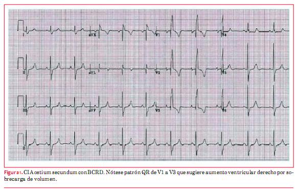
<body><![CDATA[<br> <b><span style="font-size: 10pt; font-family: Verdana; ">Patolog&iacute;as m&aacute;s frecuentes&nbsp;</span></b><span style="font-size: 10pt; font-family: Verdana; "> </span>       <o:p></o:p>           <br> <o:p>&nbsp;</o:p>           <br> <b><span style="font-size: 10pt; font-family: Verdana; ">Comunicaci&oacute;n <span class="SpellE">interauricular</span>&nbsp;</span></b><span style="font-size: 10pt; font-family: Verdana; "> </span><span style="font-size: 10pt;"><o:p></o:p></span>       </p>           <p style="margin: 0cm 0cm 0.0001pt;"> <span style="font-size: 10pt; font-family: Verdana; ">La comunicaci&oacute;n <span class="SpellE">interauricular</span> (CIA) es por lejos la CC que con mayor frecuencia alcanza la edad adulta. Los hallazgos electrocardiogr&aacute;ficos t&iacute;picos de la CIA, tipo <span class="SpellE">ostium</span> <span class="SpellE">secundum</span> (OS), el m&aacute;s com&uacute;n de estos defectos, son la desviaci&oacute;n del eje el&eacute;ctrico del QRS a derecha y el bloqueo de rama <span class="GramE">derecha<sup>(</sup></span></span><sup><span style="font-size: 10pt; font-family: Verdana; "><a name="2_"></a></span><span style="font-size: 10pt; font-family: Verdana; color: rgb(31, 26, 23);"><a href="#2" style="color: blue; text-decoration: underline; text-underline: single">2</a></span><span style="font-size: 10pt; font-family: Verdana; ">)</span></sup><span style="font-size: 10pt; font-family: Verdana; "> (</span><span style="font-size: 10pt; font-family: Verdana; color: rgb(31, 26, 23);"><a href="/img/revistas/ruc/v29n3/3a20f1.JPG" style="color: blue; text-decoration: underline; text-underline: single">figura 1</a></span><span style="font-size: 10pt; font-family: Verdana; ">). Cohen y colaboradores describen que encontrar una muesca en el sector superior de la onda R en derivaciones inferiores tiene alta especificidad para el diagn&oacute;stico de CIA <span style="font-family: Verdana;" class="GramE">OS<sup>(</sup></span></span><sup style="font-family: Verdana;"><span style="font-size: 10pt; font-family: Verdana; "><a name="3_"></a></span><span style="font-size: 10pt; font-family: Verdana; color: rgb(31, 26, 23);"><a href="#3" style="color: blue; text-decoration: underline; text-underline: single">3</a></span><span style="font-size: 10pt; font-family: Verdana; ">)</span></sup><span style="font-size: 10pt; font-family: Verdana; "><span style="font-family: Verdana;">. Esto tiene poca sensibilidad. Se pueden observar ondas P picudas en DII como elemento sugestivo de </span><span style="font-family: Verdana;" class="SpellE">auriculomegalia</span><span style="font-family: Verdana;"> derecha. Ni&ntilde;os con defecto tipo OS generalmente se presentan con ritmo sinusal, en cambio los adultos que mantienen este defecto sin reparar tienen mayor incidencia de fibrilaci&oacute;n auricular (FA) (hasta un tercio de los pacientes mayores de 40 a&ntilde;os), de </span><span style="font-family: Verdana;" class="SpellE">flutter</span><span style="font-family: Verdana;"> y de bradicardia sinusal con ritmo de escape </span><span style="font-family: Verdana;" class="SpellE">unional</span><span style="font-family: Verdana;">. El cierre quir&uacute;rgico de esta comunicaci&oacute;n disminuye la incidencia de </span><span style="font-family: Verdana;" class="SpellE">flutter</span><span style="font-family: Verdana;">, pero no de la FA.&nbsp;</span></span>    <br>     </p>            <p style="margin: 0cm 0cm 0.0001pt;">  <multicol gutter="18" cols="2"></multicol> <span style="font-size: 10pt; font-family: Verdana; ">  <span class="SpellE" style="font-family: Verdana;">En los defectos altos, CIA tipo seno venoso, se aprecia eje anormal de onda P (eje izquierdo y superior en plano frontal</span><span style="font-family: Verdana;" class="GramE SpellE">)<sup>(</sup></span></span><sup class="SpellE" style="font-family: Verdana;"><span style="font-size: 10pt; font-family: Verdana; color: rgb(31, 26, 23);"><a href="#2" style="color: blue; text-decoration: underline; text-underline: single">2</a></span><span style="font-size: 10pt; font-family: Verdana; ">,<a name="4_"></a></span><span style="font-size: 10pt; font-family: Verdana; color: rgb(31, 26, 23);"><a href="#4" style="color: blue; text-decoration: underline; text-underline: single">4</a></span><span style="font-size: 10pt; font-family: Verdana; ">,<a name="5_"></a></span><span style="font-size: 10pt; font-family: Verdana; color: rgb(31, 26, 23);"><a href="#5" style="color: blue; text-decoration: underline; text-underline: single">5</a></span><span style="font-size: 10pt; font-family: Verdana; ">)</span></sup><span style="font-size: 10pt; font-family: Verdana; "><span class="SpellE" style="font-family: Verdana;">. El eje el&eacute;ctrico a izquierda m&aacute;s el retardo en la conducci&oacute;n en la rama derecha del haz de His, que se aprecian en las CIA tipo ostium primum, se debe a la posici&oacute;n de los fasc&iacute;culos de conducci&oacute;n y no debe confundirse con un bloqueo bifascicular</span></span><sup class="SpellE" style="font-family: Verdana;"><span style="font-size: 10pt; font-family: Verdana; ">(</span><span style="font-size: 10pt; font-family: Verdana; color: rgb(31, 26, 23);"><a href="#2" style="color: blue; text-decoration: underline; text-underline: single">2</a></span><span style="font-size: 10pt; font-family: Verdana; ">)</span></sup><span style="font-size: 10pt; font-family: Verdana; "><span class="SpellE" style="font-family: Verdana;">.</span></span><o:p class="SpellE" style="font-family: Verdana;"></o:p></p>            <p class="SpellE" style="margin: 0cm 0cm 0.0001pt; font-family: Verdana;"> <span style="font-size: 10pt; ">&nbsp; </span><span style="font-size: 10pt;"><o:p></o:p></span></p>            <p class="SpellE" style="margin: 0cm 0cm 0.0001pt; font-family: Verdana;"> <span style="font-size: 10pt; ">En casos de CIA familiar puede asociarse el bloqueo <span class="SpellE">auriculoventricular</span> completo (BAVC<span class="GramE">)<sup>(</sup></span></span><sup><span style="font-size: 10pt; "><a name="6_"></a></span><span style="font-size: 10pt; color: rgb(31, 26, 23);"><a href="#6" style="color: blue; text-decoration: underline; text-underline: single">6</a></span><span style="font-size: 10pt; ">).</span><o:p></o:p></sup></p>            <p class="SpellE" style="margin: 0cm 0cm 0.0001pt; font-family: Verdana;"> <span style="font-size: 10pt; ">&nbsp; </span><span style="font-size: 10pt;"><o:p></o:p></span></p>            <p style="margin: 0cm 0cm 0.0001pt;"> <span style="font-size: 10pt; font-family: Verdana; "><span class="SpellE" style="font-family: Verdana;">La PEG es &uacute;til para evidenciar s&iacute;ntomas o evaluar la capacidad de ejercicio en pacientes </span><span style="font-family: Verdana;" class="GramE SpellE">sintom&aacute;ticos<sup>(</sup></span></span><sup class="SpellE" style="font-family: Verdana;"><span style="font-size: 10pt; font-family: Verdana; color: rgb(31, 26, 23);"><a href="#2" style="color: blue; text-decoration: underline; text-underline: single">2</a></span><span style="font-size: 10pt; font-family: Verdana; ">)</span></sup><span style="font-size: 10pt; font-family: Verdana; "><span class="SpellE">.</span>&nbsp;</span><o:p></o:p></p>            ]]></body>
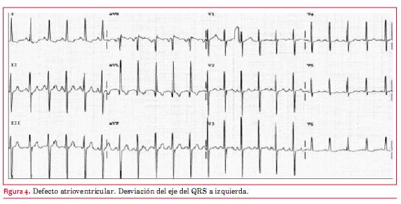
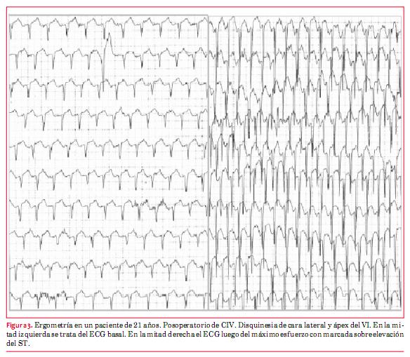
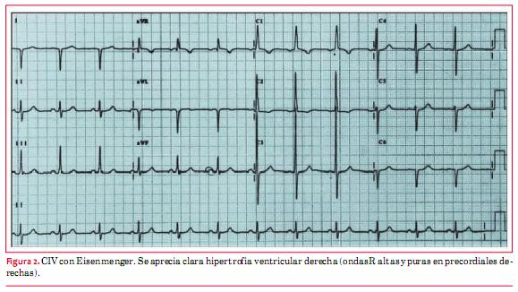
<body><![CDATA[<p style="margin: 0cm 0cm 0.0001pt;"> <span style="font-size: 10pt; font-family: Verdana; ">&nbsp;</span><span style="font-size: 10pt;"><o:p></o:p></span></p>            <p style="margin: 0cm 0cm 0.0001pt;"><b> <span style="font-size: 10pt; font-family: Verdana; ">Comunicaci&oacute;n interventricular&nbsp;</span></b><span style="font-size: 10pt; font-family: Verdana; "> </span><span style="font-size: 10pt;"><o:p></o:p></span></p>            <p style="margin: 0cm 0cm 0.0001pt;"> <span style="font-size: 10pt; font-family: Verdana; ">En la comunicaci&oacute;n interventricular (CIV) con defectos peque&ntilde;os, el ECG suele ser normal. Cuando son de tama&ntilde;o moderado puede apreciarse un patr&oacute;n de sobrecarga diast&oacute;lica caracterizado por ondas q profundas en DI, <span class="SpellE">avL</span>, V5 y <span class="GramE">V6<sup><span style="font-size: 7.5pt;">(</span></sup></span></span><sup><span style="font-size: 7.5pt; font-family: Verdana; color: rgb(31, 26, 23);"><a href="#4" style="color: blue; text-decoration: underline; text-underline: single">4</a></span><span style="font-size: 7.5pt; font-family: Verdana; ">)</span></sup><span style="font-size: 10pt; font-family: Verdana; ">. Cuando se asocia una insuficiencia a&oacute;rtica puede aparecer tard&iacute;amente una FA. En aquellos de tama&ntilde;o grande, que no fueron operados en la infancia y tienen fisiolog&iacute;a de <span class="SpellE">Eisenmenger</span> (enfermedad vascular pulmonar), se aprecia t&iacute;picamente una hipertrofia ventricular derecha en el <span class="GramE">ECG<sup><span style="font-size: 7.5pt;">(</span></sup></span></span><sup><span style="font-size: 7.5pt; font-family: Verdana; color: rgb(31, 26, 23);"><a href="#4" style="color: blue; text-decoration: underline; text-underline: single">4</a></span><span style="font-size: 7.5pt; font-family: Verdana; ">)</span></sup><span style="font-size: 10pt; font-family: Verdana; "> (</span><span style="font-size: 10pt; font-family: Verdana; color: rgb(31, 26, 23);"><a href="#fig_2" style="color: blue; text-decoration: underline; text-underline: single">figura 2</a></span><span style="font-size: 10pt; font-family: Verdana; ">).    <br>     &nbsp; </span>    <br>       <a name="fig_2"></a><img style="width: 579px; height: 326px;" alt="" src="/img/revistas/ruc/v29n3/3a20f2.JPG">    <br>     </p>               <p><span style="font-size: 10pt; font-family: Verdana; ">Los pacientes que desarrollaron bloqueo <span class="SpellE">auriculoventricular</span> (AV) <span class="SpellE">bifascicular</span> o <span class="SpellE">trifascicular</span> transitorio en el <span class="SpellE">posoperatorio</span> del cierre de la comunicaci&oacute;n presentan riesgo de BAVC y deben ser evaluados anualmente con ECG y peri&oacute;dicamente con <span class="SpellE"><span class="GramE">Holter</span></span><span class="GramE"><sup>(</sup></span></span><sup><span style="font-size: 10pt; font-family: Verdana; color: rgb(31, 26, 23);"><a href="#2" style="color: blue; text-decoration: underline; text-underline: single">2</a></span><span style="font-size: 10pt; font-family: Verdana; ">)</span></sup><span style="font-size: 10pt; font-family: Verdana; ">. El cierre con dispositivos de defectos <span class="SpellE">perimembranosos</span> se acompa&ntilde;&oacute; de bloqueo completo de rama derecha (BCRD) y <span class="SpellE">hemibloqueo</span> anterior izquierdo (HBAI) en una minor&iacute;a de pacientes. El BAVC se vio en el 1% de los <span class="GramE">casos<sup>(</sup></span></span><sup><span style="font-size: 10pt; font-family: Verdana; "><a name="7_"></a></span><span style="font-size: 10pt; font-family: Verdana; color: rgb(31, 26, 23);"><a href="#7" style="color: blue; text-decoration: underline; text-underline: single">7</a></span><span style="font-size: 10pt; font-family: Verdana; ">)</span></sup><span style="font-size: 10pt; font-family: Verdana; "> (</span><span style="font-size: 10pt; font-family: Verdana; color: rgb(31, 26, 23);"><a href="/img/revistas/ruc/v29n3/3a20f3.JPG" style="color: blue; text-decoration: underline; text-underline: single">figura 3</a></span><span style="font-size: 10pt; font-family: Verdana; ">).</span></p>        <p style="margin: 0cm 0cm 0.0001pt;"><b> <span style="font-size: 10pt; font-family: Verdana; ">Defectos del <span class="SpellE">septum</span> <span class="SpellE">atrioventricular</span>&nbsp;</span></b><span style="font-size: 10pt; font-family: Verdana; "> </span><span style="font-size: 10pt;"><o:p></o:p></span></p>            <p style="margin: 0cm 0cm 0.0001pt; font-family: Verdana;"> <span style="font-size: 10pt; ">El defecto <span class="SpellE">atrioventricular</span> es la cardiopat&iacute;a t&iacute;pica del s&iacute;ndrome de <span class="SpellE">Down</span> (aunque puede tener otras cardiopat&iacute;as o asociaciones, como la tetralog&iacute;a de <span class="SpellE">Fallot</span>). La mayor&iacute;a de los pacientes han sido intervenidos en la infancia y algunos reintervenidos por insuficiencia de la v&aacute;lvula AV o por una estenosis <span class="SpellE">suba&oacute;rtica</span> que a veces se desarrolla en la evoluci&oacute;n. Los no operados tienen invariablemente una enfermedad vascular pulmonar obstructiva. T&iacute;picamente muestran en el ECG desviaci&oacute;n del eje el&eacute;ctrico del QRS a izquierda (</span><span style="font-size: 10pt; color: rgb(31, 26, 23);"><a href="#fig_4" style="color: blue; text-decoration: underline; text-underline: single">figura 4</a></span><span style="font-size: 10pt; ">). Agrandamiento de la aur&iacute;cula izquierda e hipertrofia del ventr&iacute;culo izquierdo (HVI) pueden estar presentes si hay una importante regurgitaci&oacute;n de la v&aacute;lvula AV. Cuando se asocia una estenosis del tracto de salida del VD o en los no operados que tienen hipertensi&oacute;n pulmonar con fisiolog&iacute;a de <span class="SpellE">Eisenmenger</span> predomina la hipertrofia ventricular derecha (HVD). <span class="SpellE">Flutter</span> y FA pueden ocurrir en pacientes <span class="GramE">mayores<sup>(</sup></span></span><sup><span style="font-size: 10pt; color: rgb(31, 26, 23);"><a href="#2" style="color: blue; text-decoration: underline; text-underline: single">2</a></span><span style="font-size: 10pt; ">)</span></sup><span style="font-size: 10pt; ">. Puede verse tambi&eacute;n bloqueo AV de primer grado. Se aconseja la realizaci&oacute;n de <span class="SpellE">Holter</span> peri&oacute;dico para descartar grados mayores de bloqueo AV.&nbsp;</span></p>        <p style="margin: 0cm 0cm 0.0001pt;">&nbsp;</p>        ]]></body>
<body><![CDATA[<p style="margin: 0cm 0cm 0.0001pt;"> <span style="font-size: 10pt; font-family: Verdana; "><a name="fig_4"></a><img style="width: 577px; height: 294px;" alt="" src="/img/revistas/ruc/v29n3/3a20f4.JPG"></span></p>        <p style="margin: 0cm 0cm 0.0001pt;"><span class="SpellE"><b>  <multicol gutter="18" cols="2"></multicol> <span style="font-size: 10pt; font-family: Verdana; ">  Ductus</span></b></span><b><span style="font-size: 10pt; font-family: Verdana; "> arterioso permeable&nbsp;</span></b><span style="font-size: 10pt; font-family: Verdana; "> </span> <o:p></o:p></p>            <p style="margin: 0cm 0cm 0.0001pt;"> <span style="font-size: 10pt; font-family: Verdana; ">En ni&ntilde;os con <span class="SpellE">ductus</span> arterioso permeable (DAP) grande, con importante <span class="SpellE">shunt</span> de izquierda a derecha, se observa HVI y aumento de la AI por sobrecarga de volumen en cavidades <span class="GramE">izquierdas<sup>(</sup></span></span><sup><span style="font-size: 10pt; font-family: Verdana; color: rgb(31, 26, 23);"><a href="#5" style="color: blue; text-decoration: underline; text-underline: single">5</a></span><span style="font-size: 10pt; font-family: Verdana; ">)</span></sup><span style="font-size: 10pt; font-family: Verdana; ">.&nbsp;</span><o:p></o:p></p>            <p style="margin: 0cm 0cm 0.0001pt;"> <span style="font-size: 10pt; font-family: Verdana; ">&nbsp;</span><span style="font-size: 10pt;"><o:p></o:p></span></p>            <p style="margin: 0cm 0cm 0.0001pt;"> <span style="font-size: 10pt; font-family: Verdana; ">Un paciente con DAP grande raramente se encuentre en la edad adulta. En estos casos los pacientes tienen fisiolog&iacute;a de <span class="SpellE">Eisenmenger</span> con cortocircuito <span class="SpellE">bidireccional</span> por el <span class="SpellE">ductus</span> e hipertensi&oacute;n pulmonar y en el ECG se aprecia una HVD.&nbsp;</span><o:p></o:p></p>            <p style="margin: 0cm 0cm 0.0001pt;"> <span style="font-size: 10pt; font-family: Verdana; ">&nbsp;</span><span style="font-size: 10pt;"><o:p></o:p></span></p>            <p style="margin: 0cm 0cm 0.0001pt;"> <span style="font-size: 10pt; font-family: Verdana; ">En los <span class="SpellE">ductus</span> peque&ntilde;os, que han pasado desapercibidos, el ECG es normal.&nbsp; </span> <o:p></o:p></p>            <p style="margin: 0cm 0cm 0.0001pt;"><span style="font-size: 10pt;"><o:p>&nbsp;</o:p></span></p>            <p style="margin: 0cm 0cm 0.0001pt;"> <span style="font-size: 10pt; font-family: Verdana; ">En los de tama&ntilde;o moderado, con remodelaci&oacute;n del VI, pueden verse en el ECG signos de agrandamiento de la AI, incremento de voltaje de R en V5 y cambios inespec&iacute;ficos de ST y <span class="GramE">T<sup>(</sup></span></span><sup><span style="font-size: 10pt; font-family: Verdana; color: rgb(31, 26, 23);"><a href="#4" style="color: blue; text-decoration: underline; text-underline: single">4</a></span><span style="font-size: 10pt; font-family: Verdana; ">)</span></sup><span style="font-size: 10pt; font-family: Verdana; ">.&nbsp; </span> <o:p></o:p></p>            <p style="margin: 0cm 0cm 0.0001pt;"><span style="font-size: 10pt;"><o:p>&nbsp;</o:p></span></p>            ]]></body>
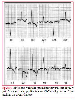
<body><![CDATA[<p style="margin: 0cm 0cm 0.0001pt;"><b> <span style="font-size: 10pt; font-family: Verdana; ">Estenosis valvular pulmonar&nbsp;</span></b><span style="font-size: 10pt; font-family: Verdana; "> </span><span style="font-size: 10pt;"><o:p></o:p></span></p>            <p style="margin: 0cm 0cm 0.0001pt;"> <span style="font-size: 10pt; font-family: Verdana; ">El ECG es &uacute;til para evaluar la severidad de la estenosis. En las formas leves (cuando la presi&oacute;n sist&oacute;lica del VD est&aacute; por debajo de 60 <span class="SpellE">mmHg</span>), el ECG es <span class="GramE">normal<sup>(</sup></span></span><sup><span style="font-size: 10pt; font-family: Verdana; color: rgb(31, 26, 23);"><a href="#2" style="color: blue; text-decoration: underline; text-underline: single">2</a></span><span style="font-size: 10pt; font-family: Verdana; ">)</span></sup><span style="font-size: 10pt; font-family: Verdana; ">. En las moderadas el eje el&eacute;ctrico del QRS se desv&iacute;a a derecha y puede aparecer un patr&oacute;n <span class="SpellE">rsR</span>&rsquo; en precordiales <span class="GramE">derechas<sup>(</sup></span></span><sup><span style="font-size: 10pt; font-family: Verdana; "><a name="8_"></a></span><span style="font-size: 10pt; font-family: Verdana; color: rgb(31, 26, 23);"><a href="#8" style="color: blue; text-decoration: underline; text-underline: single">8</a></span><span style="font-size: 10pt; font-family: Verdana; ">)</span></sup><span style="font-size: 10pt; font-family: Verdana; ">.&nbsp; </span> <o:p></o:p></p>            <p style="margin: 0cm 0cm 0.0001pt;"><span style="font-size: 10pt;"><o:p>&nbsp;</o:p></span></p>            <p style="margin: 0cm 0cm 0.0001pt;"> <span style="font-size: 10pt; font-family: Verdana; ">Cuando la obstrucci&oacute;n es de grado severo pueden visualizarse ondas R puras (R en V1 &gt; 20 <span class="SpellE">mm</span>)</span><sup><span style="font-size: 10pt; font-family: Verdana; "> (<a name="9_"></a></span><span style="font-size: 10pt; font-family: Verdana; color: rgb(31, 26, 23);"><a href="#9" style="color: blue; text-decoration: underline; text-underline: single">9</a></span><span style="font-size: 10pt; font-family: Verdana; ">)</span></sup><span style="font-size: 10pt; font-family: Verdana; ">, complejos QR o RS en V4R y V1 y ocasionalmente tambi&eacute;n patr&oacute;n de sobrecarga derecha (</span><span style="font-size: 10pt; font-family: Verdana; color: rgb(31, 26, 23);"><a href="#fig_5" style="color: blue; text-decoration: underline; text-underline: single">figura 5</a></span><span style="font-size: 10pt; font-family: Verdana; ">). Ocasionalmente se ve crecimiento auricular derecho que se visualiza como una onda P picuda en DII y V1. La altura de la onda R multiplicada por 5 da una estimaci&oacute;n de la presi&oacute;n sist&oacute;lica del VD en <span class="SpellE"><span class="GramE">mmHg</span></span><span class="GramE"><sup>(</sup></span></span><sup><span style="font-size: 10pt; font-family: Verdana; "><a name="10_"></a></span><span style="font-size: 10pt; font-family: Verdana; color: rgb(31, 26, 23);"><a href="#10" style="color: blue; text-decoration: underline; text-underline: single">10</a></span><span style="font-size: 10pt; font-family: Verdana; ">)</span></sup><span style="font-size: 10pt; font-family: Verdana; ">.&nbsp;</span></p>        <p style="margin: 0cm 0cm 0.0001pt;"><a name="fig_5"></a><img style="width: 292px; height: 387px;" alt="" src="/img/revistas/ruc/v29n3/3a20f5.JPG"></p>        <p style="margin: 0cm 0cm 0.0001pt;">&nbsp;</p>                    <p style="margin: 0cm 0cm 0.0001pt;"><b> <span style="font-size: 10pt; font-family: Verdana; ">Coartaci&oacute;n de aorta&nbsp;</span></b><span style="font-size: 10pt; font-family: Verdana; "> </span><span style="font-size: 10pt;"><o:p></o:p></span></p>            <p style="margin: 0cm 0cm 0.0001pt;"> <span style="font-size: 10pt; font-family: Verdana; ">Puede verse coartaci&oacute;n de aorta nativa en adultos con normal crecimiento y desarrollo que se identifica por soplo precordial, soplo de colaterales, hipertensi&oacute;n arterial sist&eacute;mica (HTA) con presi&oacute;n arterial diferente en miembros superiores e inferiores (mayor en miembros superiores) y disminuci&oacute;n o ausencia de pulsos en miembros inferiores. En el ECG &ndash;a diferencia del reci&eacute;n nacido&ndash; se observan signos de HVI, con o sin sobrecarga. En aquellos pacientes que fueron bien corregidos en la infancia, el ECG es habitualmente normal y en los que quedaron con una HTA residual puede verse o no <span class="GramE">HVI<sup>(</sup></span></span><sup><span style="font-size: 10pt; font-family: Verdana; color: rgb(31, 26, 23);"><a href="#2" style="color: blue; text-decoration: underline; text-underline: single">2</a></span><span style="font-size: 10pt; font-family: Verdana; ">)</span></sup><span style="font-size: 10pt; font-family: Verdana; ">.</span><o:p></o:p></p>            <p style="margin: 0cm 0cm 0.0001pt;"> <span style="font-size: 10pt; font-family: Verdana; ">&nbsp; </span><span style="font-size: 10pt;"><o:p></o:p></span></p>            <p style="margin: 0cm 0cm 0.0001pt;"><b> <span style="font-size: 10pt; font-family: Verdana; ">Estenosis valvular a&oacute;rtica&nbsp;</span></b><span style="font-size: 10pt; font-family: Verdana; "> </span><span style="font-size: 10pt;"><o:p></o:p></span></p>            ]]></body>
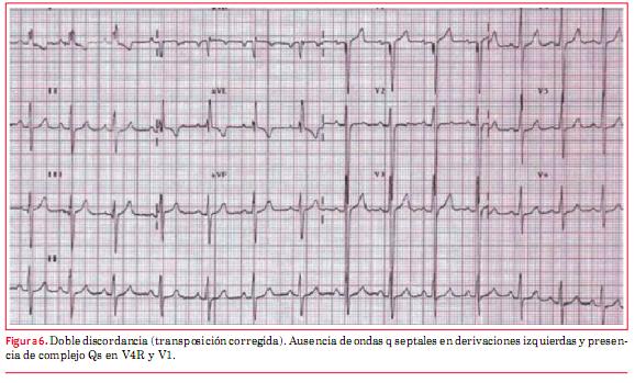
<body><![CDATA[<p>        <span style="font-size: 10pt; font-family: Verdana; ">Las anormalidades electrocardiogr&aacute;ficas est&aacute;n relacionadas habitualmente con la severidad de la estenosis. Se recomienda cl&aacute;sicamente un ECG anual.&nbsp; </span>    <o:p></o:p>    <span style="font-size: 10pt;"><o:p></o:p></span>    <br>     <br> <span style="font-size: 10pt; font-family: Verdana; ">   Cuando la obstrucci&oacute;n es de grado leve el ECG es normal. Si es moderada, es notorio un aumento del voltaje de R en precordiales izquierdas y agrandamiento de la AI. Cuando es de grado severo se puede agregar un patr&oacute;n de sobrecarga (descenso del ST en cara lateral). Pero debe reconocerse y tenerse en cuenta que en algunas estenosis severas el ECG es normal. La PEG es &uacute;til en esta patolog&iacute;a en adolescentes y adultos j&oacute;venes, ya que puede poner en evidencia la aparici&oacute;n de s&iacute;ntomas, permite evaluar la respuesta <span class="SpellE">presora</span> al esfuerzo y descartar arritmias y signos de sobrecarga ventricular izquierda. Las anomal&iacute;as en esta prueba que sugieren una estenosis significativa son una respuesta <span class="SpellE">presora</span> en platillo, un desnivel del ST mayor a 2 <span class="SpellE">mm</span> y las arritmias ventriculares. Debe recordarse que en <span class="SpellE">ergometr&iacute;as</span> realizadas a los adultos mayores portadores de estenosis a&oacute;rtica, el 80% de los pacientes muestran depresi&oacute;n del segmento ST e inversi&oacute;n de T que carecen de valor <span class="GramE">pron&oacute;stico<sup>(</sup></span></span><sup><span style="font-size: 10pt; font-family: Verdana; color: rgb(31, 26, 23);"><a href="#2" style="color: blue; text-decoration: underline; text-underline: single">2</a></span><span style="font-size: 10pt; font-family: Verdana; ">)</span></sup><span style="font-size: 10pt; font-family: Verdana; ">. En cambio, los mismos hallazgos en un adolescente o en un adulto joven pueden ser indicativos de intervenci&oacute;n, ya sea <span class="SpellE">valvuloplastia</span> o sustituci&oacute;n <span class="GramE">valvular<sup>(</sup></span></span><sup><span style="font-size: 10pt; font-family: Verdana; color: rgb(31, 26, 23);"><a href="#2" style="color: blue; text-decoration: underline; text-underline: single">2</a></span><span style="font-size: 10pt; font-family: Verdana; ">)</span></sup><span style="font-size: 10pt; font-family: Verdana; ">.&nbsp; </span>    <o:p></o:p>    </p>        <p style="margin: 0cm 0cm 0.0001pt;"><span style="font-size: 10pt;"><o:p>&nbsp;</o:p></span></p>            <p style="margin: 0cm 0cm 0.0001pt;"><b> <span style="font-size: 10pt; font-family: Verdana; ">Transposici&oacute;n corregida de las grandes arterias&nbsp;</span></b><span style="font-size: 10pt; font-family: Verdana; "> </span><span style="font-size: 10pt;"><o:p></o:p></span></p>            <p>        <span style="font-size: 10pt; font-family: Verdana; ">La transposici&oacute;n corregida de las grandes arterias (L-TGA) o discordancia en las conexiones AV y <span class="SpellE">ventriculoarterial</span> es una CC poco frecuente, pero espor&aacute;dicamente pueden verse casos en la edad adulta. La evoluci&oacute;n depende de las asociaciones <span class="SpellE">lesionales</span>: CIV, estenosis pulmonar, insuficiencia de la v&aacute;lvula AV sist&eacute;mica &ndash;que en este caso es la tric&uacute;spide&ndash;, dilataci&oacute;n y/o alteraciones de la contractilidad del ventr&iacute;culo sist&eacute;mico (VD) y BAVC.&nbsp; </span>       <o:p></o:p>           <br> <span style="font-size: 10pt;"><o:p></o:p></span>    <br> <span style="font-size: 10pt; font-family: Verdana; ">El diagn&oacute;stico de L-TGA debe sospecharse en el ECG:&nbsp; </span><span style="font-size: 10pt;"><o:p></o:p></span>       </p>           <p class="MsoNormal" style="margin-left: 35.7pt; text-indent: -26.7pt;"> <span style="font-size: 10pt; font-family: Symbol">&middot;<span style="font-family: &quot;Times New Roman&quot;; font-style: normal; font-variant: normal; font-weight: normal; font-size: 7pt; line-height: normal; font-size-adjust: none; font-stretch: normal;">&nbsp;&nbsp;&nbsp;</span></span><span style="font-size: 10pt; font-family: Verdana; ">por la ausencia de ondas q <span class="SpellE">septales</span> en DI, <span class="SpellE">avL</span> y de V4 a V6;&nbsp; </span><span style="font-size: 10pt;"><o:p></o:p></span></p>            <p class="MsoNormal" style="margin-left: 18pt; text-indent: -9pt;"> <span style="font-size: 10pt; font-family: Symbol">&middot;<span style="font-family: &quot;Times New Roman&quot;; font-style: normal; font-variant: normal; font-weight: normal; font-size: 7pt; line-height: normal; font-size-adjust: none; font-stretch: normal;">&nbsp;&nbsp;&nbsp;</span></span><span style="font-size: 10pt; font-family: Verdana; ">por la presencia de onda q en V4R y V1. La explicaci&oacute;n de estos hallazgos es que las ramas derecha e izquierda del haz de <span class="SpellE">His</span> est&aacute;n invertidas (recordar que hay inversi&oacute;n ventricular), raz&oacute;n por la cual la activaci&oacute;n <span class="SpellE">septal</span> ocurre de derecha a <span class="GramE">izquierda<sup>(</sup></span></span><sup><span style="font-size: 10pt; font-family: Verdana; color: rgb(31, 26, 23);"><a href="#1" style="color: blue; text-decoration: underline; text-underline: single">1</a></span><span style="font-size: 10pt; font-family: Verdana; ">)</span></sup><span style="font-size: 10pt; font-family: Verdana; "> (</span><span style="font-size: 10pt; font-family: Verdana; color: rgb(31, 26, 23);"><a href="#fig_6" style="color: blue; text-decoration: underline; text-underline: single">figura 6</a></span><span style="font-size: 10pt; font-family: Verdana; ">).</span></p>        ]]></body>
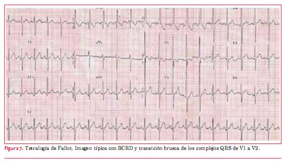
<body><![CDATA[<p>    <span style="font-size: 10pt; font-family: Verdana; ">    <br>    <a name="fig_6"></a><img style="width: 578px; height: 349px;" alt="" src="/img/revistas/ruc/v29n3/3a20f6.JPG"></span> </p>     <p class="MsoNormal" style="margin-left: 18pt; text-indent: -9pt;"><span style="font-size: 10pt;"><o:p></o:p></span></p>            <p style="margin: 0cm 0cm 0.0001pt;">  <multicol gutter="18" cols="2"></multicol> <span style="font-size: 10pt; font-family: Verdana; ">  Puede haber un nodo AV accesorio en muchos <span class="GramE">pacientes<sup>(</sup></span></span><sup><span style="font-size: 10pt; font-family: Verdana; color: rgb(31, 26, 23);"><a href="#10" style="color: blue; text-decoration: underline; text-underline: single">10</a></span><span style="font-size: 10pt; font-family: Verdana; ">)</span><o:p></o:p></sup></p>            <p style="margin: 0cm 0cm 0.0001pt;"> <span style="font-size: 10pt; font-family: Verdana; ">&nbsp; </span><span style="font-size: 10pt;"><o:p></o:p></span></p>            <p>        <span style="font-size: 10pt; font-family: Verdana; ">Es caracter&iacute;stico de esta patolog&iacute;a, como ya se mencion&oacute;, la aparici&oacute;n de un BAVC. Este puede aparecer en el curso del <span class="SpellE">posoperatorio</span> de cierre de CIV asociada (v&iacute;as de conducci&oacute;n pr&oacute;ximas al defecto) o en forma espont&aacute;nea a raz&oacute;n de un 2% por <span class="GramE">a&ntilde;o<sup>(</sup></span></span><sup><span style="font-size: 10pt; font-family: Verdana; "><a name="11_"></a></span><span style="font-size: 10pt; font-family: Verdana; color: rgb(31, 26, 23);"><a href="#11" style="color: blue; text-decoration: underline; text-underline: single">11</a></span><span style="font-size: 10pt; font-family: Verdana; ">-<a name="12_"></a><a name="13_"></a></span><span style="font-size: 10pt; font-family: Verdana; color: rgb(31, 26, 23);"><a href="#13" style="color: blue; text-decoration: underline; text-underline: single">13</a></span><span style="font-size: 10pt; font-family: Verdana; ">)</span></sup><span style="font-size: 10pt; font-family: Verdana; ">. La incidencia de bloqueo se incrementa con la edad y probablemente est&eacute; relacionado con fibrosis en las v&iacute;as de conducci&oacute;n anormalmente largas y que transcurren por trayecto <span class="GramE">inhabitual<sup>(</sup></span></span><sup><span style="font-size: 10pt; font-family: Verdana; color: rgb(31, 26, 23);"><a href="#2" style="color: blue; text-decoration: underline; text-underline: single">2</a></span><span style="font-size: 10pt; font-family: Verdana; ">)</span></sup><span style="font-size: 10pt; font-family: Verdana; ">.&nbsp; </span>       <o:p></o:p>           <br> <span style="font-size: 10pt;"><o:p></o:p></span>    <br> <b><span style="font-size: 10pt; font-family: Verdana; ">Tetralog&iacute;a de <span class="SpellE">Fallot</span>&nbsp;</span></b><span style="font-size: 10pt; font-family: Verdana; "> </span><span style="font-size: 10pt;"><o:p></o:p></span>           <br> <span style="font-size: 10pt; font-family: Verdana; ">La imagen caracter&iacute;stica del ECG en la tetralog&iacute;a de <span class="SpellE">Fallot</span> muestra una HVD con imagen de BCRD del haz de <span class="SpellE">His</span> y transici&oacute;n brusca en V1-V2 (<span class="SpellE">qR</span> o <span class="SpellE">Rs</span> a RS) (</span><span style="font-size: 10pt; font-family: Verdana; color: rgb(31, 26, 23);"><a href="/img/revistas/ruc/v29n3/3a20f7.JPG" style="color: blue; text-decoration: underline; text-underline: single">figura 7</a></span><span style="font-size: 10pt; font-family: Verdana; ">). En aquellos pacientes que fueron reparados con parche <span class="SpellE">transanular</span>, como era la norma antes de 1990, el BCRD est&aacute; casi siempre presente y puede reflejar el grado de dilataci&oacute;n del VD.</span>       <br> <span style="font-size: 10pt;"><o:p></o:p></span><!-- big --><span style="font-family: Verdana;">La duraci&oacute;n de los complejos QRS de 180 </span><span style="font-family: Verdana;" class="SpellE">ms</span><span style="font-family: Verdana;"> o m&aacute;s es un importante </span><span style="font-family: Verdana;" class="SpellE">predictor</span><span style="font-family: Verdana;"> de taquicardia ventricular (TV) sostenida y de MS. El ECG debe realizarse una vez al </span><span style="font-family: Verdana;" class="GramE">a&ntilde;o<sup>(</sup></span><sup style="font-family: Verdana;"><a href="#4" style="color: blue; text-decoration: underline; text-underline: single">4</a>,<a name="14_"></a><a href="#14" style="color: blue; text-decoration: underline; text-underline: single">14</a>)</sup><span style="font-family: Verdana;">.</span><!-- /big --><o:p></o:p>    ]]></body>
<body><![CDATA[<br>    </p>        <p style="margin: 0cm 0cm 0.0001pt;"> <span style="font-size: 10pt; font-family: Verdana; ">&nbsp; </span><span style="font-size: 10pt;"><o:p></o:p></span></p>                   <p>               <span style="font-size: 10pt; font-family: Verdana; ">Debe solicitarse <span class="SpellE">Holter</span> cuando haya sospecha de arritmias. La PEG debe hacerse en forma peri&oacute;dica para determinar capacidad funcional y detecci&oacute;n de arritmias <span class="GramE">ventriculares<sup>(</sup></span></span><sup><span style="font-size: 10pt; font-family: Verdana; color: rgb(31, 26, 23);"><a href="#2" style="color: blue; text-decoration: underline; text-underline: single">2</a></span><span style="font-size: 10pt; font-family: Verdana; ">)</span></sup><span style="font-size: 10pt; font-family: Verdana; ">.<sup>&nbsp;</sup></span>   <span style="font-size: 10pt; font-family: Verdana; ">    <br>    </span>       <br> <span style="font-size: 10pt;"><o:p></o:p></span> <span style="font-size: 10pt; font-family: Verdana; ">La incidencia de MS ha sido estimada en distintas series y es aproximadamente de 2,5% por d&eacute;cada de seguimiento. Los pacientes de mayor riesgo de MS son aquellos que han tenido:&nbsp; </span><span style="font-size: 10pt;"><o:p></o:p></span>    <br>        <span style="font-size: 10pt; font-family: Symbol">&middot;<span style="font-family: &quot;Times New Roman&quot;; font-style: normal; font-variant: normal; font-weight: normal; font-size: 7pt; line-height: normal; font-size-adjust: none; font-stretch: normal;">&nbsp;&nbsp;&nbsp;</span></span><span class="SpellE"><span style="font-size: 10pt; font-family: Verdana; ">shunts</span></span><span style="font-size: 10pt; font-family: Verdana; "> sist&eacute;mico-pulmonares por muchos a&ntilde;os;&nbsp; </span><span style="font-size: 10pt;"><o:p></o:p></span> </p>     <div style="text-align: left;">   <span style="font-size: 10pt; font-family: Symbol">&middot;<span style="font-family: &quot;Times New Roman&quot;; font-style: normal; font-variant: normal; font-weight: normal; font-size: 7pt; line-height: normal; font-size-adjust: none; font-stretch: normal;"> &nbsp; </span></span> <span style="font-size: 10pt; font-family: Verdana; ">mayor edad en el momento de la correcci&oacute;n quir&uacute;rgica;&nbsp; </span><span style="font-size: 10pt;"><o:p></o:p></span>    <br>     </div>               <p><span style="font-size: 10pt; font-family: Symbol">&middot;<span style="font-family: &quot;Times New Roman&quot;; font-style: normal; font-variant: normal; font-weight: normal; font-size: 7pt; line-height: normal; font-size-adjust: none; font-stretch: normal;">&nbsp;&nbsp;&nbsp;</span></span><span style="font-size: 10pt; font-family: Verdana; ">anormalidades hemodin&aacute;micas en el VD debidas a insuficiencia pulmonar u obstrucci&oacute;n del TSVD;</span>    <br>     <span style="font-size: 10pt; font-family: Symbol">&middot; </span><span class="SpellE"> <span style="font-size: 10pt; font-family: Verdana; "> extrasistol&iacute;a</span></span><span style="font-size: 10pt; font-family: Verdana; "> ventricular de alto grado en el <span class="SpellE">Holter</span>;&nbsp; </span><span style="font-size: 10pt;"><o:p></o:p></span>       ]]></body>
<body><![CDATA[<br>     </p>         <div style="text-align: left;">   <span style="font-size: 10pt; font-family: Symbol">&middot;<span style="font-family: &quot;Times New Roman&quot;; font-style: normal; font-variant: normal; font-weight: normal; font-size: 7pt; line-height: normal; font-size-adjust: none; font-stretch: normal;">&nbsp;&nbsp;&nbsp;</span></span><span style="font-size: 10pt; font-family: Verdana; ">taquicardia ventricular <span class="SpellE">inducible</span> en el estudio electrofisiol&oacute;gico;&nbsp; </span><span style="font-size: 10pt;"><o:p></o:p></span></div>            <p><span style="font-size: 10pt; font-family: Symbol">&middot;<span style="font-family: &quot;Times New Roman&quot;; font-style: normal; font-variant: normal; font-weight: normal; font-size: 7pt; line-height: normal; font-size-adjust: none; font-stretch: normal;">&nbsp;&nbsp;&nbsp;</span></span><span style="font-size: 10pt; font-family: Verdana; ">duraci&oacute;n de QRS de 180 <span class="SpellE">ms</span> en el ECG <span class="GramE">basal<sup>(</sup></span></span><sup><span style="font-size: 10pt; font-family: Verdana; color: rgb(31, 26, 23);"><a href="#14" style="color: blue; text-decoration: underline; text-underline: single">14</a></span><span style="font-size: 10pt; font-family: Verdana; ">-<a name="15_"></a><a name="16_"></a><a name="17_"></a><a name="18_"></a><a name="19_"></a><a name="20_"></a></span><span style="font-size: 10pt; font-family: Verdana; color: rgb(31, 26, 23);"><a href="#20" style="color: blue; text-decoration: underline; text-underline: single">20</a></span><span style="font-size: 10pt; font-family: Verdana; ">)</span></sup><span style="font-size: 10pt; font-family: Verdana; ">.&nbsp; </span><span style="font-size: 10pt;"><o:p></o:p></span>   </p>       <p style="margin: 0cm 0cm 0.0001pt;"> <span style="font-size: 10pt; font-family: Verdana; ">La presencia de <span class="SpellE">flutter</span>, FA o TV en pacientes mayores siempre revela serias dificultades <span class="GramE">hemodin&aacute;micas<sup>(</sup></span></span><sup><span style="font-size: 10pt; font-family: Verdana; color: rgb(31, 26, 23);"><a href="#2" style="color: blue; text-decoration: underline; text-underline: single">2</a></span><span style="font-size: 10pt; font-family: Verdana; ">)</span></sup><span style="font-size: 10pt; font-family: Verdana; ">.&nbsp; </span> <o:p></o:p></p>            <p style="margin: 0cm 0cm 0.0001pt;"><span style="font-size: 10pt;"><o:p>&nbsp;</o:p></span></p>            <p style="margin: 0cm 0cm 0.0001pt;"><b> <span style="font-size: 10pt; font-family: Verdana; ">Otras cardiopat&iacute;as cian&oacute;ticas&nbsp;</span></b><span style="font-size: 10pt; font-family: Verdana; "> </span></p>            <p style="margin: 0cm 0cm 0.0001pt;"> <span style="font-size: 10pt; font-family: Verdana; ">El ECG puede tener especial utilidad para distinguir obstrucciones derechas: &nbsp; </span></p>            <p class="MsoNormal" style="margin-left: 18pt; text-indent: -9pt;"> <span style="font-size: 10pt; font-family: Symbol">&middot;<span style="font-family: &quot;Times New Roman&quot;; font-style: normal; font-variant: normal; font-weight: normal; font-size: 7pt; line-height: normal; font-size-adjust: none; font-stretch: normal;">&nbsp;&nbsp;&nbsp;</span></span><span style="font-size: 10pt; font-family: Verdana; ">en la atresia pulmonar con tabique intacto &ndash;que habitualmente tiene VD poco desarrollado&ndash; el eje el&eacute;ctrico est&aacute; entre 0 y 120 grados y hay disminuci&oacute;n de potenciales del VD;&nbsp; </span></p>            <p class="MsoNormal" style="margin-left: 27pt; text-indent: -18pt;"> <span style="font-size: 10pt; font-family: Symbol">&middot;<span style="font-family: &quot;Times New Roman&quot;; font-style: normal; font-variant: normal; font-weight: normal; font-size: 7pt; line-height: normal; font-size-adjust: none; font-stretch: normal;">&nbsp;&nbsp;&nbsp;</span></span><span style="font-size: 10pt; font-family: Verdana; ">en la atresia pulmonar con CIV el eje est&aacute; a derecha y hay hipertrofia ventricular derecha (como en <span class="SpellE">Fallot</span>);&nbsp; </span><o:p></o:p></p>        <p>&nbsp; &nbsp;<span style="font-size: 10pt; font-family: Symbol">&middot;</span><small><span style="font-family: Verdana;"> &nbsp;<!-- big --><!-- big -->en la atresia<!-- /big --><!-- /big --></span><!-- big --><!-- big --><span style="font-family: Verdana;" class="SpellE">&nbsp;tricusp&iacute;dea</span><span style="font-family: Verdana;"> con vasos de salida normalmente relacionados, el eje el&eacute;ctrico est&aacute; caracter&iacute;sticamente desviado a izquierda y los potenciales derechos disminuidos, hay distintos grados de </span><span style="font-family: Verdana;" class="SpellE">hipoplasia</span><span style="font-family: Verdana;"> del </span><span style="font-family: Verdana;" class="GramE">VD<sup><span style="font-size: 7.5pt;">(</span></sup></span><sup style="font-family: Verdana;"><span style="font-size: 7.5pt; "><a name="21_"></a></span><span style="font-size: 7.5pt; color: rgb(31, 26, 23);"><a href="#21" style="color: blue; text-decoration: underline; text-underline: single">21</a></span><span style="font-size: 7.5pt; ">)</span></sup><!-- /big --><!-- /big --></small><span style="font-size: 10pt; font-family: Verdana; "><!-- big -->.<!-- /big -->&nbsp; </span><o:p></o:p>        ]]></body>
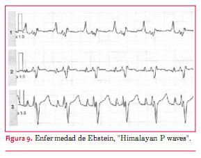
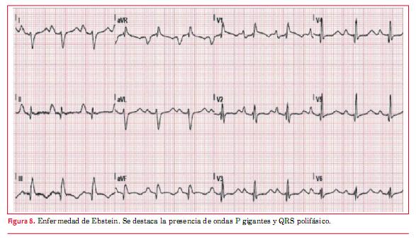
<body><![CDATA[<br> <o:p>&nbsp;</o:p>           <br> <b><span style="font-size: 10pt; font-family: Verdana; ">Enfermedad de <span class="SpellE">Ebstein</span>&nbsp;</span></b><span style="font-size: 10pt; font-family: Verdana; "> </span><span style="font-size: 10pt;"><o:p></o:p></span>       </p>           <p style="margin: 0cm 0cm 0.0001pt;"> <span style="font-size: 10pt; font-family: Verdana; ">Es una patolog&iacute;a poco frecuente con una enorme variabilidad anat&oacute;mica que condiciona el pron&oacute;stico (desde una muerte in &uacute;tero o neonatal hasta una <span class="SpellE">sobrevida</span> de 80 a&ntilde;os sin mayores sobresaltos).&nbsp; </span> <o:p></o:p></p>            <p style="margin: 0cm 0cm 0.0001pt;"><span style="font-size: 10pt;"><o:p>&nbsp;</o:p></span></p>            <p style="margin: 0cm 0cm 0.0001pt;"> <span style="font-size: 10pt; font-family: Verdana; ">El ECG muestra t&iacute;picamente un agrandamiento de AD con ondas P gigantes (<span class="SpellE">Himalayan</span> P <span class="SpellE">waves</span>) (figuras </span><span style="font-size: 10pt; font-family: Verdana; color: rgb(31, 26, 23);">  <a href="/img/revistas/ruc/v29n3/3a20f8.JPG" style="color: blue; text-decoration: underline; text-underline: single">8</a></span><span style="font-size: 10pt; font-family: Verdana; "> y </span><span style="font-size: 10pt; font-family: Verdana; color: rgb(31, 26, 23);">  <a href="#fig_9" style="color: blue; text-decoration: underline; text-underline: single">9</a></span><span style="font-size: 10pt; font-family: Verdana; ">). Tambi&eacute;n son caracter&iacute;sticos bajos voltajes de QRS y BCRD con complejos que a veces son polif&aacute;sicos (<span class="SpellE">rsr</span>&rsquo;r&rsquo;&rsquo;). Puede verse imagen de tipo QR en precordiales derechas debido a la <span class="SpellE">auriculomegalia</span> derecha que permite ver potenciales <span class="SpellE">intracavitarios</span> en dichas derivaciones. El 25% de los pacientes pueden tener un s&iacute;ndrome de <span class="SpellE">preexcitaci&oacute;n</span> tipo <span class="SpellE">Wolff</span>-Parkinson-<span class="SpellE">White</span> asociado a veces con v&iacute;as accesorias m&uacute;ltiples, la mayor&iacute;a de las cuales est&aacute;n situadas del lado derecho</span><sup><span style="font-size: 7.5pt; font-family: Verdana; ">(</span><span style="font-size: 7.5pt; font-family: Verdana; color: rgb(31, 26, 23);"><a href="#2" style="color: blue; text-decoration: underline; text-underline: single">2</a></span><span style="font-size: 7.5pt; font-family: Verdana; ">)</span></sup><span style="font-size: 10pt; font-family: Verdana; ">.    <br>    </span></p>            <p>        <a name="fig_9"></a><img style="width: 289px; height: 227px;" alt="" src="/img/revistas/ruc/v29n3/3a20f9.JPG"> </p>     <p style="margin: 0cm 0cm 0.0001pt;"><b> <span style="font-size: 10pt; font-family: Verdana; ">Procedimiento de <span class="SpellE">Fontan</span>&nbsp;</span></b><span style="font-size: 10pt; font-family: Verdana; "> </span> <o:p></o:p></p>            <p>        <!-- big --><!-- big --><span style="font-size: 10pt; font-family: Verdana; ">La cirug&iacute;a de <span class="SpellE">Fontan</span> es un procedimiento paliativo que se practica en pacientes que tienen anat&oacute;mica o funcionalmente un ventr&iacute;culo &uacute;nico.</span>           <br> <span style="font-size: 10pt; font-family: Verdana; color: rgb(31, 26, 23);"><o:p></o:p></span><!-- /big --><!-- /big -->   <small><!-- big --><!-- big --><span style="font-family: Verdana;">Consiste en derivar todo el retorno venoso sist&eacute;mico hacia las arterias pulmonares sin emplear el ventr&iacute;culo </span><span style="font-family: Verdana;" class="SpellE">subpulmonar</span><span style="font-family: Verdana;">. Dada la diferente anatom&iacute;a de las cardiopat&iacute;as que conducen a esta cirug&iacute;a, se pueden encontrar en el ECG distintos patrones, con grados variables de hipertrofia, trastornos de la conducci&oacute;n </span><span style="font-family: Verdana;" class="SpellE">intraventricular</span><span style="font-family: Verdana;"> y anormalidades del segmento ST y la onda T. Pero lo que se busca fundamentalmente son los trastornos del ritmo. Las arritmias t&iacute;picas son las taquicardias por reentrada </span><span style="font-family: Verdana;" class="SpellE">intraatrial</span><span style="font-family: Verdana;"> &ndash;IART&ndash; (llamados tambi&eacute;n </span><span style="font-family: Verdana;" class="SpellE">flutter</span><span style="font-family: Verdana;"> at&iacute;pico o </span><span style="font-family: Verdana;" class="SpellE">flutter</span><span style="font-family: Verdana;"> lento) y la FA. Son arritmias mal toleradas </span><span style="font-family: Verdana;" class="GramE">debido</span><span style="font-family: Verdana;"> a que se produce un aumento de la presi&oacute;n en la aur&iacute;cula sist&eacute;mica y por consiguiente en todo el sistema </span><span style="font-family: Verdana;" class="SpellE">cavopulmonar</span><span style="font-family: Verdana;">. Los pacientes operados con la t&eacute;cnica de anastomosis directa de AD con arteria pulmonar tienen mayor tendencia a las arritmias que los corregidos con t&uacute;nel lateral o t&uacute;nel </span><span style="font-family: Verdana;" class="SpellE">extracard&iacute;aco</span><!-- /big --><!-- /big --><span style="font-family: Verdana;"><!-- big --><!-- big -->.<!-- /big --><!-- /big -->&nbsp;</span></small> <o:p></o:p>    ]]></body>
<body><![CDATA[<br>    </p>            <p style="margin: 0cm 0cm 0.0001pt;">  <multicol gutter="18" cols="2"></multicol> <span style="font-size: 10pt; font-family: Verdana; ">  En el ECG t&iacute;pico las ondas P son visibles y entre ellas hay per&iacute;odos con l&iacute;nea de base <span class="SpellE">isoel&eacute;ctrica</span>. Las longitudes de ciclo pueden variar ampliamente pero son m&aacute;s lentas que en el <span class="SpellE">flutter</span> t&iacute;pico. Pueden llegar hasta 600 <span class="SpellE">ms</span> y permitir conducci&oacute;n AV 1:1</span><sup><span style="font-size: 10pt; font-family: Verdana; ">(<a name="22_"></a></span><span style="font-size: 10pt; font-family: Verdana; color: rgb(31, 26, 23);"><a href="#22" style="color: blue; text-decoration: underline; text-underline: single">22</a></span><span style="font-size: 10pt; font-family: Verdana; ">)</span></sup><span style="font-size: 10pt; font-family: Verdana; ">.&nbsp; </span> <o:p></o:p></p>            <p style="margin: 0cm 0cm 0.0001pt;"><span style="font-size: 10pt;"><o:p>&nbsp;</o:p></span></p>            <p style="margin: 0cm 0cm 0.0001pt;"><b> <span style="font-size: 10pt; font-family: Verdana; ">Transposici&oacute;n cl&aacute;sica de las grandes arterias&nbsp;</span></b><span style="font-size: 10pt; font-family: Verdana; "> </span><span style="font-size: 10pt;"><o:p></o:p></span></p>            <p style="margin: 0cm 0cm 0.0001pt;"> <span style="font-size: 10pt; font-family: Verdana; ">El ECG depende del tipo de operaci&oacute;n a la que fueron sometidos los pacientes. En la d&eacute;cada de 1980 gan&oacute; aceptaci&oacute;n el <span class="SpellE">switch</span> auricular &ndash;cirug&iacute;a de <span class="SpellE">Mustard</span> o de <span class="SpellE"><span class="GramE">Senning</span></span><span class="GramE"><sup>(</sup></span></span><sup><span style="font-size: 10pt; font-family: Verdana; color: rgb(31, 26, 23);"><a href="#22" style="color: blue; text-decoration: underline; text-underline: single">22</a></span><span style="font-size: 10pt; font-family: Verdana; ">)</span></sup><span style="font-size: 10pt; font-family: Verdana; ">&ndash; (correcci&oacute;n fisiol&oacute;gica). En estos procedimientos se coloca parche auricular que redirige el flujo venoso sist&eacute;mico hacia la mitral (el VI se deja conectado con la arteria pulmonar). Dado que el VD queda como ventr&iacute;culo sist&eacute;mico y persiste conectado a la aorta, en el ECG se observa HVD con desviaci&oacute;n del eje a <span class="GramE">derecha<sup>(</sup></span></span><sup><span style="font-size: 10pt; font-family: Verdana; color: rgb(31, 26, 23);"><a href="#2" style="color: blue; text-decoration: underline; text-underline: single">2</a></span><span style="font-size: 10pt; font-family: Verdana; ">)</span></sup><span style="font-size: 10pt; font-family: Verdana; ">. Tambi&eacute;n puede verse bradicardia sinusal o ritmo de la uni&oacute;n AV por enfermedad del n&oacute;dulo sinusal (que puede quedar afectado por las m&uacute;ltiples l&iacute;neas de sutura en la aur&iacute;cula al colocar el parche).&nbsp;</span></p>        <p style="margin: 0cm 0cm 0.0001pt;">&nbsp;</p>        <p>    <span style="font-size: 10pt; font-family: Verdana; ">Por esta raz&oacute;n se aconseja controlar peri&oacute;dicamente con <span class="SpellE">Holter</span> y PEG para evaluar capacidad funcional, respuesta <span class="SpellE">cronotr&oacute;pica</span> y descartar arritmias &ndash;IART&ndash; </span>       <sup>    <span style="font-size: 10pt; font-family: Verdana; ">(</span><span style="font-size: 10pt; font-family: Verdana; color: rgb(31, 26, 23);"><a href="#2" style="color: blue; text-decoration: underline; text-underline: single">2</a></span><span style="font-size: 10pt; font-family: Verdana; ">)</span></sup><span style="font-size: 10pt; font-family: Verdana; ">. El ECG en los pacientes operados con la t&eacute;cnica de <span class="SpellE">switch</span> arterial (desde 1990 en adelante) suele ser normal si no hay defectos residuales significativos. De todas formas, deben realizarse PEG en forma peri&oacute;dica, cada tres a&ntilde;os, a efectos de descartar cambios <span class="SpellE">isqu&eacute;micos</span> relacionados con el reimplante de las arterias coronarias o estenosis a nivel de los <span class="SpellE">ostium</span> de las <span class="GramE">mismas<sup>(</sup></span></span><sup><span style="font-size: 10pt; font-family: Verdana; color: rgb(31, 26, 23);"><a href="#2" style="color: blue; text-decoration: underline; text-underline: single">2</a></span><span style="font-size: 10pt; font-family: Verdana; ">)</span></sup><span style="font-size: 10pt; font-family: Verdana; ">. Tambi&eacute;n puede observarse HVI o HVD por estenosis <span class="SpellE">supravalvular</span> a&oacute;rtica o pulmonar, respectivamente, referida a los sitios de sutura quir&uacute;rgica en la <span class="SpellE">translocaci&oacute;n</span> de las arterias aorta y pulmonar.&nbsp; </span>       <o:p></o:p>           <br> <span style="font-size: 10pt;"><o:p>&nbsp;</o:p></span>           <br> <o:p>&nbsp;</o:p>           <br> <b><span style="font-size: 10pt; font-family: Verdana; ">Bibliograf&iacute;a&nbsp;</span></b><span style="font-size: 10pt; font-family: Verdana; "> </span>       <o:p></o:p>       </p>           ]]></body>
<body><![CDATA[<p style="margin: 0cm 0cm 0.0001pt;"><span style="font-size: 10pt;"><o:p>&nbsp;</o:p></span></p>            <p style="margin: 0cm 0cm 0.0001pt;"> <span style="font-size: 10pt; font-family: Verdana; "><a name="1"></a> </span><span style="font-size: 10pt; font-family: Verdana; color: rgb(31, 26, 23);"> <a href="#1_" style="color: blue; text-decoration: underline; text-underline: single">1</a></span><span style="font-size: 10pt; font-family: Verdana; ">.&nbsp;&nbsp;&nbsp;&nbsp;<span class="SpellE"><b>Koyak</b></span><b> Z, <span class="SpellE">Harris</span> L, De <span class="SpellE">Groot</span> JR, <span class="SpellE">Silversides</span> CK, <span class="SpellE">Oechslin</span> EN, <span class="SpellE">Bouma</span> BJ, et al. </b><span class="SpellE">Sudden</span> <span class="SpellE">cardiac</span> <span class="SpellE">death</span> in <span class="SpellE">adult</span> <span class="SpellE">congenital</span> <span class="SpellE">heart</span> <span class="SpellE">disease</span>. <span class="SpellE">Circulation</span> 2012; 16:1944&ndash;54.&nbsp; </span><span style="font-size: 10pt;"><o:p></o:p></span></p>            <!-- ref --><p style="margin: 0cm 0cm 0.0001pt;"> <span style="font-size: 10pt; font-family: Verdana; "><a name="2"></a> </span><span style="font-size: 10pt; font-family: Verdana; color: rgb(31, 26, 23);"> <a href="#2_" style="color: blue; text-decoration: underline; text-underline: single">2</a></span><span style="font-size: 10pt; font-family: Verdana; ">.&nbsp;&nbsp;&nbsp;&nbsp;<span class="SpellE"><b>Warnes</b></span><b> CA, <span class="SpellE">Williams</span> RG, <span class="SpellE">Bashore</span> TM, <span class="SpellE">Child</span> JS, <span class="SpellE">Connolly</span> HM, <span class="SpellE">Dearani</span> JA, et al.</b> ACC/AHA 2008 <span class="SpellE">Guidelines</span> <span class="SpellE">for</span> <span class="SpellE">the</span> <span class="SpellE">management</span> <span class="SpellE">of</span> <span class="SpellE">adults</span> <span class="SpellE">with</span> <span class="SpellE">congenital</span> <span class="SpellE">heart</span> <span class="SpellE">disease</span>. <span class="SpellE">Circulation</span> 2008; 118: 2395-451.    &nbsp; </span><span style="font-size: 10pt;"><o:p></o:p></span></p>            <!-- ref --><p style="margin: 0cm 0cm 0.0001pt;"> <span style="font-size: 10pt; font-family: Verdana; "><a name="3"></a> </span><span style="font-size: 10pt; font-family: Verdana; color: rgb(31, 26, 23);"> <a href="#3_" style="color: blue; text-decoration: underline; text-underline: single">3</a></span><span style="font-size: 10pt; font-family: Verdana; ">.&nbsp;&nbsp;&nbsp;&nbsp;<b>Cohen JS, <span class="SpellE">Patton</span> DJ, <span class="SpellE">Giuffre</span> RM.</b> <span class="SpellE">The</span> <span class="SpellE">crochetage</span> <span class="SpellE">pattern</span> in <span class="SpellE">electrocardiograms</span> <span class="SpellE">of</span> <span class="SpellE">pediatric</span> <span class="SpellE">atrial</span> <span class="SpellE">septal</span> <span class="SpellE">defect</span> <span class="SpellE">patients</span>. Can J <span class="SpellE">Cardiol</span> 2000; 16:1241-7.    &nbsp; </span><span style="font-size: 10pt;"><o:p></o:p></span></p>            <!-- ref --><p style="margin: 0cm 0cm 0.0001pt;"> <span style="font-size: 10pt; font-family: Verdana; "><a name="4"></a> </span><span style="font-size: 10pt; font-family: Verdana; color: rgb(31, 26, 23);"> <a href="#4_" style="color: blue; text-decoration: underline; text-underline: single">4</a></span><span style="font-size: 10pt; font-family: Verdana; ">.&nbsp;&nbsp;&nbsp;&nbsp;<span class="SpellE"><b>Gersony</b></span><b> WM, <span class="SpellE">Rosembaum</span> MS.</b> <span class="SpellE">Congenital</span> <span class="SpellE">heart</span> <span class="SpellE">disease</span> in <span class="SpellE">the</span> <span class="SpellE">adult</span>. <span class="SpellE">New</span> <span class="SpellE">York</span>: <span class="SpellE">McGraw</span>-Hill, 2002; 310 p.    &nbsp; </span><span style="font-size: 10pt;"><o:p></o:p></span></p>            <p style="margin: 0cm 0cm 0.0001pt;"> <span style="font-size: 10pt; font-family: Verdana; "><a name="5"></a> </span><span style="font-size: 10pt; font-family: Verdana; color: rgb(31, 26, 23);"> <a href="#5_" style="color: blue; text-decoration: underline; text-underline: single">5</a></span><span style="font-size: 10pt; font-family: Verdana; ">.&nbsp;&nbsp;&nbsp;&nbsp;<span class="SpellE"><b>Surawicz</b></span><b> B, <span class="SpellE">Knilans</span> T.</b> <span class="SpellE">Chou</span>&rsquo;s <span class="SpellE">Electrocardiography</span> in <span class="SpellE">clinical</span> <span class="SpellE">practice</span>. 6th <span class="SpellE">ed</span>. <span class="SpellE">Philadelphia</span>: <span class="SpellE">Sounders</span> <span class="SpellE">Elsevier</span>, 2010.&nbsp; </span><span style="font-size: 10pt;"><o:p></o:p></span></p>            <!-- ref --><p style="margin: 0cm 0cm 0.0001pt;"> <span style="font-size: 10pt; font-family: Verdana; "><a name="6"></a> </span><span style="font-size: 10pt; font-family: Verdana; color: rgb(31, 26, 23);"> <a href="#6_" style="color: blue; text-decoration: underline; text-underline: single">6</a></span><span style="font-size: 10pt; font-family: Verdana; ">.&nbsp;&nbsp;&nbsp;&nbsp;<b>Bizarro RO, <span class="SpellE">Callahan</span> JA, <span class="SpellE">Feldt</span> RH, <span class="SpellE">Kurland</span> LT, <span class="SpellE">Gordon</span> H, <span class="SpellE">Brandenburg</span> RO. </b>Familial <span class="SpellE">atrial</span> <span class="SpellE">septal</span> <span class="SpellE">defect</span> <span class="SpellE">with</span> <span class="SpellE">prolonged</span> <span class="SpellE">atrioventricular</span> <span class="SpellE">conduction</span>. A <span class="SpellE">syndrome</span> <span class="SpellE">showing</span> <span class="SpellE">the</span> <span class="SpellE">autosomal</span> <span class="SpellE">dominant</span> <span class="SpellE">pattern</span> <span class="SpellE">of</span> <span class="SpellE">inheritance</span>. <span class="SpellE">Circulation</span> 1970; 41:677-83.    &nbsp; </span><span style="font-size: 10pt;"><o:p></o:p></span></p>            <!-- ref --><p style="margin: 0cm 0cm 0.0001pt;"> <span style="font-size: 10pt; font-family: Verdana; "><a name="7"></a> </span><span style="font-size: 10pt; font-family: Verdana; color: rgb(31, 26, 23);"> <a href="#7_" style="color: blue; text-decoration: underline; text-underline: single">7</a></span><span style="font-size: 10pt; font-family: Verdana; ">.&nbsp;&nbsp;&nbsp;&nbsp;<span class="SpellE"><b>Masura</b></span><b> J, Gao W, <span class="SpellE">Gavora</span> P, <span class="SpellE">Sun</span> K, <span class="SpellE">Zhou</span> AQ, <span class="SpellE">Jiang</span> S, et al. </b><span class="SpellE">Percutaneous</span> <span class="SpellE">closure</span> <span class="SpellE">of</span> <span class="SpellE">perimembranous</span> ventricular <span class="SpellE">septal</span> <span class="SpellE">defects</span> <span class="SpellE">with</span> <span class="SpellE">the</span> <span class="SpellE">eccentric</span> <span class="SpellE">Amplatzer</span> <span class="SpellE">device</span>: <span class="SpellE">multicentre</span> <span class="SpellE">follow</span>-up <span class="SpellE">study</span>. <span class="SpellE">Pediatr</span> <span class="SpellE">Cardiol</span> 2005; 26:216-9.    &nbsp; </span><span style="font-size: 10pt;"><o:p></o:p></span></p>            <!-- ref --><p style="margin: 0cm 0cm 0.0001pt;"> <span style="font-size: 10pt; font-family: Verdana; "><a name="8"></a> </span><span style="font-size: 10pt; font-family: Verdana; color: rgb(31, 26, 23);"> <a href="#8_" style="color: blue; text-decoration: underline; text-underline: single">8</a></span><span style="font-size: 10pt; font-family: Verdana; ">.&nbsp;&nbsp;&nbsp;&nbsp;<span class="SpellE"><b>Chou</b></span><b> TC, <span class="SpellE">Knilans</span> TK. </b><span class="SpellE">Congenital</span> <span class="SpellE">heart</span> <span class="SpellE">disease</span> in <span class="SpellE">adults</span>. In: <span class="SpellE">Electrocardiography</span> in <span class="SpellE">Clinical</span> <span class="SpellE">Practice</span>. <span class="SpellE">Philadelphia</span>: <span class="SpellE">W.B</span>. <span class="SpellE">Saunders</span>, 1996; p. 296-318.    &nbsp; </span><span style="font-size: 10pt;"><o:p></o:p></span></p>            <!-- ref --><p style="margin: 0cm 0cm 0.0001pt;"> <span style="font-size: 10pt; font-family: Verdana; "><a name="9"></a> </span><span style="font-size: 10pt; font-family: Verdana; color: rgb(31, 26, 23);"> <a href="#9_" style="color: blue; text-decoration: underline; text-underline: single">9</a></span><span style="font-size: 10pt; font-family: Verdana; ">.&nbsp;&nbsp;&nbsp;&nbsp;<span class="SpellE"><b>Ardura</b></span><b> J, <span class="SpellE">Gonzalez</span> C, <span class="SpellE">Andres</span> J.</b> <span class="SpellE">Does</span> <span class="SpellE">mild</span> <span class="SpellE">pulmonary</span> <span class="SpellE">stenosis</span> <span class="SpellE">progress</span> <span class="SpellE">during</span> <span class="SpellE">childhood</span>? A <span class="SpellE">study</span> <span class="SpellE">of</span> <span class="SpellE">its</span> natural <span class="SpellE">course</span>. <span class="SpellE">Clin</span> <span class="SpellE">Cardiol</span> 2004; 27:519-22.    &nbsp; </span><span style="font-size: 10pt;"><o:p></o:p></span></p>            <!-- ref --><p style="margin: 0cm 0cm 0.0001pt;"> <span style="font-size: 10pt; font-family: Verdana; "><a name="10"></a> </span><span style="font-size: 10pt; font-family: Verdana; color: rgb(31, 26, 23);"> <a href="#10_" style="color: blue; text-decoration: underline; text-underline: single">10</a></span><span style="font-size: 10pt; font-family: Verdana; ">.&nbsp;&nbsp;&nbsp;&nbsp;<span class="SpellE"><b>Mehran</b></span><b>-<span class="SpellE">Pou</span> M, <span class="SpellE">Whitney</span> A, <span class="SpellE">Liebman</span> J, <span class="SpellE">Borkat</span> G. </b><span class="SpellE">Quantification</span> <span class="SpellE">of</span> <span class="SpellE">the</span> <span class="SpellE">Frank</span> <span class="SpellE">and</span> <span class="SpellE">McFee</span>-<span class="SpellE">Parungao</span> <span class="SpellE">orthogonal</span> <span class="SpellE">electrocardiogram</span> in valvular <span class="SpellE">pulmonic</span> <span class="SpellE">stenosis</span>. <span class="SpellE">Correlations</span> <span class="SpellE">with</span> <span class="SpellE">hemodynamic</span> <span class="SpellE">measurement</span>. J <span class="SpellE">Electrocardiol</span> 1979; 12:69-76.    &nbsp; </span><span style="font-size: 10pt;"><o:p></o:p></span></p>            <!-- ref --><p style="margin: 0cm 0cm 0.0001pt;"> <span style="font-size: 10pt; font-family: Verdana; "><a name="11"></a> </span><span style="font-size: 10pt; font-family: Verdana; color: rgb(31, 26, 23);"> <a href="#11_" style="color: blue; text-decoration: underline; text-underline: single">11</a></span><span style="font-size: 10pt; font-family: Verdana; ">.&nbsp;&nbsp;&nbsp;&nbsp;<span class="SpellE"><b>Anderson</b></span><b> RH, <span class="SpellE">Becker</span> AE, <span class="SpellE">Arnold</span> R, <span class="SpellE">Wilkinson</span> JL. </b><span class="SpellE">The</span> <span class="SpellE">conduction</span> <span class="SpellE">tissues</span> in <span class="SpellE">congenitally</span> <span class="SpellE">corrected</span> <span class="SpellE">transpositition</span>. <span class="SpellE">Circulation</span> 1974; 50:911-23.    &nbsp; </span><span style="font-size: 10pt;"><o:p></o:p></span></p>            <!-- ref --><p style="margin: 0cm 0cm 0.0001pt;"> <span style="font-size: 10pt; font-family: Verdana; "><a name="12"></a> </span><span style="font-size: 10pt; font-family: Verdana; color: rgb(31, 26, 23);"> <a href="#12_" style="color: blue; text-decoration: underline; text-underline: single">12</a></span><span style="font-size: 10pt; font-family: Verdana; ">.&nbsp;&nbsp;&nbsp;&nbsp;<span class="SpellE"><b>Huhta</b></span><b> JC, <span class="SpellE">Maloney</span> JD, <span class="SpellE">Ritter</span> DG, <span class="SpellE">Ilstrup</span> DM, <span class="SpellE">Feld</span> RH. </b>Complete <span class="SpellE">atrioventricular</span> <span class="SpellE">block</span> in <span class="SpellE">patients</span> <span class="SpellE">with</span> <span class="SpellE">atrioventricular</span> <span class="SpellE">discordance</span>. <span class="SpellE">Circulation</span>. 1983; 67:1374-7.    &nbsp; </span><span style="font-size: 10pt;"><o:p></o:p></span></p>            <!-- ref --><p style="margin: 0cm 0cm 0.0001pt;"> <span style="font-size: 10pt; font-family: Verdana; "><a name="13"></a> </span><span style="font-size: 10pt; font-family: Verdana; color: rgb(31, 26, 23);"> <a href="#13_" style="color: blue; text-decoration: underline; text-underline: single">13</a></span><span style="font-size: 10pt; font-family: Verdana; ">.&nbsp;&nbsp;&nbsp;&nbsp;<span class="SpellE"><b>Bharati</b></span><b> S, <span class="SpellE">Mc</span> <span class="SpellE">Cue</span> CM, <span class="SpellE">Tingelsted</span> JB, <span class="SpellE">Mantakas</span> M, <span class="SpellE">Shiel</span> F, Lev M.</b> <span class="SpellE">Lack</span> <span class="SpellE">of</span> <span class="SpellE">connection</span> <span class="SpellE">between</span> <span class="SpellE">the</span> <span class="SpellE">atrial</span> <span class="SpellE">and</span> <span class="SpellE">the</span> <span class="SpellE">periphereal</span> <span class="SpellE">conduction</span> <span class="SpellE">system</span> in a case <span class="SpellE">of</span> <span class="SpellE">corrected</span> <span class="SpellE">transposition</span> <span class="SpellE">with</span> <span class="SpellE">congenital</span> <span class="SpellE">atrioventricular</span> <span class="SpellE">block</span>. <span class="SpellE">Am</span> J <span class="SpellE">Cardiol</span> 1978; 42:47-53.    &nbsp; </span><span style="font-size: 10pt;"><o:p></o:p></span></p>            <!-- ref --><p style="margin: 0cm 0cm 0.0001pt;"> <span style="font-size: 10pt; font-family: Verdana; "><a name="14"></a> </span><span style="font-size: 10pt; font-family: Verdana; color: rgb(31, 26, 23);"> <a href="#14_" style="color: blue; text-decoration: underline; text-underline: single">14</a></span><span style="font-size: 10pt; font-family: Verdana; ">.&nbsp;&nbsp;&nbsp;&nbsp;<span class="SpellE"><b>Gatzoulis</b></span><b> MA, <span class="SpellE">Till</span> JA, <span class="SpellE">Somerville</span> J, <span class="SpellE">Redinghton</span> AN. </b><span class="SpellE">Mechanoelectrical</span> <span class="SpellE">interaction</span> in <span class="SpellE">tetralogy</span> <span class="SpellE">of</span> <span class="SpellE">Fallot</span>. QRS <span class="SpellE">prolongation</span> relates <span class="SpellE">to</span> <span class="SpellE">right</span> ventricular <span class="SpellE">size</span> <span class="SpellE">and</span> <span class="SpellE">predicts</span> <span class="SpellE">malignant</span> ventricular <span class="SpellE">arrhythmias</span> <span class="SpellE">and</span> <span class="SpellE">sudden</span> <span class="SpellE">death</span>. <span class="SpellE">Circulation</span> 1995; 92:231-7.    &nbsp; </span><span style="font-size: 10pt;"><o:p></o:p></span></p>            <!-- ref --><p style="margin: 0cm 0cm 0.0001pt;"> <span style="font-size: 10pt; font-family: Verdana; "><a name="15"></a> </span><span style="font-size: 10pt; font-family: Verdana; color: rgb(31, 26, 23);"> <a href="#15_" style="color: blue; text-decoration: underline; text-underline: single">15</a></span><span style="font-size: 10pt; font-family: Verdana; ">.&nbsp;&nbsp;&nbsp;&nbsp;<span class="SpellE"><b>Walsh</b></span><b> EP, <span class="SpellE">Rockenmacher</span> S, <span class="SpellE">Keane</span> JF, <span class="SpellE">HougenTJ</span>, <span class="SpellE">Lock</span> JE, <span class="SpellE">Castaneda</span> AR.</b> Late <span class="SpellE">results</span> in <span class="SpellE">patients</span> <span class="SpellE">with</span> <span class="SpellE">tetralogy</span> <span class="SpellE">of</span> <span class="SpellE">Fallot</span> <span class="SpellE">repaired</span> <span class="SpellE">during</span> <span class="SpellE">infancy</span>. <span class="SpellE">Circulation</span> 1988; 77:1062-7.    </span><span style="font-size: 10pt; font-family: &quot;Courier New&quot;; ">&nbsp;</span><span style="font-size: 10pt; font-family: Verdana; "> </span><span style="font-size: 10pt;"><o:p></o:p></span></p>            <!-- ref --><p style="margin: 0cm 0cm 0.0001pt;"> <span style="font-size: 10pt; font-family: Verdana; "><a name="16"></a> </span><span style="font-size: 10pt; font-family: Verdana; color: rgb(31, 26, 23);"> <a href="#16_" style="color: blue; text-decoration: underline; text-underline: single">16</a></span><span style="font-size: 10pt; font-family: Verdana; ">.&nbsp;&nbsp;&nbsp;&nbsp;<span class="SpellE"><b>Chandar</b></span><b> JS, <span class="SpellE">Wolff</span> GS, <span class="SpellE">Garson</span> A <span class="SpellE">Jr</span>, <span class="SpellE">Bell</span> TJ, <span class="SpellE">Beder</span> SD, <span class="SpellE">Bink</span>-<span class="SpellE">Boelkens</span> M, et al.</b> Ventricular <span class="SpellE">arrhythmias</span> in <span class="SpellE">postoperative</span> <span class="SpellE">tetralogy</span> <span class="SpellE">of</span> <span class="SpellE">Fallot</span>. <span class="SpellE">Am</span> J <span class="SpellE">Cardiol</span> 1990; 65:655-61.    &nbsp; </span><span style="font-size: 10pt;"><o:p></o:p></span></p>            <!-- ref --><p style="margin: 0cm 0cm 0.0001pt;"> <span style="font-size: 10pt; font-family: Verdana; "><a name="17"></a> </span><span style="font-size: 10pt; font-family: Verdana; color: rgb(31, 26, 23);"> <a href="#17_" style="color: blue; text-decoration: underline; text-underline: single">17</a></span><span style="font-size: 10pt; font-family: Verdana; ">.&nbsp;&nbsp;&nbsp;&nbsp;<span class="SpellE"><b>Gatzoulis</b></span><b> MA, <span class="SpellE">Balaji</span> S, <span class="SpellE">Webber</span> SA, <span class="SpellE">Siu</span> SC, <span class="SpellE">Hokanson</span> JS, <span class="SpellE">Poile</span> C, et al.</b> <span class="SpellE">Risk</span> <span class="SpellE">factors</span> <span class="SpellE">for</span> <span class="SpellE">arrhythmia</span> <span class="SpellE">and</span> <span class="SpellE">sudden</span> <span class="SpellE">cardiac</span> <span class="SpellE">death</span> late <span class="SpellE">after</span> <span class="SpellE">repair</span> <span class="SpellE">of</span> <span class="SpellE">tetralogy</span> <span class="SpellE">of</span> <span class="SpellE">Fallot</span>: a <span class="SpellE">multicenter</span> <span class="SpellE">study</span>. <span class="SpellE">Lancet</span> 2000; 356:975-81.    &nbsp; </span><span style="font-size: 10pt;"><o:p></o:p></span></p>            <!-- ref --><p style="margin: 0cm 0cm 0.0001pt;"> <span style="font-size: 10pt; font-family: Verdana; "><a name="18"></a> </span><span style="font-size: 10pt; font-family: Verdana; color: rgb(31, 26, 23);"> <a href="#18_" style="color: blue; text-decoration: underline; text-underline: single">18</a></span><span style="font-size: 10pt; font-family: Verdana; ">.&nbsp;&nbsp;&nbsp;&nbsp;<span class="SpellE"><b>Therrien</b></span><b> J, <span class="SpellE">Siu</span> SC, <span class="SpellE">Harris</span> L, Dore A, <span class="SpellE">Niwa</span> K, <span class="SpellE">Janousek</span> J, et al.</b> <span class="SpellE">Impact</span> <span class="SpellE">of</span> <span class="SpellE">pulmonary</span> <span class="SpellE">valve</span> <span class="SpellE">replacement</span> <span class="SpellE">on</span> <span class="SpellE">arrhythmia</span> <span class="SpellE">propensity</span> late <span class="SpellE">after</span> <span class="SpellE">repair</span> <span class="SpellE">of</span> <span class="SpellE">tetralogy</span> <span class="SpellE">of</span> <span class="SpellE">Fallot</span>. <span class="SpellE">Circulation</span> 2001; 103:2489-94.    &nbsp; </span><span style="font-size: 10pt;"><o:p></o:p></span></p>              <!-- ref --><p style="margin: 0cm 0cm 0.0001pt;"> <span style="font-size: 10pt; font-family: Verdana; ">  <a name="19"></a> </span><span style="font-size: 10pt; font-family: Verdana; color: rgb(31, 26, 23);">  <a href="#19_" style="color: blue; text-decoration: underline; text-underline: single">19</a></span><span style="font-size: 10pt; font-family: Verdana; ">.&nbsp;&nbsp;&nbsp;&nbsp;<span class="SpellE"><b>Daliento</b></span><b> L, <span class="SpellE">Rizzoli</span> G, <span class="SpellE">Menti</span> L, <span class="SpellE">Baratella</span> MC, <span class="SpellE">Turrini</span> P, <span class="SpellE">Nava</span> A, et al.</b> <span class="SpellE">Accuracy</span> <span class="SpellE">of</span> <span class="SpellE">electrocardiographic</span> <span class="SpellE">and</span> <span class="SpellE">ecocardiographic</span> <span class="SpellE">indices</span> in <span class="SpellE">predicting</span> <span class="SpellE">life</span> <span class="SpellE">threatening</span> ventricular <span class="SpellE">arrhythmias</span> in <span class="SpellE">patients</span> <span class="SpellE">operated</span> <span class="SpellE">for</span> <span class="SpellE">tetralogy</span> <span class="SpellE">of</span> <span class="SpellE">Fallot</span>. <span class="SpellE">Heart</span> 1999; 81:650-5.    &nbsp; </span><span style="font-size: 10pt;"><o:p></o:p></span></p>            <!-- ref --><p style="margin: 0cm 0cm 0.0001pt;"> <span style="font-size: 10pt; font-family: Verdana; "><a name="20"></a> </span><span style="font-size: 10pt; font-family: Verdana; color: rgb(31, 26, 23);"> <a href="#20_" style="color: blue; text-decoration: underline; text-underline: single">20</a></span><span style="font-size: 10pt; font-family: Verdana; ">.&nbsp;&nbsp;&nbsp;&nbsp;<span class="SpellE"><b>Garson</b></span><b> A <span class="SpellE">Jr</span>, <span class="SpellE">Randall</span> DC, Gillette PC, <span class="SpellE">Smith</span> RT, <span class="SpellE">Moak</span> JP, <span class="SpellE">McVey</span> P, et al. </b><span class="SpellE">Prevention</span> <span class="SpellE">of</span> <span class="SpellE">sudden</span> <span class="SpellE">death</span> <span class="SpellE">after</span> <span class="SpellE">repair</span> <span class="SpellE">of</span> <span class="SpellE">tetralogy</span> <span class="SpellE">of</span> <span class="SpellE">Fallot</span>: <span class="SpellE">treatment</span> <span class="SpellE">of</span> ventricular <span class="SpellE">arrhythmias</span>. J <span class="SpellE">Am</span> <span class="SpellE">Coll</span> <span class="SpellE">Cardiol</span>. 1985; 6:221-7.    &nbsp; </span><span style="font-size: 10pt;"><o:p></o:p></span></p>            <p style="margin: 0cm 0cm 0.0001pt;"> <span style="font-size: 10pt; font-family: Verdana; "><a name="21"></a> </span><span style="font-size: 10pt; font-family: Verdana; color: rgb(31, 26, 23);"> <a href="#21_" style="color: blue; text-decoration: underline; text-underline: single">21</a></span><span style="font-size: 10pt; font-family: Verdana; ">.&nbsp;&nbsp;&nbsp;&nbsp;<span class="SpellE"><b>Keane</b></span><b> JF, <span class="SpellE">Lock</span> JE, <span class="SpellE">Fyler</span> DC.</b> Nadas&rsquo; <span class="SpellE">Pediatric</span> <span class="SpellE">Cardiology</span>. 2nd <span class="SpellE">ed</span>. <span class="SpellE">Philadelphia</span>: <span class="SpellE">Elsevier</span>, 2006.&nbsp; </span><span style="font-size: 10pt;"><o:p></o:p></span></p>            ]]></body>
<body><![CDATA[<!-- ref --><p style="margin: 0cm 0cm 0.0001pt;"> <span style="font-size: 10pt; font-family: Verdana; "><a name="22"></a> </span><span style="font-size: 10pt; font-family: Verdana; color: rgb(31, 26, 23);"> <a href="#22_" style="color: blue; text-decoration: underline; text-underline: single">22</a></span><span style="font-size: 10pt; font-family: Verdana; ">.&nbsp;&nbsp;&nbsp;&nbsp;<span class="SpellE"><b>Walsh</b></span><b> EP, <span class="SpellE">Saul</span> JP, <span class="SpellE">Triedman</span> JK.</b> <span class="SpellE">Cardiac</span> <span class="SpellE">arrhythmias</span>. In: <span class="SpellE">Children</span> <span class="SpellE">and</span> <span class="SpellE">young</span> <span class="SpellE">adults</span> <span class="SpellE">with</span> <span class="SpellE">congenital</span> <span class="SpellE">heart</span> <span class="SpellE">disease</span>. <span class="SpellE">Philadelphia</span>: <span class="SpellE">Lippincot</span>-<span class="SpellE">Williams</span> <span class="SpellE">and</span> <span class="SpellE">Wilkins</span>,  2001.    </span></p>               ]]></body><back>
<ref-list>
<ref id="B1">
<label>1</label><nlm-citation citation-type="journal">
<person-group person-group-type="author">
<name>
<surname><![CDATA[Koyak]]></surname>
<given-names><![CDATA[Z]]></given-names>
</name>
<name>
<surname><![CDATA[Harris]]></surname>
<given-names><![CDATA[L]]></given-names>
</name>
<name>
<surname><![CDATA[De Groot]]></surname>
<given-names><![CDATA[JR]]></given-names>
</name>
<name>
<surname><![CDATA[Silversides]]></surname>
<given-names><![CDATA[CK]]></given-names>
</name>
<name>
<surname><![CDATA[Oechslin]]></surname>
<given-names><![CDATA[EN]]></given-names>
</name>
<name>
<surname><![CDATA[Bouma]]></surname>
<given-names><![CDATA[BJ]]></given-names>
</name>
</person-group>
<article-title xml:lang="en"><![CDATA[Sudden cardiac death in adult congenital heart disease]]></article-title>
<source><![CDATA[Circulation]]></source>
<year>2012</year>
<volume>16</volume>
<page-range>1944-54</page-range></nlm-citation>
</ref>
<ref id="B2">
<label>2</label><nlm-citation citation-type="journal">
<person-group person-group-type="author">
<name>
<surname><![CDATA[Warnes]]></surname>
<given-names><![CDATA[CA]]></given-names>
</name>
<name>
<surname><![CDATA[Williams]]></surname>
<given-names><![CDATA[RG]]></given-names>
</name>
<name>
<surname><![CDATA[Bashore]]></surname>
<given-names><![CDATA[TM]]></given-names>
</name>
<name>
<surname><![CDATA[Child]]></surname>
<given-names><![CDATA[JS]]></given-names>
</name>
<name>
<surname><![CDATA[Connolly]]></surname>
<given-names><![CDATA[HM]]></given-names>
</name>
<name>
<surname><![CDATA[Dearani]]></surname>
<given-names><![CDATA[JA]]></given-names>
</name>
</person-group>
<article-title xml:lang="en"><![CDATA[ACC/AHA 2008 Guidelines for the management of adults with congenital heart disease]]></article-title>
<source><![CDATA[Circulation]]></source>
<year>2008</year>
<volume>118</volume>
<page-range>2395-451</page-range></nlm-citation>
</ref>
<ref id="B3">
<label>3</label><nlm-citation citation-type="journal">
<person-group person-group-type="author">
<name>
<surname><![CDATA[Cohen]]></surname>
<given-names><![CDATA[JS]]></given-names>
</name>
<name>
<surname><![CDATA[Patton]]></surname>
<given-names><![CDATA[DJ]]></given-names>
</name>
<name>
<surname><![CDATA[Giuffre]]></surname>
<given-names><![CDATA[RM]]></given-names>
</name>
</person-group>
<article-title xml:lang="en"><![CDATA[The crochetage pattern in electrocardiograms of pediatric atrial septal defect patients]]></article-title>
<source><![CDATA[Can J Cardiol]]></source>
<year>2000</year>
<volume>16</volume>
<page-range>1241-7</page-range></nlm-citation>
</ref>
<ref id="B4">
<label>4</label><nlm-citation citation-type="book">
<person-group person-group-type="author">
<name>
<surname><![CDATA[Gersony]]></surname>
<given-names><![CDATA[WM]]></given-names>
</name>
<name>
<surname><![CDATA[Rosembaum]]></surname>
<given-names><![CDATA[MS]]></given-names>
</name>
</person-group>
<source><![CDATA[Congenital heart disease in the adult]]></source>
<year>2002</year>
<page-range>310</page-range><publisher-loc><![CDATA[New York ]]></publisher-loc>
<publisher-name><![CDATA[McGraw-Hill]]></publisher-name>
</nlm-citation>
</ref>
<ref id="B5">
<label>5</label><nlm-citation citation-type="book">
<person-group person-group-type="author">
<name>
<surname><![CDATA[Surawicz]]></surname>
<given-names><![CDATA[B]]></given-names>
</name>
<name>
<surname><![CDATA[Knilans]]></surname>
<given-names><![CDATA[T]]></given-names>
</name>
</person-group>
<source><![CDATA[Chou&rsquo;s Electrocardiography in clinical practice]]></source>
<year>2010</year>
<edition>6th</edition>
<publisher-loc><![CDATA[Philadelphia ]]></publisher-loc>
<publisher-name><![CDATA[Sounders Elsevier]]></publisher-name>
</nlm-citation>
</ref>
<ref id="B6">
<label>6</label><nlm-citation citation-type="journal">
<person-group person-group-type="author">
<name>
<surname><![CDATA[Bizarro]]></surname>
<given-names><![CDATA[RO]]></given-names>
</name>
<name>
<surname><![CDATA[Callahan]]></surname>
<given-names><![CDATA[JA]]></given-names>
</name>
<name>
<surname><![CDATA[Feldt]]></surname>
<given-names><![CDATA[RH]]></given-names>
</name>
<name>
<surname><![CDATA[Kurland]]></surname>
<given-names><![CDATA[LT]]></given-names>
</name>
<name>
<surname><![CDATA[Gordon]]></surname>
<given-names><![CDATA[H]]></given-names>
</name>
<name>
<surname><![CDATA[Brandenburg]]></surname>
<given-names><![CDATA[RO]]></given-names>
</name>
</person-group>
<article-title xml:lang="en"><![CDATA[Familial atrial septal defect with prolonged atrioventricular conduction: A syndrome showing the autosomal dominant pattern of inheritance]]></article-title>
<source><![CDATA[Circulation]]></source>
<year>1970</year>
<volume>41</volume>
<page-range>677-83</page-range></nlm-citation>
</ref>
<ref id="B7">
<label>7</label><nlm-citation citation-type="journal">
<person-group person-group-type="author">
<name>
<surname><![CDATA[Masura]]></surname>
<given-names><![CDATA[J]]></given-names>
</name>
<name>
<surname><![CDATA[Gao]]></surname>
<given-names><![CDATA[W]]></given-names>
</name>
<name>
<surname><![CDATA[Gavora]]></surname>
<given-names><![CDATA[P]]></given-names>
</name>
<name>
<surname><![CDATA[Sun]]></surname>
<given-names><![CDATA[K]]></given-names>
</name>
<name>
<surname><![CDATA[Zhou]]></surname>
<given-names><![CDATA[AQ]]></given-names>
</name>
<name>
<surname><![CDATA[Jiang]]></surname>
<given-names><![CDATA[S]]></given-names>
</name>
</person-group>
<article-title xml:lang="en"><![CDATA[Percutaneous closure of perimembranous ventricular septal defects with the eccentric Amplatzer device: multicentre follow-up study]]></article-title>
<source><![CDATA[Pediatr Cardiol]]></source>
<year>2005</year>
<volume>26</volume>
<page-range>216-9</page-range></nlm-citation>
</ref>
<ref id="B8">
<label>8</label><nlm-citation citation-type="book">
<person-group person-group-type="author">
<name>
<surname><![CDATA[Chou]]></surname>
<given-names><![CDATA[TC]]></given-names>
</name>
<name>
<surname><![CDATA[Knilans]]></surname>
<given-names><![CDATA[TK]]></given-names>
</name>
</person-group>
<article-title xml:lang="en"><![CDATA[Congenital heart disease in adults]]></article-title>
<source><![CDATA[Electrocardiography in Clinical Practice]]></source>
<year>1996</year>
<page-range>296-318</page-range><publisher-loc><![CDATA[Philadelphia ]]></publisher-loc>
<publisher-name><![CDATA[W.B. Saunders]]></publisher-name>
</nlm-citation>
</ref>
<ref id="B9">
<label>9</label><nlm-citation citation-type="journal">
<person-group person-group-type="author">
<name>
<surname><![CDATA[Ardura]]></surname>
<given-names><![CDATA[J]]></given-names>
</name>
<name>
<surname><![CDATA[Gonzalez]]></surname>
<given-names><![CDATA[C]]></given-names>
</name>
<name>
<surname><![CDATA[Andres]]></surname>
<given-names><![CDATA[J]]></given-names>
</name>
</person-group>
<article-title xml:lang="en"><![CDATA[Does mild pulmonary stenosis progress during childhood?: A study of its natural course]]></article-title>
<source><![CDATA[Clin Cardiol]]></source>
<year>2004</year>
<volume>27</volume>
<page-range>519-22</page-range></nlm-citation>
</ref>
<ref id="B10">
<label>10</label><nlm-citation citation-type="journal">
<person-group person-group-type="author">
<name>
<surname><![CDATA[Mehran-Pou]]></surname>
<given-names><![CDATA[M]]></given-names>
</name>
<name>
<surname><![CDATA[Whitney]]></surname>
<given-names><![CDATA[A]]></given-names>
</name>
<name>
<surname><![CDATA[Liebman]]></surname>
<given-names><![CDATA[J]]></given-names>
</name>
<name>
<surname><![CDATA[Borkat]]></surname>
<given-names><![CDATA[G]]></given-names>
</name>
</person-group>
<article-title xml:lang="en"><![CDATA[Quantification of the Frank and McFee-Parungao orthogonal electrocardiogram in valvular pulmonic stenosis: Correlations with hemodynamic measurement]]></article-title>
<source><![CDATA[J Electrocardiol]]></source>
<year>1979</year>
<volume>12</volume>
<page-range>69-76</page-range></nlm-citation>
</ref>
<ref id="B11">
<label>11</label><nlm-citation citation-type="journal">
<person-group person-group-type="author">
<name>
<surname><![CDATA[Anderson]]></surname>
<given-names><![CDATA[RH]]></given-names>
</name>
<name>
<surname><![CDATA[Becker]]></surname>
<given-names><![CDATA[AE]]></given-names>
</name>
<name>
<surname><![CDATA[Arnold]]></surname>
<given-names><![CDATA[R]]></given-names>
</name>
<name>
<surname><![CDATA[Wilkinson]]></surname>
<given-names><![CDATA[JL]]></given-names>
</name>
</person-group>
<article-title xml:lang="en"><![CDATA[The conduction tissues in congenitally corrected transpositition]]></article-title>
<source><![CDATA[Circulation]]></source>
<year>1974</year>
<volume>50</volume>
<page-range>911-23</page-range></nlm-citation>
</ref>
<ref id="B12">
<label>12</label><nlm-citation citation-type="journal">
<person-group person-group-type="author">
<name>
<surname><![CDATA[Huhta]]></surname>
<given-names><![CDATA[JC]]></given-names>
</name>
<name>
<surname><![CDATA[Maloney]]></surname>
<given-names><![CDATA[JD]]></given-names>
</name>
<name>
<surname><![CDATA[Ritter]]></surname>
<given-names><![CDATA[DG]]></given-names>
</name>
<name>
<surname><![CDATA[Ilstrup]]></surname>
<given-names><![CDATA[DM]]></given-names>
</name>
<name>
<surname><![CDATA[Feld]]></surname>
<given-names><![CDATA[RH]]></given-names>
</name>
</person-group>
<article-title xml:lang="en"><![CDATA[Complete atrioventricular block in patients with atrioventricular discordance]]></article-title>
<source><![CDATA[Circulation]]></source>
<year>1983</year>
<volume>67</volume>
<page-range>1374-7</page-range></nlm-citation>
</ref>
<ref id="B13">
<label>13</label><nlm-citation citation-type="journal">
<person-group person-group-type="author">
<name>
<surname><![CDATA[Bharati]]></surname>
<given-names><![CDATA[S]]></given-names>
</name>
<name>
<surname><![CDATA[Mc Cue]]></surname>
<given-names><![CDATA[CM]]></given-names>
</name>
<name>
<surname><![CDATA[Tingelsted]]></surname>
<given-names><![CDATA[JB]]></given-names>
</name>
<name>
<surname><![CDATA[Mantakas]]></surname>
<given-names><![CDATA[M]]></given-names>
</name>
<name>
<surname><![CDATA[Shiel]]></surname>
<given-names><![CDATA[F]]></given-names>
</name>
<name>
<surname><![CDATA[Lev]]></surname>
<given-names><![CDATA[M]]></given-names>
</name>
</person-group>
<article-title xml:lang="en"><![CDATA[Lack of connection between the atrial and the periphereal conduction system in a case of corrected transposition with congenital atrioventricular block]]></article-title>
<source><![CDATA[Am J Cardiol]]></source>
<year>1978</year>
<volume>42</volume>
<page-range>47-53</page-range></nlm-citation>
</ref>
<ref id="B14">
<label>14</label><nlm-citation citation-type="journal">
<person-group person-group-type="author">
<name>
<surname><![CDATA[Gatzoulis]]></surname>
<given-names><![CDATA[MA]]></given-names>
</name>
<name>
<surname><![CDATA[Till]]></surname>
<given-names><![CDATA[JA]]></given-names>
</name>
<name>
<surname><![CDATA[Somerville]]></surname>
<given-names><![CDATA[J]]></given-names>
</name>
<name>
<surname><![CDATA[Redinghton]]></surname>
<given-names><![CDATA[AN]]></given-names>
</name>
</person-group>
<article-title xml:lang="en"><![CDATA[Mechanoelectrical interaction in tetralogy of Fallot: QRS prolongation relates to right ventricular size and predicts malignant ventricular arrhythmias and sudden death]]></article-title>
<source><![CDATA[Circulation]]></source>
<year>1995</year>
<volume>92</volume>
<page-range>231-7</page-range></nlm-citation>
</ref>
<ref id="B15">
<label>15</label><nlm-citation citation-type="journal">
<person-group person-group-type="author">
<name>
<surname><![CDATA[Walsh]]></surname>
<given-names><![CDATA[EP]]></given-names>
</name>
<name>
<surname><![CDATA[Rockenmacher]]></surname>
<given-names><![CDATA[S]]></given-names>
</name>
<name>
<surname><![CDATA[Keane]]></surname>
<given-names><![CDATA[JF]]></given-names>
</name>
<name>
<surname><![CDATA[Hougen]]></surname>
<given-names><![CDATA[TJ]]></given-names>
</name>
<name>
<surname><![CDATA[Lock]]></surname>
<given-names><![CDATA[JE]]></given-names>
</name>
<name>
<surname><![CDATA[Castaneda]]></surname>
<given-names><![CDATA[AR]]></given-names>
</name>
</person-group>
<article-title xml:lang="en"><![CDATA[Late results in patients with tetralogy of Fallot repaired during infancy]]></article-title>
<source><![CDATA[Circulation]]></source>
<year>1988</year>
<volume>77</volume>
<page-range>1062-7</page-range></nlm-citation>
</ref>
<ref id="B16">
<label>16</label><nlm-citation citation-type="journal">
<person-group person-group-type="author">
<name>
<surname><![CDATA[Chandar]]></surname>
<given-names><![CDATA[JS]]></given-names>
</name>
<name>
<surname><![CDATA[Wolff]]></surname>
<given-names><![CDATA[GS]]></given-names>
</name>
<name>
<surname><![CDATA[Garson]]></surname>
<given-names><![CDATA[AJr]]></given-names>
</name>
<name>
<surname><![CDATA[Bell]]></surname>
<given-names><![CDATA[TJ]]></given-names>
</name>
<name>
<surname><![CDATA[Beder]]></surname>
<given-names><![CDATA[SD]]></given-names>
</name>
<name>
<surname><![CDATA[Bink-Boelkens]]></surname>
<given-names><![CDATA[M]]></given-names>
</name>
</person-group>
<article-title xml:lang="en"><![CDATA[Ventricular arrhythmias in postoperative tetralogy of Fallot]]></article-title>
<source><![CDATA[Am J Cardiol]]></source>
<year>1990</year>
<volume>65</volume>
<page-range>655-61</page-range></nlm-citation>
</ref>
<ref id="B17">
<label>17</label><nlm-citation citation-type="journal">
<person-group person-group-type="author">
<name>
<surname><![CDATA[Gatzoulis]]></surname>
<given-names><![CDATA[MA]]></given-names>
</name>
<name>
<surname><![CDATA[Balaji]]></surname>
<given-names><![CDATA[S]]></given-names>
</name>
<name>
<surname><![CDATA[Webber]]></surname>
<given-names><![CDATA[SA]]></given-names>
</name>
<name>
<surname><![CDATA[Siu]]></surname>
<given-names><![CDATA[SC]]></given-names>
</name>
<name>
<surname><![CDATA[Hokanson]]></surname>
<given-names><![CDATA[JS]]></given-names>
</name>
<name>
<surname><![CDATA[Poile]]></surname>
<given-names><![CDATA[C]]></given-names>
</name>
</person-group>
<article-title xml:lang="en"><![CDATA[Risk factors for arrhythmia and sudden cardiac death late after repair of tetralogy of Fallot: a multicenter study]]></article-title>
<source><![CDATA[Lancet]]></source>
<year>2000</year>
<volume>356</volume>
<page-range>975-81</page-range></nlm-citation>
</ref>
<ref id="B18">
<label>18</label><nlm-citation citation-type="journal">
<person-group person-group-type="author">
<name>
<surname><![CDATA[Therrien]]></surname>
<given-names><![CDATA[J]]></given-names>
</name>
<name>
<surname><![CDATA[Siu]]></surname>
<given-names><![CDATA[SC]]></given-names>
</name>
<name>
<surname><![CDATA[Harris]]></surname>
<given-names><![CDATA[L]]></given-names>
</name>
<name>
<surname><![CDATA[Dore]]></surname>
<given-names><![CDATA[A]]></given-names>
</name>
<name>
<surname><![CDATA[Niwa]]></surname>
<given-names><![CDATA[K]]></given-names>
</name>
<name>
<surname><![CDATA[Janousek]]></surname>
<given-names><![CDATA[J]]></given-names>
</name>
</person-group>
<article-title xml:lang="en"><![CDATA[Impact of pulmonary valve replacement on arrhythmia propensity late after repair of tetralogy of Fallot]]></article-title>
<source><![CDATA[Circulation]]></source>
<year>2001</year>
<volume>103</volume>
<page-range>2489-94</page-range></nlm-citation>
</ref>
<ref id="B19">
<label>19</label><nlm-citation citation-type="journal">
<person-group person-group-type="author">
<name>
<surname><![CDATA[Daliento]]></surname>
<given-names><![CDATA[L]]></given-names>
</name>
<name>
<surname><![CDATA[Rizzoli]]></surname>
<given-names><![CDATA[G]]></given-names>
</name>
<name>
<surname><![CDATA[Menti]]></surname>
<given-names><![CDATA[L]]></given-names>
</name>
<name>
<surname><![CDATA[Baratella]]></surname>
<given-names><![CDATA[MC]]></given-names>
</name>
<name>
<surname><![CDATA[Turrini]]></surname>
<given-names><![CDATA[P]]></given-names>
</name>
<name>
<surname><![CDATA[Nava]]></surname>
<given-names><![CDATA[A]]></given-names>
</name>
</person-group>
<article-title xml:lang="en"><![CDATA[Accuracy of electrocardiographic and ecocardiographic indices in predicting life threatening ventricular arrhythmias in patients operated for tetralogy of Fallot]]></article-title>
<source><![CDATA[Heart]]></source>
<year>1999</year>
<volume>81</volume>
<page-range>650-5</page-range></nlm-citation>
</ref>
<ref id="B20">
<label>20</label><nlm-citation citation-type="journal">
<person-group person-group-type="author">
<name>
<surname><![CDATA[Garson]]></surname>
<given-names><![CDATA[AJr]]></given-names>
</name>
<name>
<surname><![CDATA[Randall]]></surname>
<given-names><![CDATA[DC]]></given-names>
</name>
<name>
<surname><![CDATA[Gillette]]></surname>
<given-names><![CDATA[PC]]></given-names>
</name>
<name>
<surname><![CDATA[Smith]]></surname>
<given-names><![CDATA[RT]]></given-names>
</name>
<name>
<surname><![CDATA[Moak]]></surname>
<given-names><![CDATA[JP]]></given-names>
</name>
<name>
<surname><![CDATA[McVey]]></surname>
<given-names><![CDATA[P]]></given-names>
</name>
</person-group>
<article-title xml:lang="en"><![CDATA[Prevention of sudden death after repair of tetralogy of Fallot: treatment of ventricular arrhythmias]]></article-title>
<source><![CDATA[J Am Coll Cardiol]]></source>
<year>1985</year>
<volume>6</volume>
<page-range>221-7</page-range></nlm-citation>
</ref>
<ref id="B21">
<label>21</label><nlm-citation citation-type="book">
<person-group person-group-type="author">
<name>
<surname><![CDATA[Keane]]></surname>
<given-names><![CDATA[JF]]></given-names>
</name>
<name>
<surname><![CDATA[Lock]]></surname>
<given-names><![CDATA[JE]]></given-names>
</name>
<name>
<surname><![CDATA[Fyler]]></surname>
<given-names><![CDATA[DC]]></given-names>
</name>
</person-group>
<source><![CDATA[Nadas&rsquo; Pediatric Cardiology]]></source>
<year>2006</year>
<edition>2nd</edition>
<publisher-loc><![CDATA[Philadelphia ]]></publisher-loc>
<publisher-name><![CDATA[Elsevier]]></publisher-name>
</nlm-citation>
</ref>
<ref id="B22">
<label>22</label><nlm-citation citation-type="book">
<person-group person-group-type="author">
<name>
<surname><![CDATA[Walsh]]></surname>
<given-names><![CDATA[EP]]></given-names>
</name>
<name>
<surname><![CDATA[Saul]]></surname>
<given-names><![CDATA[JP]]></given-names>
</name>
<name>
<surname><![CDATA[Triedman]]></surname>
<given-names><![CDATA[JK]]></given-names>
</name>
</person-group>
<article-title xml:lang="en"><![CDATA[Cardiac arrhythmias]]></article-title>
<source><![CDATA[Children and young adults with congenital heart disease]]></source>
<year>2001</year>
<publisher-loc><![CDATA[Philadelphia ]]></publisher-loc>
<publisher-name><![CDATA[Lippincot-Williams and Wilkins]]></publisher-name>
</nlm-citation>
</ref>
</ref-list>
</back>
</article>