<?xml version="1.0" encoding="ISO-8859-1"?><article xmlns:mml="http://www.w3.org/1998/Math/MathML" xmlns:xlink="http://www.w3.org/1999/xlink" xmlns:xsi="http://www.w3.org/2001/XMLSchema-instance">
<front>
<journal-meta>
<journal-id>1688-0420</journal-id>
<journal-title><![CDATA[Revista Uruguaya de Cardiología]]></journal-title>
<abbrev-journal-title><![CDATA[Rev.Urug.Cardiol.]]></abbrev-journal-title>
<issn>1688-0420</issn>
<publisher>
<publisher-name><![CDATA[Sociedad Uruguaya de Cardiología]]></publisher-name>
</publisher>
</journal-meta>
<article-meta>
<article-id>S1688-04202014000300015</article-id>
<title-group>
<article-title xml:lang="es"><![CDATA[Nuevos procedimientos intervencionistas cardíacos no coronarios]]></article-title>
</title-group>
<contrib-group>
<contrib contrib-type="author">
<name>
<surname><![CDATA[Meier]]></surname>
<given-names><![CDATA[Pascal]]></given-names>
</name>
<xref ref-type="aff" rid="A01"/>
</contrib>
<contrib contrib-type="author">
<name>
<surname><![CDATA[Franzen]]></surname>
<given-names><![CDATA[Olaf]]></given-names>
</name>
<xref ref-type="aff" rid="A03"/>
</contrib>
<contrib contrib-type="author">
<name>
<surname><![CDATA[Lansky]]></surname>
<given-names><![CDATA[Alexandra J.]]></given-names>
</name>
<xref ref-type="aff" rid="A02"/>
</contrib>
</contrib-group>
<aff id="A02">
<institution><![CDATA[,Facultad de Medicina de Yale Departamento de Cardiología ]]></institution>
<addr-line><![CDATA[Connecticut ]]></addr-line>
<country>EEUU</country>
</aff>
<aff id="A03">
<institution><![CDATA[,Hospital Universitario Rigshopitalet Departamento de Cardiología ]]></institution>
<addr-line><![CDATA[Copenhague ]]></addr-line>
<country>Dinamarca</country>
</aff>
<aff id="A01">
<institution><![CDATA[,University College London Hospitals The Heart Hospital Yale-Programa de Investigación Cardiovascular de UCL]]></institution>
<addr-line><![CDATA[Londres ]]></addr-line>
</aff>
<pub-date pub-type="pub">
<day>00</day>
<month>12</month>
<year>2014</year>
</pub-date>
<pub-date pub-type="epub">
<day>00</day>
<month>12</month>
<year>2014</year>
</pub-date>
<volume>29</volume>
<numero>3</numero>
<fpage>391</fpage>
<lpage>404</lpage>
<copyright-statement/>
<copyright-year/>
<self-uri xlink:href="http://www.scielo.edu.uy/scielo.php?script=sci_arttext&amp;pid=S1688-04202014000300015&amp;lng=en&amp;nrm=iso"></self-uri><self-uri xlink:href="http://www.scielo.edu.uy/scielo.php?script=sci_abstract&amp;pid=S1688-04202014000300015&amp;lng=en&amp;nrm=iso"></self-uri><self-uri xlink:href="http://www.scielo.edu.uy/scielo.php?script=sci_pdf&amp;pid=S1688-04202014000300015&amp;lng=en&amp;nrm=iso"></self-uri><abstract abstract-type="short" xml:lang="es"><p><![CDATA[Resumen Las recientes innovaciones en el área de la cardiología intervencionista han ampliado drásticamente las opciones terapéuticas aplicables a los pacientes portadores de afecciones cardíacas. La cardiología intervencionista ya no queda limitada al tratamiento de las coronariopatías, sino que permite también tratar valvulopatías, prevenir accidentes cerebrovasculares, hipertensión, etcétera. Una de las principales opciones nuevas es el tratamiento percutáneo de la estenosis de válvula aórtica (implantación de la válvula aórtica transcatéter o percutánea); la valvulopatía aórtica es un problema bastante común en pacientes añosos, muchos de los cuales presentan un alto riesgo quirúrgico. De igual manera, la insuficiencia mitral a menudo se asocia con comorbilidades que determinan un alto riesgo quirúrgico. MitraClip es una promisoria alternativa percutánea que sustituiría la reparación o el reemplazo valvular quirúrgico. Otros procedimientos analizados en esta revisión son el cierre percutáneo de la orejuela de la aurícula izquierda como terapia no farmacológica para evitar accidentes cerebrovasculares y la denervación renal para la hipertensión resistente. Esta revisión explica los principios básicos de estos procedimientos, la evidencia clínica más importante, y también aporta datos clínicos recientes adicionales sobre cada uno de ellos.]]></p></abstract>
</article-meta>
</front><body><![CDATA[ <p><b>  <multicol gutter="18" cols="2"></multicol><span style="font-family: Verdana; ">  Art&iacute;culo seleccionado&nbsp;</span></b></p>            <p><span style="font-size: 11pt; font-family: Verdana; ">Almanac 2013: las Revistas de las Sociedades Nacionales presentan investigaciones seleccionadas que han determinado recientes avances en la cardiolog&iacute;a cl&iacute;nica&nbsp; </span><span style="font-size: 11pt;"><o:p></o:p></span></p>            <p><b style=""><span style="font-size: 14pt; font-family: Verdana; ">Nuevos procedimientos intervencionistas card&iacute;acos no coronarios&nbsp; </span><span style="font-size: 14pt;"><o:p></o:p></span></b></p>            <p><span style="font-size: 10pt; font-family: Verdana; ">Pascal Meier</span><sup><span style="font-size: 10pt; font-family: Verdana; "><a name="1aa"></a></span><span style="font-size: 10pt; font-family: Verdana; color: rgb(218, 37, 29);"><a href="#1a" style="color: blue; text-decoration: underline; text-underline: single">1</a></span><span class="GramE"><span style="font-size: 10pt; font-family: Verdana; ">,<a name="2aa"></a></span><span style="font-size: 10pt; font-family: Verdana; color: rgb(218, 37, 29);"><a href="#2a" style="color: blue; text-decoration: underline; text-underline: single">2</a></span></span></sup><span style="font-size: 10pt; font-family: Verdana; ">, Olaf Franzen</span><sup><span style="font-size: 10pt; font-family: Verdana; "><a name="3aa"></a></span><span style="font-size: 10pt; font-family: Verdana; color: rgb(218, 37, 29);"><a href="#3a" style="color: blue; text-decoration: underline; text-underline: single">3</a></span></sup><span style="font-size: 10pt; font-family: Verdana; ">, Alexandra J. Lansky</span><sup><span style="font-size: 10pt; font-family: Verdana; color: rgb(218, 37, 29);"><a href="#2a" style="color: blue; text-decoration: underline; text-underline: single">2</a></span><span style="font-size: 10pt; font-family: Verdana; ">&nbsp;</span></sup><span style="font-size: 10pt; font-family: Verdana; "> </span><span style="font-size: 10pt;"><o:p></o:p></span></p>            <p><span style="font-size: 10pt; font-family: Verdana; "><a name="1a"></a>    </span><span style="font-size: 10pt; font-family: Verdana; color: rgb(31, 26, 23);"> <a href="#1aa" style="color: blue; text-decoration: underline; text-underline: single">1</a></span><span style="font-size: 10pt; font-family: Verdana; ">. Yale-Programa de Investigaci&oacute;n Cardiovascular de UCL, The Heart Hospital, University College London Hospitals UCLH, Londres, Reino Unido.    <br>     <a name="2a"></a>    </span><span style="font-size: 10pt; font-family: Verdana; color: rgb(31, 26, 23);">  <a href="#2aa" style="color: blue; text-decoration: underline; text-underline: single">2</a></span><span style="font-size: 10pt; font-family: Verdana; ">. Departamento de Cardiolog&iacute;a, Facultad de Medicina de Yale, New Haven, Connecticut, EEUU.    <br>     <a name="3a"></a>    </span><span style="font-size: 10pt; font-family: Verdana; color: rgb(31, 26, 23);">  <a href="#3aa" style="color: blue; text-decoration: underline; text-underline: single">3</a></span><span style="font-size: 10pt; font-family: Verdana; ">. Departamento de Cardiolog&iacute;a, Hospital Universitario Rigshopitalet, Copenhague, Dinamarca.    <br>       Correspondencia: Dr. Pascal Meier. Yale-Programa de Investigaci&oacute;n Cardiovascular de UCL, The Heart Hospital, University College London Hospitals, UCLH16-18 Westmoreland Street, Londres W1G 8PH, RU. Correo electr&oacute;nico:    </span><span style="font-size: 10pt; font-family: Verdana; color: rgb(31, 26, 23);"> <a href="mailto:pascalmeier74@gmail.com" style="color: blue; text-decoration: underline; text-underline: single">pascalmeier74@gmail.com</a></span><span style="font-size: 10pt; font-family: Verdana; ">    <br>    </span></p>        <p><span style="font-size: 10pt; font-family: Verdana; ">   Recibido el 10 de junio de 2013.    ]]></body>
<body><![CDATA[<br>       Revisado el 27 de junio de 2013.    <br>       Aceptado el 30 de junio de 2013.&nbsp; </span><span style="font-size: 10pt;"><o:p></o:p></span></p>            <p style="margin: 0cm 0cm 0.0001pt;"> <span style="font-size: 10pt; font-family: Verdana; ">Este art&iacute;culo fue publicado en Heart 2013<span class="GramE">;99:1309</span>&ndash;1316. <span class="GramE">doi:</span>10.1136/heartjnl-2013-304430, y es reproducido y traducido con autorizaci&oacute;n</span><o:p></o:p></p>            <p style="margin: 0cm 0cm 0.0001pt;"> <span style="font-size: 10pt; font-family: Verdana; "><o:p>&nbsp;</o:p></span></p>            <p style="margin: 0cm 0cm 0.0001pt;"> <span style="font-size: 10pt; font-family: Verdana; ">&nbsp; </span> <o:p></o:p></p>            <p style="margin: 0cm 0cm 0.0001pt;"><b> <span style="font-size: 10pt; font-family: Verdana; ">Resumen</span><o:p></o:p></b></p>            <p style="margin: 0cm 0cm 0.0001pt;"><b> <span style="font-size: 10pt; font-family: Verdana; ">&nbsp;</span></b><span style="font-size: 10pt; font-family: Verdana; "> </span></p>            <p style="margin: 0cm 0cm 0.0001pt;"> <span style="font-size: 10pt; font-family: Verdana; ">Las recientes innovaciones en el &aacute;rea de la cardiolog&iacute;a intervencionista han ampliado dr&aacute;sticamente las opciones terap&eacute;uticas aplicables a los pacientes portadores de afecciones card&iacute;acas. La cardiolog&iacute;a intervencionista ya no queda limitada al tratamiento de las coronariopat&iacute;as, sino que permite tambi&eacute;n tratar valvulopat&iacute;as, prevenir accidentes cerebrovasculares, hipertensi&oacute;n, etc&eacute;tera. Una de las principales opciones nuevas es el tratamiento percut&aacute;neo de la estenosis de v&aacute;lvula a&oacute;rtica (implantaci&oacute;n de la v&aacute;lvula a&oacute;rtica transcat&eacute;ter o percut&aacute;nea); la valvulopat&iacute;a a&oacute;rtica es un problema bastante com&uacute;n en pacientes a&ntilde;osos, muchos de los cuales presentan un alto riesgo quir&uacute;rgico. De igual manera, la insuficiencia mitral a menudo se asocia con comorbilidades que determinan un alto riesgo quir&uacute;rgico. MitraClip es una promisoria alternativa percut&aacute;nea que sustituir&iacute;a la reparaci&oacute;n o el reemplazo valvular quir&uacute;rgico. Otros procedimientos analizados en esta revisi&oacute;n son el cierre percut&aacute;neo de la orejuela de la aur&iacute;cula izquierda como terapia no farmacol&oacute;gica para evitar accidentes cerebrovasculares y la denervaci&oacute;n renal para la hipertensi&oacute;n resistente. Esta revisi&oacute;n explica los principios b&aacute;sicos de estos procedimientos, la evidencia cl&iacute;nica m&aacute;s importante, y tambi&eacute;n aporta datos cl&iacute;nicos recientes adicionales sobre cada uno de ellos.&nbsp; </span><span style="font-size: 10pt;"><o:p></o:p></span></p>            <p style="margin: 0cm 0cm 0.0001pt;"><span style="font-size: 10pt;"><o:p>&nbsp;</o:p></span></p>            <p class="MsoNormal"><span style="font-size: 10pt;"><o:p>&nbsp;</o:p></span></p>            ]]></body>
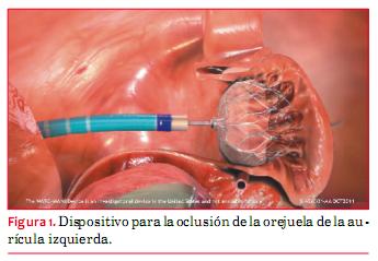
<body><![CDATA[<p>        <b><span style="font-size: 10pt; font-family: Verdana; ">Introducci&oacute;n&nbsp;</span></b><span style="font-size: 10pt; font-family: Verdana; "> </span>       <o:p></o:p>           <br> <span style="font-size: 10pt;"><o:p>&nbsp;</o:p></span>           <br> <span style="font-size: 10pt; font-family: Verdana; ">Luego de los trabajos pioneros de angioplastia con bal&oacute;n de Andreas Gruentzig, las intervenciones coronarias percut&aacute;neas pasaron a ser el puntal de la cardiolog&iacute;a en las d&eacute;cadas <span class="GramE">siguientes<sup>(</sup></span></span><sup><span style="font-size: 10pt; font-family: Verdana; "><a name="1."></a></span><span style="font-size: 10pt; font-family: Verdana; color: rgb(31, 26, 23);"><a href="#1" style="color: blue; text-decoration: underline; text-underline: single">1</a></span><span style="font-size: 10pt; font-family: Verdana; ">)</span></sup><span style="font-size: 10pt; font-family: Verdana; ">; esto fue as&iacute; hasta hace muy poco, cuando la cardiolog&iacute;a adopt&oacute; innovaciones que pueden ser consideradas tan revolucionarias como la angioplastia de Gruentzig. Ante todo, en el desarrollo de las opciones de tratamiento percut&aacute;neo para la estenosis de la v&aacute;lvula a&oacute;rtica. La implantaci&oacute;n transcat&eacute;ter de la v&aacute;lvula a&oacute;rtica (TAVI) ha mejorado las opciones de tratamiento para los pacientes a&ntilde;osos con estenosis de la v&aacute;lvula a&oacute;rtica. Otros desarrollos importantes son las opciones de tratamiento percut&aacute;neo para la insuficiencia mitral (IM) (MitraClip), la terapia no farmacol&oacute;gica para evitar la embolizaci&oacute;n cerebral en pacientes con fibrilaci&oacute;n auricular (FA), tal como el cierre de la orejuela de la aur&iacute;cula izquierda (OAI) y el cierre del foramen oval permeable y la denervaci&oacute;n renal para tratar la hipertensi&oacute;n resistente.&nbsp; </span><span style="font-size: 10pt;"><o:p></o:p></span>           </p>               <p style="margin: 0cm 0cm 0.0001pt;"><b>  <multicol gutter="18" cols="2"></multicol> <span style="font-size: 10pt; font-family: Verdana; ">  Cierre de la orejuela de la aur&iacute;cula izquierda</span><o:p></o:p></b></p>            <p style="margin: 0cm 0cm 0.0001pt;"><b> <span style="font-size: 10pt; font-family: Verdana; ">&nbsp;</span></b><span style="font-size: 10pt; font-family: Verdana; "> </span><span style="font-size: 10pt;"><o:p></o:p></span></p>            <p>        <span style="font-size: 10pt; font-family: Verdana; ">La FA es una patolog&iacute;a muy prevalente y constituye la principal causa de accidentes cerebrovasculares. El riesgo de presentar una FA en el curso de la vida es de aproximadamente 1 en 4</span><sup><span style="font-size: 10pt; font-family: Verdana; ">(<a name="2."></a></span><span style="font-size: 10pt; font-family: Verdana; color: rgb(31, 26, 23);"><a href="#1" style="color: blue; text-decoration: underline; text-underline: single">2</a></span><span style="font-size: 10pt; font-family: Verdana; ">)</span></sup><span style="font-size: 10pt; font-family: Verdana; ">. Dada la dificultad de detectar una FA parox&iacute;stica, es probable que la verdadera prevalencia est&eacute; subestimada. Es probable que los pacientes con FA parox&iacute;stica tengan un riesgo de accidente cerebrovascular parecido al de los pacientes con FA <span class="GramE">persistente<sup>(</sup></span></span><sup><span style="font-size: 10pt; font-family: Verdana; "><a name="3."></a></span><span style="font-size: 10pt; font-family: Verdana; color: rgb(31, 26, 23);"><a href="#3" style="color: blue; text-decoration: underline; text-underline: single">3</a></span><span style="font-size: 10pt; font-family: Verdana; ">)</span></sup><span style="font-size: 10pt; font-family: Verdana; ">.</span><sup><span style="font-size: 10pt; font-family: Verdana; ">&nbsp;</span><o:p></o:p></sup>           <br> <span style="font-size: 10pt; font-family: Verdana; ">&nbsp;</span><span style="font-size: 10pt;"><o:p></o:p></span>           <br> <span style="font-size: 10pt; font-family: Verdana; ">La anticoagulaci&oacute;n oral siempre ha sido el tratamiento de primera l&iacute;nea para evitar los accidentes cerebrovasculares, pero entra&ntilde;a riesgos importantes. La estrecha ventana terap&eacute;utica de la warfarina obliga a un equilibrio delicado entre la falta de eficacia y el riesgo muy elevado de sangrado, requiriendo por lo tanto ex&aacute;menes de sangre frecuentes, adem&aacute;s de sus numerosas interacciones con alimentos y medicamentos que tienen un importante impacto en la vida cotidiana de los pacientes. Hasta 40% de los pacientes con FA tienen contraindicaciones para la anticoagulaci&oacute;n. Incluso en condiciones de estudios cl&iacute;nicos, una importante proporci&oacute;n de pacientes est&aacute; recibiendo un tratamiento con warfarina o bien excesivo o insuficiente. En un estudio que incluy&oacute; a 41.900 pacientes con FA cr&oacute;nica, solo 70% de los pacientes tratados con warfarina manten&iacute;an ese tratamiento al a&ntilde;o, destacando aun m&aacute;s las dificultades que plantea la anticoagulaci&oacute;n.&nbsp; </span>       <o:p></o:p>       </p>           <p style="margin: 0cm 0cm 0.0001pt;"><span style="font-size: 10pt;"><o:p>&nbsp;</o:p></span></p>            <p style="margin: 0cm 0cm 0.0001pt;"> <span style="font-size: 10pt; font-family: Verdana; ">Entre los pacientes con FA no valvular, la gran mayor&iacute;a de los trombos evolucionan a partir de la OAI. La OAI fibrilante es un callej&oacute;n sin salida que crea las condiciones que favorecen la estasis sangu&iacute;nea y la formaci&oacute;n de trombos. Por lo tanto, uno podr&iacute;a esperar que la exclusi&oacute;n de la OAI de la circulaci&oacute;n redujera el riesgo de accidente cerebrovascular. Se han desarrollado varios m&eacute;todos: ligadura o amputaci&oacute;n quir&uacute;rgica y oclusi&oacute;n con cat&eacute;ter percut&aacute;neo con dispositivos especialmente dise&ntilde;ados para la oclusi&oacute;n (</span><span style="font-size: 10pt; font-family: Verdana; color: rgb(31, 26, 23);"><a href="#fig_1" style="color: blue; text-decoration: underline; text-underline: single">figura 1</a></span><span style="font-size: 10pt; font-family: Verdana; ">).</span></p>     ]]></body>
<body><![CDATA[<p style="margin: 0cm 0cm 0.0001pt;"><span style="font-size: 10pt;"><o:p>&nbsp;</o:p></span></p>     <p style="margin: 0cm 0cm 0.0001pt;"> <span style="font-size: 10pt; font-family: Verdana; "><a name="fig_1"></a><img style="width: 345px; height: 239px;" alt="" src="/img/revistas/ruc/v29n3/3a15f1.JPG">    <br>      </span></p>     <p style="margin: 0cm 0cm 0.0001pt;"><span style="font-size: 10pt;"><o:p>&nbsp;</o:p></span></p>     <p> <span style="font-size: 10pt; font-family: Verdana; ">La ligadura o amputaci&oacute;n quir&uacute;rgica ha sido utilizada durante muchos a&ntilde;os aun cuando existe muy poca evidencia en cuanto a su <span class="GramE">eficacia<sup>(<a name="5."></a></sup></span></span><sup><span style="font-size: 10pt; font-family: Verdana; color: rgb(31, 26, 23);"><a href="#5" style="color: blue; text-decoration: underline; text-underline: single">5</a></span><span style="font-size: 10pt; font-family: Verdana; ">)</span></sup><span style="font-size: 10pt; font-family: Verdana; ">.<sup> </sup>Por supuesto que se realiza solo como una ampliaci&oacute;n de otra operaci&oacute;n (&lsquo;bystander&rsquo;) en oportunidad, por ejemplo, de una cirug&iacute;a v&aacute;lvular, pero no como un procedimiento <span class="GramE">independiente<sup>(</sup></span></span><sup><span style="font-size: 10pt; font-family: Verdana; "><a name="6."></a></span><span style="font-size: 10pt; font-family: Verdana; color: rgb(31, 26, 23);"><a href="#6" style="color: blue; text-decoration: underline; text-underline: single">6</a></span><span style="font-size: 10pt; font-family: Verdana; ">)</span></sup><span style="font-size: 10pt; font-family: Verdana; ">.</span><o:p></o:p>           <br>     <br>        <span style="font-size: 10pt; font-family: Verdana; ">Los m&eacute;todos percut&aacute;neos se han ido desarrollando desde 2002. Se han completado ya los estudios preliminares de dos sistemas espec&iacute;ficamente dise&ntilde;ados para esta finalidad (oclusi&oacute;n percut&aacute;nea transcat&eacute;ter de la OAI [PLAATO] y sistemas Watchman)</span><sup><span style="font-size: 10pt; font-family: Verdana; "> (<a name="7."></a></span><span style="font-size: 10pt; font-family: Verdana; color: rgb(31, 26, 23);"><a href="#7" style="color: blue; text-decoration: underline; text-underline: single">7</a></span><span style="font-size: 10pt; font-family: Verdana; ">)</span></sup><span style="font-size: 10pt; font-family: Verdana; ">.<sup> </sup>Estos dispositivos se despliegan a trav&eacute;s de un acceso venoso, pasando hacia la aur&iacute;cula izquierda (AI) atravesando el tabique. Estos dispositivos han sido aprobados para su uso en la Comunidad Europea (CE) pero no est&aacute;n aprobados todav&iacute;a para el uso cl&iacute;nico por la Administraci&oacute;n de Alimentos y Medicamentos de EEUU (FDA).</span><o:p></o:p>           <br> <span style="font-size: 10pt; font-family: Verdana; ">&nbsp; </span><span style="font-size: 10pt;"><o:p></o:p></span>           <br> <span style="font-size: 10pt; font-family: Verdana; ">Adem&aacute;s de estos dos sistemas tambi&eacute;n se cuenta con el tap&oacute;n card&iacute;aco de Amplatzer y el Lariat (dispositivo con asa).</span> </p>     <p style="margin: 0cm 0cm 0.0001pt;"> <span style="font-size: 10pt; font-family: Verdana; ">&nbsp; </span><span style="font-size: 10pt;"><o:p></o:p></span></p>                   ]]></body>
<body><![CDATA[<p style="margin: 0cm 0cm 0.0001pt;"> <span style="font-size: 10pt; font-family: Verdana; "><span style="font-weight: bold;">Sistema PLAATO</span>&nbsp;</span><o:p></o:p></p>            <p style="margin: 0cm 0cm 0.0001pt;"> <span style="font-size: 10pt; font-family: Verdana; ">&nbsp;</span><span style="font-size: 10pt;"><o:p></o:p></span></p>            <p style="margin: 0cm 0cm 0.0001pt;">El sistema PLAATO era un dispositivo que se colocaba en la OAI mediante un cat&eacute;ter que atravesaba el septum. Ten&iacute;a una armaz&oacute;n autoexpandible de nitinol <span style="font-size: 10pt; font-family: Verdana;">cubierto por un revestimiento impermeable a la sangre, sellando as&iacute; la OAI e impidiendo la formaci&oacute;n o el desprendimiento de trombos. Sin embargo, durante el seguimiento hubo efectos adversos, por ejemplo, derrame peric&aacute;rdico en ocho pacientes, dos accidentes cerebrovasculares, dos accidentes isqu&eacute;micos pasajeros, y tres muertes no relacionadas con los <span class="GramE">procedimientos<sup>(<a name="8."></a></sup></span><sup><a href="#8" style="color: blue; text-decoration: underline; text-underline: single">8</a>)</sup>.<sup> </sup>El fabricante ha interrumpido el desarrollo del dispositivo PLAATO.</span>&nbsp; <br style="">       </p>     <p> <span style="font-size: 10pt;"><o:p></o:p></span>             <br> <b><span style="font-size: 10pt; font-family: Verdana; ">Dispositivo Watchman</span><o:p></o:p></b> </p>           <p style="margin: 0cm 0cm 0.0001pt;"><b> <span style="font-size: 10pt; font-family: Verdana; ">&nbsp;</span></b><span style="font-size: 10pt; font-family: Verdana; "> </span> <b><o:p></o:p></b></p>            <p style="margin: 0cm 0cm 0.0001pt;"> <span style="font-size: 10pt; font-family: Verdana; ">El dispositivo Watchman tambi&eacute;n consiste en un dispositivo expandible que se despliega en la OAI utilizando un cat&eacute;ter transeptal. El dispositivo implantado tiene un marco de nitinol autoexpandible para fijarlo al interior de la OAI. A diferencia del dispositivo PLAATO, la malla del dispositivo Watchman es permeable a la <span class="GramE">sangre<sup>(<a name="9."></a></sup></span></span><sup><span style="font-size: 10pt; font-family: Verdana; color: rgb(31, 26, 23);"><a href="#9" style="color: blue; text-decoration: underline; text-underline: single">9</a></span><span style="font-size: 10pt; font-family: Verdana; ">)</span></sup><span style="font-size: 10pt; font-family: Verdana; ">.<sup> </sup>Por esta raz&oacute;n, los pacientes necesitan profilaxis tromboemb&oacute;lica convencional con warfarina hasta que el dispositivo se haya endotelizado (por ejemplo, por lo menos 45 d&iacute;as despu&eacute;s del implante), momento en el cual se realiza una ecocardiograf&iacute;a transesof&aacute;gica para garantizar la endotelizaci&oacute;n. Adem&aacute;s, todos los pacientes reciben tanto aspirina (81-325 mg) como clopidogrel (75 mg) diariamente durante seis meses.&nbsp; </span> <o:p></o:p></p>            <p style="margin: 0cm 0cm 0.0001pt;"><span style="font-size: 10pt; font-family: Verdana;"><o:p>&nbsp;</o:p></span></p>            <p style="margin: 0cm 0cm 0.0001pt;"> <span style="font-size: 10pt; font-family: Verdana; ">El dispositivo Watchman fue evaluado en el estudio PROTECT FA (Watchman Left Atrial Appendage System for Embolic Protection in Patients with AF) en el que se distribuy&oacute; aleatoriamente a m&aacute;s de 700 pacientes con FA no valvular, con una relaci&oacute;n 2 a 1, a recibir el dispositivo (con el esquema antitromb&oacute;tico arriba descrito) o warfarina por un per&iacute;odo prolongado (INR de 2,0 a 3,0)</span><sup><span style="font-size: 10pt; font-family: Verdana; "> (<a name="10."></a></span><span style="font-size: 10pt; font-family: Verdana; color: rgb(31, 26, 23);"><a href="#10" style="color: blue; text-decoration: underline; text-underline: single">10</a></span><span style="font-size: 10pt; font-family: Verdana; ">)</span></sup><span style="font-size: 10pt; font-family: Verdana; ">. El dise&ntilde;o del ensayo era de no inferioridad. Los criterios de inclusi&oacute;n permit&iacute;an el ingreso de pacientes con FA parox&iacute;stica, persistente, o permanente, y todos los pacientes ten&iacute;an una puntuaci&oacute;n CHADS<sub>2</sub> &sup3;1.&nbsp; </span> <o:p></o:p></p>            <p style="margin: 0cm 0cm 0.0001pt;"><span style="font-size: 10pt; font-family: Verdana;"><o:p>&nbsp;</o:p></span></p>            ]]></body>
<body><![CDATA[<p style="margin: 0cm 0cm 0.0001pt;"> <span style="font-size: 10pt; font-family: Verdana; ">El ensayo confirm&oacute; la no inferioridad de la oclusi&oacute;n de la orejuela auricular comparado con la terapia warfar&iacute;nica en lo que concierne al punto final primario compuesto de accidente cerebrovascular, embolia sist&eacute;mica y muerte cardiovascular, con un cociente de riesgo de 0,62 (IC 95% 0,35 a 1,25<span class="GramE">)<sup>(</sup></span></span><sup><span style="font-size: 10pt; font-family: Verdana; "><a name="11."></a></span><span style="font-size: 10pt; font-family: Verdana; color: rgb(31, 26, 23);"><a href="#11" style="color: blue; text-decoration: underline; text-underline: single">11</a></span><span style="font-size: 10pt; font-family: Verdana; ">)</span></sup><span style="font-size: 10pt; font-family: Verdana; ">. Sin embargo, el punto final primario de seguridad evaluado (combinaci&oacute;n de sangrado mayor, derrame peric&aacute;rdico, accidente cerebrovascular relacionado con el procedimiento y embolizaci&oacute;n del dispositivo) estuvo aumentada en el grupo del dispositivo (7,4 vs 4,4 eventos cada 100 a&ntilde;os-pacientes, respectivamente). La mayor&iacute;a de los eventos del grupo del dispositivo fueron precoces. De estos, alrededor del 50% fueron derrames peric&aacute;rdicos que necesitaron drenaje.&nbsp;</span><o:p></o:p></p>            <p style="margin: 0cm 0cm 0.0001pt;"> <span style="font-size: 10pt; font-family: Verdana; ">&nbsp;</span><span style="font-size: 10pt; font-family: Verdana;"><o:p></o:p></span></p>            <p style="margin: 0cm 0cm 0.0001pt;"> <span style="font-size: 10pt; font-family: Verdana; ">Dos registros ulteriores mostraron una mayor seguridad de este dispositivo, probablemente debido al efecto de la curva de aprendizaje. La tasa de complicaciones dentro de los siete d&iacute;as fue de 3,7% comparado con 7,7%, cifra observada en el ensayo aleatorizado <span class="GramE">inicial<sup>(</sup></span></span><sup><span style="font-size: 10pt; font-family: Verdana; color: rgb(31, 26, 23);"><a href="#11" style="color: blue; text-decoration: underline; text-underline: single">11</a></span><span style="font-size: 10pt; font-family: Verdana; ">,<a name="12."></a></span><span style="font-size: 10pt; font-family: Verdana; color: rgb(31, 26, 23);"><a href="#12" style="color: blue; text-decoration: underline; text-underline: single">12</a></span><span style="font-size: 10pt; font-family: Verdana; ">)</span></sup><span style="font-size: 10pt; font-family: Verdana; ">.&nbsp; </span><span style="font-size: 10pt; font-family: Verdana;"><o:p></o:p></span></p>            <p>&nbsp; </p>           <p style="margin: 0cm 0cm 0.0001pt;"><b> <span style="font-size: 10pt; font-family: Verdana; ">Oclusor septal de Amplatzer y tap&oacute;n card&iacute;aco de Amplatzer&nbsp;</span><o:p></o:p></b></p>            <p style="margin: 0cm 0cm 0.0001pt;"> <span style="font-size: 10pt; font-family: Verdana; ">&nbsp;</span><span style="font-size: 10pt; font-family: Verdana;"><o:p></o:p></span></p>            <p style="margin: 0cm 0cm 0.0001pt;"> <span style="font-size: 10pt; font-family: Verdana; ">El oclusor septal de Amplatzer utiliza una t&eacute;cnica m&aacute;s sencilla que la t&eacute;cnica PLAATO. En vez de recurrir a la anestesia general, como se hace en el procedimiento de PLAATO, a menudo se implanta bajo anestesia local. Se presentaron los resultados de una serie de casos en los que se estudiaron 16 pacientes a los que se coloc&oacute; el oclusor septal de Amplatzer para cerrar la OAI bajo anestesia local sin gu&iacute;a ecocardiogr&aacute;fica</span><sup><span style="font-size: 10pt; font-family: Verdana; ">(<a name="13."></a></span><span style="font-size: 10pt; font-family: Verdana; color: rgb(31, 26, 23);"><a href="#13" style="color: blue; text-decoration: underline; text-underline: single">13</a></span><span style="font-size: 10pt; font-family: Verdana; ">)</span></sup><span style="font-size: 10pt; font-family: Verdana; ">. El tap&oacute;n card&iacute;aco de Amplatzer es otro dispositivo dise&ntilde;ado espec&iacute;ficamente para el cierre de la OAI y est&aacute; siendo estudiado en ensayos <span class="GramE">cl&iacute;nicos<sup>(</sup></span></span><sup><span style="font-size: 10pt; font-family: Verdana; color: rgb(31, 26, 23);"><a href="#14" style="color: blue; text-decoration: underline; text-underline: single">14</a></span><span style="font-size: 10pt; font-family: Verdana; ">)</span></sup><span style="font-size: 10pt; font-family: Verdana; ">.<sup> </sup>Este dispositivo est&aacute; hecho de nitinol y consiste en un disco que se coloca en la aur&iacute;cula izquierda y un tap&oacute;n distal conectado al disco por una cintura corta. El tap&oacute;n distal contiene seis pares de pinchos para aumentar la estabilidad dentro de la orejuela. Este dispositivo es m&aacute;s corto que el dispositivo de Watchman y puede ser m&aacute;s ventajoso en individuos con orejuelas de morfolog&iacute;a variable. Se han publicado datos de estudios en animales demostrando la falta de complicaciones de la colocaci&oacute;n del dispositivo, alcanzando una oclusi&oacute;n completa de la orejuela a los 30 a 60 <span class="GramE">d&iacute;as<sup>(</sup></span></span><sup><span style="font-size: 10pt; font-family: Verdana; "><a name="14."></a></span><span style="font-size: 10pt; font-family: Verdana; color: rgb(31, 26, 23);"><a href="#14" style="color: blue; text-decoration: underline; text-underline: single">14</a></span><span style="font-size: 10pt; font-family: Verdana; ">,<a name="15."></a></span><span style="font-size: 10pt; font-family: Verdana; color: rgb(31, 26, 23);"><a href="#15" style="color: blue; text-decoration: underline; text-underline: single">15</a></span><span style="font-size: 10pt; font-family: Verdana; ">)</span></sup><span style="font-size: 10pt; font-family: Verdana; ">.<sup>&nbsp;</sup> </span> <o:p></o:p></p>            <p style="margin: 0cm 0cm 0.0001pt;"><span style="font-size: 10pt;"><o:p>&nbsp;</o:p></span></p>            <p style="margin: 0cm 0cm 0.0001pt;"> <span style="font-size: 10pt; font-family: Verdana; ">El rol futuro de este procedimiento tambi&eacute;n depender&aacute; de otras alternativas a la terapia warfar&iacute;nica, como los nuevos anticoagulantes. Hasta ahora no existe una fuerte evidencia que sugiera que sean superiores a la warfarina, a excepci&oacute;n de rivaroxab&aacute;n -que mostr&oacute; un menor riesgo de sangrado- pero son una alternativa promisoria mucho m&aacute;s f&aacute;cil de usar que la <span class="GramE">warfarina<sup>(</sup></span></span><sup><span style="font-size: 10pt; font-family: Verdana; "><a name="16."></a></span><span style="font-size: 10pt; font-family: Verdana; color: rgb(31, 26, 23);"><a href="#16" style="color: blue; text-decoration: underline; text-underline: single">16</a></span><span style="font-size: 10pt; font-family: Verdana; ">)</span></sup><span style="font-size: 10pt; font-family: Verdana; ">.&nbsp; </span> <o:p></o:p></p>            <p style="margin: 0cm 0cm 0.0001pt;"><span style="font-size: 10pt;"><o:p>&nbsp;</o:p></span></p>            ]]></body>
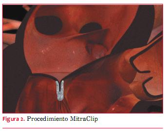
<body><![CDATA[<p style="margin: 0cm 0cm 0.0001pt;"> <span style="font-size: 10pt; font-family: Verdana; ">La actualizaci&oacute;n de las gu&iacute;as de la Sociedad Europea de Cardiolog&iacute;a (ESC) para el manejo de la FA, publicada en 2012, concluye en una recomendaci&oacute;n d&eacute;bil para el uso del cierre intervencionista de la OAI por v&iacute;a percut&aacute;nea en pacientes con alto riesgo de accidente cerebrovascular y contraindicaciones para anticoagulaci&oacute;n a largo <span class="GramE">plazo<sup>(</sup></span></span><sup><span style="font-size: 10pt; font-family: Verdana; "><a name="17."></a></span><span style="font-size: 10pt; font-family: Verdana; color: rgb(31, 26, 23);"><a href="#17" style="color: blue; text-decoration: underline; text-underline: single">17</a></span><span style="font-size: 10pt; font-family: Verdana; ">)</span></sup><span style="font-size: 10pt; font-family: Verdana; ">. En conclusi&oacute;n, el cierre percut&aacute;neo de la OAI parece tan efectivo como la warfarina, seg&uacute;n surge de un ensayo aleatorizado, pero entra&ntilde;a complicaciones en el per&iacute;odo pr&oacute;ximo al procedimiento (como derrame peric&aacute;rdico).&nbsp; </span> <o:p></o:p></p>              <p style="margin: 0cm 0cm 0.0001pt;"><b>  <multicol gutter="18" cols="2"></multicol> <span style="font-size: 10pt; font-family: Verdana; ">  Cierre de la orejuela de la aur&iacute;cula izquierda&nbsp;</span></b><span style="font-size: 10pt; font-family: Verdana; "> </span> <o:p></o:p></p>            <p style="margin: 0cm 0cm 0.0001pt;"> <span style="font-size: 10pt; font-family: Verdana; ">El cierre percut&aacute;neo de OAI es una alternativa promisoria a la terapia warfar&iacute;nica en pacientes con FA con un alto riesgo de accidente cerebrovascular. La informaci&oacute;n es escasa y el procedimiento debe limitarse a los pacientes que tengan contraindicaciones claras para warfarina. </span> <o:p></o:p></p>            <p style="margin: 0cm 0cm 0.0001pt;"> <span style="font-size: 10pt; font-family: Verdana; ">Los nuevos anticoagulantes (por ejemplo, rivaroxab&aacute;n) representan otra alternativa para los pacientes con una contraindicaci&oacute;n para la warfarina.&nbsp; </span><span style="font-size: 10pt;"><o:p></o:p></span></p>     <p>     <br> <b><span style="font-size: 10pt; font-family: Verdana; ">Intervenciones sobre la v&aacute;lvula mitral    <br> &nbsp;</span></b>    <br> <span style="font-size: 10pt; font-family: Verdana; ">La prevalencia de la IM moderada o severa supera el 10% en mayores de 75 a&ntilde;os y la evoluci&oacute;n natural es a menudo <span class="GramE">fatal<sup>(<a name="18."></a></sup></span></span><sup><span style="font-size: 10pt; font-family: Verdana; color: rgb(31, 26, 23);"><a href="#18" style="color: blue; text-decoration: underline; text-underline: single">18</a></span><span style="font-size: 10pt; font-family: Verdana; ">)</span></sup><span style="font-size: 10pt; font-family: Verdana; ">. No obstante ello, los pacientes con IM cr&oacute;nica severa a menudo sufren de otras comorbilidades que aumentan su riesgo al someterse a cirug&iacute;a card&iacute;aca. Urge desarrollar un abordaje percut&aacute;neo menos agresivo y son varios los que se han analizado. En la actualidad, el m&aacute;s promisorio es el sistema MitraClip (Abbott Laboratories, Abbott Park, Illinois, EEUU). Se basa en la t&eacute;cnica quir&uacute;rgica de suturas de Alfieri, una reparaci&oacute;n de borde a borde (</span><span style="font-size: 10pt; font-family: Verdana; color: rgb(31, 26, 23);"><a href="#fig_2" style="color: blue; text-decoration: underline; text-underline: single">figura 2</a></span><span style="font-size: 10pt; font-family: Verdana; ">)</span><sup><span style="font-size: 7.5pt; font-family: Verdana; "> (<a name="19."></a></span><span style="font-size: 7.5pt; font-family: Verdana; color: rgb(31, 26, 23);"><a href="#19" style="color: blue; text-decoration: underline; text-underline: single">19</a></span><span style="font-size: 7.5pt; font-family: Verdana; ">)</span></sup><span style="font-size: 10pt; font-family: Verdana; ">.&nbsp;</span> </p>     <p style="margin: 0cm 0cm 0.0001pt;"><span style="font-size: 10pt;"><o:p>&nbsp;</o:p></span></p>     <p style="margin: 0cm 0cm 0.0001pt;"> <span style="font-size: 10pt; font-family: Verdana; "><a name="fig_2"></a><img style="width: 341px; height: 269px;" alt="" src="/img/revistas/ruc/v29n3/3a15f2.JPG"> </span></p>     ]]></body>
<body><![CDATA[<p>     <br>        </p>            <p style="margin: 0cm 0cm 0.0001pt;"><span style="font-size: 10pt;"><o:p></o:p></span> <span style="font-size: 10pt; font-family: Verdana; ">Casi siempre bajo anestesia general, se introduce el cat&eacute;ter gu&iacute;a a trav&eacute;s de la vena femoral para llegar a la AI mediante un abordaje transeptal. Por esta v&iacute;a se introduce y despliega el MitraClip. El sistema de entrega del clip alinea el MitraClip con la l&iacute;nea de coaptaci&oacute;n, se toman los tejidos de la valva mitral, y el clip se cierra parcialmente a alrededor de 60&deg;. La longitud ideal para la coaptaci&oacute;n es por lo menos de 2 mm. Se utiliza la ecocardiograf&iacute;a transesof&aacute;gica (ETE) para guiar en el despliegue del clip, y luego para evaluar la reducci&oacute;n de la IM. T&eacute;cnicas de imagen periprocedimiento son claves, como deber&iacute;a ser el caso para la reparaci&oacute;n quir&uacute;rgica de la v&aacute;lvula mitral, que es una estructura relativamente <span class="GramE">compleja<sup>(</sup></span></span><sup><span style="font-size: 10pt; font-family: Verdana; "><a name="20."></a></span><span style="font-size: 10pt; font-family: Verdana; color: rgb(31, 26, 23);"><a href="#20" style="color: blue; text-decoration: underline; text-underline: single">20</a></span><span style="font-size: 10pt; font-family: Verdana; ">)</span></sup><span style="font-size: 10pt; font-family: Verdana; ">. Si se coloca la valva correctamente a 60&deg; del cierre del clip, este se sigue cerrando para lograr la aposici&oacute;n de las valvas e inducir una coaptaci&oacute;n y reducir al m&aacute;ximo la IM tanto en los pacientes con IM degenerativa como funcional. Para poder evitar la estenosis luego de colocar el MitraClip, el &aacute;rea inicial de la v&aacute;lvula mitral debe ser &gt;4 cm<sup>2</sup>.&nbsp; </span> <o:p></o:p></p>     <p>     <br> </p>     <p style="margin: 0cm 0cm 0.0001pt;"> <span style="font-size: 10pt; font-family: Verdana; ">La experiencia cl&iacute;nica inicial con MitraClip fue estudiada en seres humanos en el estudio fase I EVEREST (Endovascular Valve Edge-to-Edge Repair Study: Estudio de reparaci&oacute;n valvular endovascular borde a borde)</span><sup><span style="font-size: 10pt; font-family: Verdana; "> (<a name="21."></a></span><span style="font-size: 10pt; font-family: Verdana; color: rgb(31, 26, 23);"><a href="#21" style="color: blue; text-decoration: underline; text-underline: single">21</a></span><span style="font-size: 10pt; font-family: Verdana; ">)</span></sup><span style="font-size: 10pt; font-family: Verdana; ">. El estudio incluy&oacute; a 27 pacientes. Catorce de los pacientes tuvieron una reducci&oacute;n de la IM &pound;2+ a los seis meses. El estudio mostr&oacute; que es un procedimiento promisorio para los pacientes con IM con alto riesgo para la cirug&iacute;a convencional.&nbsp; </span> <o:p></o:p></p>     <p>     <br> </p>     <p style="margin: 0cm 0cm 0.0001pt;"> <span style="font-size: 10pt; font-family: Verdana; ">El ensayo fundamental ha sido el EVEREST II, que distribuy&oacute; aleatoriamente a 279 pacientes con una IM cr&oacute;nica moderadamente severa o severa (grado 3+ o 4+) a someterse ya sea a la colocaci&oacute;n percut&aacute;nea de MitraClip o cirug&iacute;a de la v&aacute;lvula mitral en una relaci&oacute;n 2:1</span><sup><span style="font-size: 10pt; font-family: Verdana; ">(<a name="22."></a></span><span style="font-size: 10pt; font-family: Verdana; color: rgb(31, 26, 23);"><a href="#22" style="color: blue; text-decoration: underline; text-underline: single">22</a></span><span style="font-size: 10pt; font-family: Verdana; ">)</span></sup><span style="font-size: 10pt; font-family: Verdana; ">. Si bien antes del alta del hospital la terapia percut&aacute;nea fue menos efectiva para reducir la IM, las tasas de reducci&oacute;n de IM a los 12 y 24 meses fueron similares en ambos grupos. No obstante ello, la terapia percut&aacute;nea result&oacute; ser m&aacute;s segura, con una menor tasa de eventos adversos mayores a los 30 d&iacute;as. El estudio mostr&oacute; una mejora cl&iacute;nica sostenida, evaluada por la calidad de vida, el grado de insuficiencia card&iacute;aca y la funci&oacute;n del ventr&iacute;culo izquierdo.</span></p>     <p style="margin: 0cm 0cm 0.0001pt;"> <span style="font-size: 10pt; font-family: Verdana; ">&nbsp; </span><span style="font-size: 10pt; font-family: Verdana;"><o:p></o:p></span></p>            ]]></body>
<body><![CDATA[<p>        <span style="font-size: 10pt; font-family: Verdana; ">El estudio EVEREST II no se concentr&oacute; espec&iacute;ficamente en pacientes de muy alto riesgo. La media de edad fue de 66 a&ntilde;os, la media de la fracci&oacute;n de eyecci&oacute;n fue de 60%, y fueron raras la comorbilidades mayores. En el estudio de alto riesgo (HRS) EVEREST II, sin embargo, los pacientes tuvieron IM severa (3-4+), una media de edad de 77 a&ntilde;os, y una mortalidad quir&uacute;rgica estimada en &sup3;12%</span><sup><span style="font-size: 10pt; font-family: Verdana; ">(<a name="23."></a></span><span style="font-size: 10pt; font-family: Verdana; color: rgb(31, 26, 23);"><a href="#23" style="color: blue; text-decoration: underline; text-underline: single">23</a></span><span style="font-size: 10pt; font-family: Verdana; ">)</span></sup><span style="font-size: 10pt; font-family: Verdana; ">. M&aacute;s de 50% de estos pacientes hab&iacute;an sido sometidos a cirug&iacute;a card&iacute;aca previa. Se los compar&oacute; retrospectivamente con un grupo de pacientes que fueron estudiados al mismo tiempo pero que no fueron inscritos en el estudio EVEREST II. Estos pacientes hab&iacute;an sido tratados con terapia m&eacute;dica est&aacute;ndar.&nbsp; </span><span style="font-size: 10pt; font-family: Verdana;"><o:p></o:p></span>             <br>  <multicol gutter="18" cols="2"></multicol> <span style="font-size: 10pt; font-family: Verdana; ">  La tasa de mortalidad relacionada con el procedimiento a los 30 d&iacute;as fue similar en ambos grupos (7,7% en el HRS contra 8,3% en el grupo de comparaci&oacute;n). La supervivencia a los 12 meses fue m&aacute;s elevada en el grupo HRS (76% vs 55%). En los pacientes que sobrevivieron se compararon los datos basales y a los 12 meses. El grado de la IM se hab&iacute;a reducido a &pound;2+ en 78% de los pacientes. Hubo una mejora en el volumen del ventr&iacute;culo izquierdo al final de la di&aacute;stole (172-140 ml), en el volumen telesist&oacute;lico (82-73 ml), la clase funcional de la Asociaci&oacute;n del Coraz&oacute;n de Nueva York (III/IV a nivel basal en 89%, a clase I/II en 74%; p &lt; 0,0001), calidad de vida y puntuaci&oacute;n del componente vital y mental a los 12 meses.&nbsp; </span><span style="font-size: 10pt; font-family: Verdana;"><o:p></o:p></span>              </p>                  <p><small><span style="font-family: Verdana;">Feldman y colaboradores</span><sup style="font-family: Verdana;"> (<a name="24.."></a><a href="#24" style="color: blue; text-decoration: underline; text-underline: single">24</a>) </sup><span style="font-family: Verdana;">comunicaron los resultados cl&iacute;nicos del uso del clip en una cohorte de los primeros 107 pacientes, seguidos hasta tres a&ntilde;os. Eran 55 pacientes tratados en el ensayo de factibilidad fase I EVEREST, y 52 pacientes tratados en el ensayo pivote EVEREST II que representaban la experiencia inicial pre-randomizaci&oacute;n. De los pacientes tratados con &eacute;xito, 66% alcanzaron el punto final primario de ausencia de muerte, cirug&iacute;a de la v&aacute;lvula mitral, o IM &gt;2+ a los 12 meses. La mayor&iacute;a de los pacientes se manten&iacute;a </span><span style="font-family: Verdana;" class="GramE">vivo</span><span style="font-family: Verdana;">, libre de cirug&iacute;a o de recurrencia de la IM incluso a tres a&ntilde;os.&nbsp;</span></small> <span style="font-size: 10pt; font-family: Verdana;"><o:p></o:p></span>    <br> </p>     <p> <span style="font-size: 10pt; font-family: Verdana; ">El dispositivo MitraClip ha recibido la aprobaci&oacute;n de la CE y ya ha sido utilizado en miles de pacientes en todo el <span class="GramE">mundo<sup>(</sup></span></span><sup><span style="font-size: 10pt; font-family: Verdana; "><a name="25."></a></span><span style="font-size: 10pt; font-family: Verdana; color: rgb(31, 26, 23);"><a href="#25" style="color: blue; text-decoration: underline; text-underline: single">25</a></span><span style="font-size: 10pt; font-family: Verdana; ">,<a name="26."></a></span><span style="font-size: 10pt; font-family: Verdana; color: rgb(31, 26, 23);"><a href="#26" style="color: blue; text-decoration: underline; text-underline: single">26</a></span><span style="font-size: 10pt; font-family: Verdana; ">)</span></sup><span style="font-size: 10pt; font-family: Verdana; ">. Hasta que sea aprobado por la FDA, en EEUU solo se puede acceder al clip ingresando a trav&eacute;s del registro del estudio REALISM. Los pacientes asentados en este registro &lsquo;de la vida real&rsquo; son asignados ya sea al brazo de alto riesgo o al brazo sin alto riesgo. Se les hace un seguimiento a 30 d&iacute;as, 6 meses y 12 meses.</span>    <br> <span style="font-size: 10pt; font-family: Verdana; ">&nbsp; </span><span style="font-size: 10pt; font-family: Verdana;"><o:p></o:p></span>           <br> <span style="font-size: 10pt; font-family: Verdana; ">Los resultados provisorios del estudio EVEREST II REALISM se presentaron en las sesiones cient&iacute;ficas de la Sociedad para Angiograf&iacute;a e Intervenciones Cardiovasculares (SCAI) en 2011</span><sup><span style="font-size: 10pt; font-family: Verdana; ">(<a name="27."></a></span><span style="font-size: 10pt; font-family: Verdana; color: rgb(31, 26, 23);"><a href="#27" style="color: blue; text-decoration: underline; text-underline: single">27</a></span><span style="font-size: 10pt; font-family: Verdana; ">-<a name="28."></a><a name="29."></a></span><span style="font-size: 10pt; font-family: Verdana; color: rgb(31, 26, 23);"><a href="#29" style="color: blue; text-decoration: underline; text-underline: single">29</a></span><span style="font-size: 10pt; font-family: Verdana; ">)</span></sup><span style="font-size: 10pt; font-family: Verdana; ">. En promedio, la edad de los pacientes en el registro fue de m&aacute;s de 70 a&ntilde;os. El procedimiento MitraClip fue seguro, con una mortalidad a los 30 d&iacute;as de 3,8%</span><sup><span style="font-size: 10pt; font-family: Verdana; ">(</span><span style="font-size: 10pt; font-family: Verdana; color: rgb(31, 26, 23);"><a href="#28" style="color: blue; text-decoration: underline; text-underline: single">28</a></span><span style="font-size: 10pt; font-family: Verdana; ">)</span></sup><span style="font-size: 10pt; font-family: Verdana; ">. Al a&ntilde;o, 83% de los pacientes en el grupo de alto riesgo ten&iacute;an solo IM leve a moderada (1+ o 2+). Antes del procedimiento, las puntuaciones de calidad de vida fueron peores para la IM funcional que para la IM <span class="GramE">degenerativa<sup>(</sup></span></span><sup><span style="font-size: 10pt; font-family: Verdana; color: rgb(31, 26, 23);"><a href="#29" style="color: blue; text-decoration: underline; text-underline: single">29</a></span><span style="font-size: 10pt; font-family: Verdana; ">)</span></sup><span style="font-size: 10pt; font-family: Verdana; ">, pero tambi&eacute;n tuvieron una significativa mejora despu&eacute;s del procedimiento.    <br> &nbsp; </span><span style="font-size: 10pt; font-family: Verdana;"><o:p></o:p></span>           <br> <span style="font-size: 10pt; font-family: Verdana; ">Un an&aacute;lisis de los primeros 100 pacientes en el registro MitraSwiss de Suiza mostr&oacute; que la insuficiencia card&iacute;aca congestiva previa al implante del clip y el antecedente de cirug&iacute;a coronaria (CABG) fueron predictivos de malos resultados con el procedimiento <span class="GramE">MitraClip<sup>(</sup></span></span><sup><span style="font-size: 10pt; font-family: Verdana; color: rgb(31, 26, 23);"><a href="#26" style="color: blue; text-decoration: underline; text-underline: single">26</a></span><span style="font-size: 10pt; font-family: Verdana; ">)</span></sup><span style="font-size: 10pt; font-family: Verdana; ">. Una reducci&oacute;n de IM a &pound;2+ durante el procedimiento y un bajo grado de IM al alta <span class="GramE">predijeron</span> una mejor supervivencia a mediano plazo. Gaemperli y <span class="GramE">colaboradores<sup>(<a name="30."></a></sup></span></span><sup><span style="font-size: 10pt; font-family: Verdana; color: rgb(31, 26, 23);"><a href="#30" style="color: blue; text-decoration: underline; text-underline: single">30</a></span><span style="font-size: 10pt; font-family: Verdana; ">)</span></sup><span style="font-size: 10pt; font-family: Verdana; "> constataron una mejora del perfil hemodin&aacute;mico inmediatamente despu&eacute;s del procedimiento. Esta mejora hemodin&aacute;mica se asoci&oacute; a un resultado favorable a mediano plazo.    <br> &nbsp; </span><span style="font-size: 10pt; font-family: Verdana;"><o:p></o:p></span>       </p>           ]]></body>
<body><![CDATA[<p style="margin: 0cm 0cm 0.0001pt;"> <span style="font-size: 10pt; font-family: Verdana; ">En conclusi&oacute;n, el implante de MitraClip es inferior a la reparaci&oacute;n quir&uacute;rgica de la v&aacute;lvula mitral para reducir el grado de IM, pero probablemente sea m&aacute;s seguro en los pacientes de m&aacute;s alto riesgo. Aunque la reducci&oacute;n de la IM es menor que con la cirug&iacute;a, la mejora subjetiva de los s&iacute;ntomas es comparable. Los pacientes m&aacute;s adecuados son aquellos con alto riesgo quir&uacute;rgico y particularmente aquellos que presentan IM funcional. No queda claro cu&aacute;l ser&iacute;a puntualmente el momento &oacute;ptimo para el procedimiento, pero en los pacientes sometidos a cirug&iacute;a los datos indican que probablemente sea mejor proceder cuanto <span class="GramE">antes<sup>(</sup></span></span><sup><span style="font-size: 10pt; font-family: Verdana; "><a name="31."></a></span><span style="font-size: 10pt; font-family: Verdana; color: rgb(31, 26, 23);"><a href="#31" style="color: blue; text-decoration: underline; text-underline: single">31</a></span><span style="font-size: 10pt; font-family: Verdana; ">)</span></sup><span style="font-size: 10pt; font-family: Verdana; ">. Esto tiene sentido, ya que la cirug&iacute;a se ha hecho m&aacute;s segura, modific&aacute;ndose las consideraciones de riesgo/beneficio. Dado que el abordaje percut&aacute;neo ha seguido reduciendo el riesgo en torno al procedimiento, aqu&iacute; tambi&eacute;n se deber&iacute;a aplicar la misma consideraci&oacute;n.&nbsp; </span><span style="font-size: 10pt; font-family: Verdana;"><o:p></o:p></span></p>            <p class="MsoNormal"><span style="font-size: 10pt; font-family: Verdana;"><o:p>&nbsp;</o:p></span></p>            <p>        <b><span style="font-size: 10pt; font-family: Verdana; ">Intervenciones de la v&aacute;lvula mitral&nbsp;</span></b><span style="font-size: 10pt; font-family: Verdana; "> </span>       <o:p></o:p>           <br> <o:p>&nbsp;</o:p>           <br> <b><span style="font-size: 10pt; font-family: Verdana; ">Puntos clave&nbsp;</span></b><span style="font-size: 10pt; font-family: Verdana; "> </span><o:p></o:p>  </p>     <ul>            <li><span style="font-size: 10pt; font-family: Verdana; ">Los datos para el procedimiento del sistema de      MitraClip son muy limitados y provienen fundamentalmente del ensayo      EVEREST II.&nbsp; </span></li>          </ul>          <ul style="margin-top: 0cm;" type="disc">        <li class="MsoNormal" style="">  <span style="font-size: 10pt; font-family: Verdana; ">Si bien el      ensayo EVEREST no incluy&oacute; pacientes de muy alto riesgo, el      procedimiento deber&iacute;a limitarse a aquellos de alto riesgo      quir&uacute;rgico debido al efecto limitado.&nbsp; </span><span style="font-size: 10pt;"><o:p></o:p></span></li>           </ul>            <p class="MsoNormal" style="margin-left: 36pt;"><span style="font-size: 10pt;"><o:p>&nbsp;</o:p></span></p>            <p>        <b><span style="font-size: 10pt; font-family: Verdana; ">Intervenciones sobre la v&aacute;lvula a&oacute;rtica    ]]></body>
<body><![CDATA[<br> &nbsp;</span><o:p></o:p></b>           <br> <span style="font-size: 10pt;"><o:p></o:p></span>       <span style="font-size: 10pt; font-family: Verdana; ">La estenosis de la v&aacute;lvula a&oacute;rtica (EA) es una patolog&iacute;a muy com&uacute;n, aumentando su prevalencia al incrementar la <span class="GramE">edad<sup>(</sup></span></span><sup><span style="font-size: 10pt; font-family: Verdana; "><a name="32."></a></span><span style="font-size: 10pt; font-family: Verdana; color: rgb(31, 26, 23);"><a href="#32" style="color: blue; text-decoration: underline; text-underline: single">32</a></span><span style="font-size: 10pt; font-family: Verdana; ">,<a name="33."></a></span><span style="font-size: 10pt; font-family: Verdana; color: rgb(31, 26, 23);"><a href="#33" style="color: blue; text-decoration: underline; text-underline: single">33</a></span><span style="font-size: 10pt; font-family: Verdana; ">)</span></sup><span style="font-size: 10pt; font-family: Verdana; ">. En pacientes correctamente seleccionados, el reemplazo de la v&aacute;lvula a&oacute;rtica (RVA) mejora sustancialmente los s&iacute;ntomas y aumenta la expectativa de vida. Sin embargo, para los pacientes con comorbilidades mayores, como suele suceder en este grupo de pacientes a&ntilde;osos, la cirug&iacute;a de RVA tal vez no sea apropiada a causa de los riesgos que implica. Un medio menos invasivo es la valvuloplastia a&oacute;rtica percut&aacute;nea, pero tiene un efecto limitado y habitualmente transitorio. El reemplazo de la v&aacute;lvula a&oacute;rtica a trav&eacute;s de un cat&eacute;ter (RVAT o TAVI, por sus siglas en ingl&eacute;s) ofrece una alternativa para tratar a los pacientes con EA con alto riesgo quir&uacute;rgico. Tambi&eacute;n es la opci&oacute;n preferida en los pacientes que puedan tener alg&uacute;n problema t&eacute;cnico con la cirug&iacute;a, por ejemplo, la aorta en porcelana o antes de irradiaci&oacute;n mediast&iacute;nica, o cirug&iacute;a con adherencias densas, o infecci&oacute;n esternal previa con reconstrucci&oacute;n compleja, o un injerto permeable de mamaria interna izquierda.&nbsp; </span><span style="font-size: 10pt; font-family: Verdana;"><o:p></o:p></span>       </p>           <p><span style="font-size: 10pt; font-family: Verdana; ">Las directrices europeas m&aacute;s recientes de 2012 recomiendan TAVI en los pacientes con EA sintom&aacute;tica severa a los que el &lsquo;equipo card&iacute;aco&rsquo; local (equipo multidisciplinario) considera inadecuados para cirug&iacute;a convencional debido a comorbilidades severas. Entre los pacientes de alto riesgo que sean posibles candidatos para cirug&iacute;a, la decisi&oacute;n deber&iacute;a individualizarse y analizarse en el seno de un &lsquo;equipo card&iacute;aco&rsquo;. Los scores de riesgo podr&iacute;an ayudar a tomar la decisi&oacute;n cl&iacute;nica. En esta situaci&oacute;n se utiliza tambi&eacute;n a menudo el EuroSCORE log&iacute;stico &sup3;20%, que est&aacute; muy bien establecido; se ha propuesto una puntuaci&oacute;n &sup3;20% para definir el alto riesgo. Sin embargo, generalmente sobreestima la mortalidad operatoria. No se contabilizan los factores como hipertensi&oacute;n pulmonar o disfunci&oacute;n del ventr&iacute;culo derecho. Es probable que el m&aacute;s reciente EuroScore II sea m&aacute;s &uacute;til; otra posibilidad es usar la puntuaci&oacute;n de la Sociedad de Cirujanos de T&oacute;rax (STS) con un valor &sup3;10% que indica un alto riesgo (se utiliz&oacute; un umbral &sup3;8% en el ensayo PARTNER A). Tenemos que ser conscientes que a&uacute;n no se ha establecido una puntuaci&oacute;n de riesgo TAVI &lsquo;perfecta&rsquo;; los scores fueron desarrollados para pacientes sometidos a cirug&iacute;a card&iacute;aca, predominantemente CABG. Estas puntuaciones no consideran factores que son importantes cuando se debate la conveniencia de TAVI contra el RVA quir&uacute;rgico, como la fragilidad, aorta de porcelana, antecedentes de irradiaci&oacute;n tor&aacute;cica o injertos de puentes coronarios permeables. Por lo tanto, la decisi&oacute;n se debe basar en un juicio cl&iacute;nico muy completo. Es importante destacar que actualmente se debe evitar el TAVI en pacientes con riesgo quir&uacute;rgico intermedio hasta que se disponga de m&aacute;s informaci&oacute;n para este grupo de pacientes, por ejemplo la que surja del estudio en curso SURTAVI (Surgery and Transcatheter Aortic Valve Implantation) (NCT01586910)</span><sup><span style="font-size: 10pt; font-family: Verdana; "> (<a name="34."></a></span><span style="font-size: 10pt; font-family: Verdana; color: rgb(31, 26, 23);"><a href="#34" style="color: blue; text-decoration: underline; text-underline: single">34</a></span><span style="font-size: 10pt; font-family: Verdana; ">)</span></sup><span style="font-size: 10pt; font-family: Verdana; ">.<sup>&nbsp;</sup> </span><span style="font-size: 10pt; font-family: Verdana;"><o:p></o:p></span></p>            <p><span style="font-size: 10pt; font-family: Verdana; ">Las directrices de 2012 del Colegio Americano de Cardiolog&iacute;a (ACC) son muy parecidas y tambi&eacute;n abogan por la actuaci&oacute;n de un equipo multidisciplinario para los pacientes de alto riesgo quir&uacute;rgico, definiendo &lsquo;riesgo quir&uacute;rgico demasiado alto&rsquo; como un riesgo de muerte o de morbilidad irreversible estimado como &sup3;50% a los 30 <span class="GramE">d&iacute;as<sup>(<a name="35."></a></sup></span></span><sup><span style="font-size: 10pt; font-family: Verdana; color: rgb(31, 26, 23);"><a href="#35" style="color: blue; text-decoration: underline; text-underline: single">35</a></span><span style="font-size: 10pt; font-family: Verdana; ">)</span></sup><span style="font-size: 10pt; font-family: Verdana; ">. Aunque estas directrices sean muy recientes, al ir aumentando la experiencia en algunos pa&iacute;ses la indicaci&oacute;n para TAVI va ampli&aacute;ndose. En ciertos centros seleccionados en Alemania, TAVI representa m&aacute;s de un tercio de todos los procedimientos de RVA. TAVI es un tratamiento costo-efectivo para aquellos pacientes que no son elegibles para RVA <span class="GramE">quir&uacute;rgico<sup>(</sup></span></span><sup><span style="font-size: 10pt; font-family: Verdana; "><a name="36."></a></span><span style="font-size: 10pt; font-family: Verdana; color: rgb(31, 26, 23);"><a href="#36" style="color: blue; text-decoration: underline; text-underline: single">36</a></span><span style="font-size: 10pt; font-family: Verdana; ">)</span></sup><span style="font-size: 10pt; font-family: Verdana; ">. En EEUU, la introducci&oacute;n de TAVI ha sido algo m&aacute;s lenta debido principalmente a razones <span class="GramE">regulatorias<sup>(</sup></span></span><sup><span style="font-size: 10pt; font-family: Verdana; color: rgb(31, 26, 23);"><a href="#35" style="color: blue; text-decoration: underline; text-underline: single">35</a></span><span style="font-size: 10pt; font-family: Verdana; ">,<a name="37."></a></span><span style="font-size: 10pt; font-family: Verdana; color: rgb(31, 26, 23);"><a href="#37" style="color: blue; text-decoration: underline; text-underline: single">37</a></span><span style="font-size: 10pt; font-family: Verdana; ">)</span></sup><span style="font-size: 10pt; font-family: Verdana; ">.<sup> </sup>Es interesante destacar que en la mayor&iacute;a de los centros la introducci&oacute;n de TAVI se ha asociado tambi&eacute;n a un aumento de la actividad quir&uacute;rgica convencional de <span class="GramE">RVA<sup>(</sup></span></span><sup><span style="font-size: 10pt; font-family: Verdana; "><a name="38."></a></span><span style="font-size: 10pt; font-family: Verdana; color: rgb(31, 26, 23);"><a href="#38" style="color: blue; text-decoration: underline; text-underline: single">38</a></span><span style="font-size: 10pt; font-family: Verdana; ">,<a name="39."></a></span><span style="font-size: 10pt; font-family: Verdana; color: rgb(31, 26, 23);"><a href="#39" style="color: blue; text-decoration: underline; text-underline: single">39</a></span><span style="font-size: 10pt; font-family: Verdana; ">)</span></sup><span style="font-size: 10pt; font-family: Verdana; ">. Actualmente hay dos v&aacute;lvulas de uso difundido y que tienen la aprobaci&oacute;n de la FDA: la colocaci&oacute;n por cateterismo de la v&aacute;lvula de Edwards Sapien expandible con bal&oacute;n (THV) (Edwards Lifesciences, Irvine, California, EEUU) y la v&aacute;lvula autoexpandible CoreValve (Medtronic Inc, Minneapolis, Minnesota, EEUU).&nbsp; </span><span style="font-size: 10pt; font-family: Verdana;"><o:p></o:p></span></p>              <p>  <multicol gutter="18" cols="2"></multicol> <span style="font-size: 10pt; font-family: Verdana; ">  Los ensayos emblem&aacute;ticos para TAVI, los ensayos PARTNER (Placement of Aortic Transcatheter Valve) fueron realizados con la v&aacute;lvula Edwards Sapien. Un registro posterior, el SOURCE (Sapien Aortic Bioprosthesis European Outcome) valor&oacute; los resultados del uso de la v&aacute;lvula Edwards Sapien en pacientes consecutivos en Europa, con tasas de &eacute;xito de hasta 93,8% y una baja incidencia de complicaciones relacionadas con los <span class="GramE">procedimientos<sup>(</sup></span></span><sup><span style="font-size: 10pt; font-family: Verdana; "><a name="40."></a></span><span style="font-size: 10pt; font-family: Verdana; color: rgb(31, 26, 23);"><a href="#40" style="color: blue; text-decoration: underline; text-underline: single">40</a></span><span style="font-size: 10pt; font-family: Verdana; ">)</span></sup><span style="font-size: 10pt; font-family: Verdana; ">. La incidencia de accidentes cerebrovasculares fue similar (2,5%) tanto con el abordaje transfemoral como transapical. La mortalidad a los 30 d&iacute;as fue m&aacute;s baja (6,3% vs 10,3%) y la supervivencia al a&ntilde;o fue superior (81,1% vs 72,1%) en los pacientes con abordaje <span class="GramE">transfemoral<sup>(</sup></span></span><sup><span style="font-size: 10pt; font-family: Verdana; color: rgb(31, 26, 23);"><a href="#40" style="color: blue; text-decoration: underline; text-underline: single">40</a></span><span style="font-size: 10pt; font-family: Verdana; ">,<a name="41."></a></span><span style="font-size: 10pt; font-family: Verdana; color: rgb(31, 26, 23);"><a href="#41" style="color: blue; text-decoration: underline; text-underline: single">41</a></span><span style="font-size: 10pt; font-family: Verdana; ">)</span></sup><span style="font-size: 10pt; font-family: Verdana; ">. Sin embargo, hubo una tasa mucho m&aacute;s alta de complicaciones vasculares con la TAVI transfemoral (22,9% vs 4,7%), probablemente debido al mayor di&aacute;metro de la vaina para su introducci&oacute;n.&nbsp; </span><span style="font-size: 10pt; font-family: Verdana;"><o:p></o:p></span></p>            <p><span style="font-size: 10pt; font-family: Verdana; ">La mayor&iacute;a de los equipos que realizan TAVI prefieren el abordaje transfemoral, ya que evita la manipulaci&oacute;n quir&uacute;rgica del t&oacute;rax y reduce el dolor <span class="GramE">posoperatorio<sup>(<a name="42."></a></sup></span></span><sup><span style="font-size: 10pt; font-family: Verdana; color: rgb(31, 26, 23);"><a href="#42" style="color: blue; text-decoration: underline; text-underline: single">42</a></span><span style="font-size: 10pt; font-family: Verdana; ">)</span></sup><span style="font-size: 10pt; font-family: Verdana; ">. Tambi&eacute;n es el m&eacute;todo menos <span class="GramE">invasivo<sup>(</sup></span></span><sup><span style="font-size: 10pt; font-family: Verdana; "><a name="43."></a></span><span style="font-size: 10pt; font-family: Verdana; color: rgb(31, 26, 23);"><a href="#43" style="color: blue; text-decoration: underline; text-underline: single">43</a></span><span style="font-size: 10pt; font-family: Verdana; ">)</span></sup><span style="font-size: 10pt; font-family: Verdana; ">. Sin embargo, la analgesia epidural tor&aacute;cica realizada durante el TAVI transapical puede reducir significativamente el dolor y las complicaciones respiratorias que rodean el <span class="GramE">procedimiento<sup>(</sup></span></span><sup><span style="font-size: 10pt; font-family: Verdana; "><a name="44.."></a></span><span style="font-size: 10pt; font-family: Verdana; color: rgb(31, 26, 23);"><a href="#44." style="color: blue; text-decoration: underline; text-underline: single">44</a></span><span style="font-size: 10pt; font-family: Verdana; ">)</span></sup><span style="font-size: 10pt; font-family: Verdana; ">.&nbsp; </span><span style="font-size: 10pt; font-family: Verdana;"><o:p></o:p></span></p>            <p><span style="font-size: 10pt; font-family: Verdana; ">La v&aacute;lvula de Edwards Sapien XT tiene un marco de cromo cobalto con montantes m&aacute;s finos y con una estructura m&aacute;s <span class="GramE">abierta<sup>(</sup></span></span><sup><span style="font-size: 10pt; font-family: Verdana; "><a name="45."></a></span><span style="font-size: 10pt; font-family: Verdana; color: rgb(31, 26, 23);"><a href="#45" style="color: blue; text-decoration: underline; text-underline: single">45</a></span><span style="font-size: 10pt; font-family: Verdana; ">)</span></sup><span style="font-size: 10pt; font-family: Verdana; ">. Un ensayo con 120 pacientes mostr&oacute; que a corto plazo se comportaba igual que la v&aacute;lvula anterior de SAPIEN, pero se asoci&oacute; con un riesgo tres veces menor de complicaciones vasculares <span class="GramE">mayores<sup>(</sup></span></span><sup><span style="font-size: 10pt; font-family: Verdana; "><a name="46."></a></span><span style="font-size: 10pt; font-family: Verdana; color: rgb(31, 26, 23);"><a href="#46" style="color: blue; text-decoration: underline; text-underline: single">46</a></span><span style="font-size: 10pt; font-family: Verdana; ">)</span></sup><span style="font-size: 10pt; font-family: Verdana; ">.&nbsp; </span><span style="font-size: 10pt; font-family: Verdana;"><o:p></o:p></span></p>            <p style="margin: 0cm 0cm 0.0001pt;"> <span style="font-size: 10pt; font-family: Verdana; ">El ensayo PARTNER fue el primer estudio controlado, aleatorizado, prospectivo para TAVI. Su dise&ntilde;o incluy&oacute; dos brazos:&nbsp; </span><span style="font-size: 10pt; font-family: Verdana;"><o:p></o:p></span></p>        <ul style="margin-top: 0cm;" type="disc">        <li class="MsoNormal" style="">  <span style="font-size: 10pt; font-family: Verdana; ">PARTNER A      distribuy&oacute; aleatoriamente a 699 pacientes de alto riesgo      quir&uacute;rgico a someterse a TAVI o a RVA quir&uacute;rgico.&nbsp; </span><span style="font-size: 10pt; font-family: Verdana;"><o:p></o:p></span></li>        <li class="MsoNormal" style="">  <span style="font-size: 10pt; font-family: Verdana; ">PARTNER B distribuy&oacute;      aleatoriamente a 358 pacientes inoperables a recibir TAVI o tratamiento      m&eacute;dico est&aacute;ndar.&nbsp; </span><span style="font-size: 10pt; font-family: Verdana;"><o:p></o:p></span></li>           </ul>            <p><span style="font-size: 10pt; font-family: Verdana; ">La mortalidad a los 30 d&iacute;as fue superior en los pacientes sometidos a TAVI que en quienes recibieron cuidados m&eacute;dicos est&aacute;ndar (5% vs 2,8%, p=0,41), pero menor para TAVI en aquellos sometidos a RVA a cirug&iacute;a abierta (3,4% vs 6,5%, p=0,07)</span><sup><span style="font-size: 10pt; font-family: Verdana; "> (<a name="47."></a></span><span style="font-size: 10pt; font-family: Verdana; color: rgb(31, 26, 23);"><a href="#47" style="color: blue; text-decoration: underline; text-underline: single">47</a></span><span style="font-size: 10pt; font-family: Verdana; ">)</span></sup><span style="font-size: 10pt; font-family: Verdana; ">.&nbsp; </span><span style="font-size: 10pt; font-family: Verdana;"><o:p></o:p></span></p>            ]]></body>
<body><![CDATA[<p><span style="font-size: 10pt; font-family: Verdana; ">En la cohorte de PARTNER A, los pacientes sometidos a TAVI tuvieron una mayor incidencia de accidentes cerebrovasculares mayores (3,8% vs 2,1% a los 30 d&iacute;as; 5,1% vs 2,4% al a&ntilde;o) y complicaciones vasculares mayores (11,0% vs 3,2% a los 30 d&iacute;as; 11,3% vs 3,5% al a&ntilde;o). Los tratados con RVA quir&uacute;rgica tuvieron una mayor incidencia de sangrado mayor (19,5% vs 9,3% a los 30 d&iacute;as; 25,7% vs 14,7% al a&ntilde;o) y nueva FA (16,0% vs 8,6% a los 30 d&iacute;as; 17,1% vs 12,1% al a&ntilde;o).&nbsp; </span><span style="font-size: 10pt; font-family: Verdana;"><o:p></o:p></span></p>            <p><span style="font-size: 10pt; font-family: Verdana; ">Tanto el RVA quir&uacute;rgico como TAVI llevaron a una disminuci&oacute;n de los gradientes de la v&aacute;lvula a&oacute;rtica y a un aumento del &aacute;rea efectiva del orificio (AEO) (p &lt;0,0001) los que se mantuvieron estables durante dos a&ntilde;os. TAVI se acompa&ntilde;&oacute; de un &iacute;ndice AEO m&aacute;s elevado, una menor discordancia entre la pr&oacute;tesis y el paciente, y m&aacute;s insuficiencia a&oacute;rtica (IA<span class="GramE">)<sup>(<a name="48."></a></sup></span></span><sup><span style="font-size: 10pt; font-family: Verdana; color: rgb(31, 26, 23);"><a href="#47" style="color: blue; text-decoration: underline; text-underline: single">48</a></span><span style="font-size: 10pt; font-family: Verdana; ">) </span> </sup><span style="font-size: 10pt; font-family: Verdana; ">. Si bien la funci&oacute;n del ventr&iacute;culo derecho se reduce tras la RVA quir&uacute;rgica, ese efecto no se observa con <span class="GramE">TAVI<sup>(<a name="49."></a></sup></span></span><sup><span style="font-size: 10pt; font-family: Verdana; color: rgb(31, 26, 23);"><a href="#49" style="color: blue; text-decoration: underline; text-underline: single">49</a></span><span style="font-size: 10pt; font-family: Verdana; ">,<a name="50."></a></span><span style="font-size: 10pt; font-family: Verdana; color: rgb(31, 26, 23);"><a href="#50" style="color: blue; text-decoration: underline; text-underline: single">50</a></span><span style="font-size: 10pt; font-family: Verdana; ">)</span></sup><span style="font-size: 10pt; font-family: Verdana; ">.&nbsp; </span><span style="font-size: 10pt; font-family: Verdana;"><o:p></o:p></span></p>            <p><span style="font-size: 10pt; font-family: Verdana; ">Un estudio reciente ha demostrado que hay disfunci&oacute;n sist&oacute;lica y diast&oacute;lica pasajera dentro de las primeras 24 horas de un TAVI <span class="GramE">exitoso<sup>(</sup></span></span><sup><span style="font-size: 10pt; font-family: Verdana; "><a name="51."></a></span><span style="font-size: 10pt; font-family: Verdana; color: rgb(31, 26, 23);"><a href="#51" style="color: blue; text-decoration: underline; text-underline: single">51</a></span><span style="font-size: 10pt; font-family: Verdana; ">)</span></sup><span style="font-size: 10pt; font-family: Verdana; ">. Esto se acompa&ntilde;&oacute; de un aumento de los marcadores s&eacute;ricos de lesi&oacute;n y disfunci&oacute;n mioc&aacute;rdica, sugiriendo que la disfunci&oacute;n posterior al procedimiento es debida a atontamiento del miocardio y lesi&oacute;n del miocardio periprocedimiento.&nbsp; </span><span style="font-size: 10pt; font-family: Verdana;"><o:p></o:p></span></p>            <p><span style="font-size: 10pt; font-family: Verdana; ">La IA paravalvular que aparece despu&eacute;s del procedimiento es m&aacute;s com&uacute;n luego de TAVI que despu&eacute;s de <span class="GramE">cirug&iacute;a<sup>(</sup></span></span><sup><span style="font-size: 10pt; font-family: Verdana; "><a name="52."></a></span><span style="font-size: 10pt; font-family: Verdana; color: rgb(31, 26, 23);"><a href="#52" style="color: blue; text-decoration: underline; text-underline: single">52</a></span><span style="font-size: 10pt; font-family: Verdana; ">,<a name="53."></a></span><span style="font-size: 10pt; font-family: Verdana; color: rgb(31, 26, 23);"><a href="#53" style="color: blue; text-decoration: underline; text-underline: single">53</a></span><span style="font-size: 10pt; font-family: Verdana; ">)</span></sup><span style="font-size: 10pt; font-family: Verdana; ">. Hemos aprendido de la informaci&oacute;n de los ensayos PARTNER, confirmada por estudios ulteriores, que existe una asociaci&oacute;n de fuga paravalvular pos TAVI y un aumento de la mortalidad. Seg&uacute;n el registro alem&aacute;n TAVI, la angiograf&iacute;a mostr&oacute; aparici&oacute;n de una IA importante inmediatamente despu&eacute;s de TAVI en 17,2% de los casos. Esta poblaci&oacute;n consist&iacute;a en 84% de sistemas CoreValve de Medtronic y 16% de v&aacute;lvulas Edwards Sapien. El riesgo mortalidad intrahospitalaria aument&oacute; alrededor de 2,5 veces en pacientes con IA <span class="GramE">importante<sup>(</sup></span></span><sup><span style="font-size: 10pt; font-family: Verdana; color: rgb(31, 26, 23);"><a href="#52" style="color: blue; text-decoration: underline; text-underline: single">52</a></span><span style="font-size: 10pt; font-family: Verdana; ">)</span></sup><span style="font-size: 10pt; font-family: Verdana; ">. Sin embargo, no queda claro si la IA es una causa de mortalidad o simplemente un marcador de pacientes de alto riesgo (calcificaci&oacute;n severa, v&aacute;lvulas m&aacute;s cerradas). En este estudio, la IA fue un predictor independiente de mortalidad en un an&aacute;lisis ajustado, pero por supuesto que los ajustes rara vez logran eliminar por completo los elementos de confusi&oacute;n.&nbsp; </span><span style="font-size: 10pt; font-family: Verdana;"><o:p></o:p></span></p>              <p>  <multicol gutter="18" cols="2"></multicol> <span style="font-size: 10pt; font-family: Verdana; ">  La supervivencia en ambos ensayos, PARTNER A y PARTNER B, fue extraordinariamente buena. Sin embargo, los casos de accidente cerebrovascular y fuga perivalvular asociados con la v&aacute;lvula Sapien requirieron un seguimiento ulterior del dispositivo.&nbsp; </span><span style="font-size: 10pt; font-family: Verdana;"><o:p></o:p></span></p>            <p><span style="font-size: 10pt; font-family: Verdana; ">A la fecha se est&aacute;n realizando dos grandes estudios con el dispositivo Sapien XT: el estudio PARTNER II y el ensayo ARTE (Aspirin versus Aspirin and Clopidogrel Following Transcatheter Aortic Valve Implantation). Este &uacute;ltimo estudio est&aacute; comparando la eficacia de la aspirina contra una combinaci&oacute;n de aspirina y clopidogrel luego de TAVI para la prevenci&oacute;n de eventos isqu&eacute;micos <span class="GramE">mayores<sup>(</sup></span></span><sup><span style="font-size: 10pt; font-family: Verdana; color: rgb(31, 26, 23);"><a href="#45" style="color: blue; text-decoration: underline; text-underline: single">45</a></span><span style="font-size: 10pt; font-family: Verdana; ">)</span></sup><span style="font-size: 10pt; font-family: Verdana; ">.&nbsp; </span><span style="font-size: 10pt; font-family: Verdana;"><o:p></o:p></span></p>            <p><span style="font-size: 10pt; font-family: Verdana; ">El estudio PARTNER II acaba de <span class="GramE">comenzar<sup>(</sup></span></span><sup><span style="font-size: 10pt; font-family: Verdana; color: rgb(31, 26, 23);"><a href="#45" style="color: blue; text-decoration: underline; text-underline: single">45</a></span><span style="font-size: 10pt; font-family: Verdana; ">)</span></sup><span style="font-size: 10pt; font-family: Verdana; ">. Consiste en dos brazos: las cohortes A y B. La cohorte A tendr&aacute; 2.000 pacientes con una puntuaci&oacute;n de riesgo STS &sup3;4. Ser&aacute;n asignados aleatoriamente siguiendo una relaci&oacute;n 1:1 a TAVI con la v&aacute;lvula Edwards Sapien XT o a RVA quir&uacute;rgico. Se har&aacute; una subestratificaci&oacute;n de acuerdo a su coronariopat&iacute;a. Los pacientes con coronariopat&iacute;a ser&aacute;n asignados aleatoriamente en una relaci&oacute;n 1:1 a TAVI m&aacute;s intervenci&oacute;n coronaria percut&aacute;nea versus RVA quir&uacute;rgica m&aacute;s CABG. Se har&aacute; una valoraci&oacute;n detallada antes y despu&eacute;s del procedimiento en todos los pacientes. Tambi&eacute;n se recoger&aacute;n datos para un subestudio de fragilidad. La cohorte B tendr&aacute; 500 pacientes inoperables que ser&aacute;n distribuidos aleatoriamente en una relaci&oacute;n 1:1 a TAVI con los dispositivos Edwards Sapien THV y Edwards Sapien XT. Se comparar&aacute;n la seguridad y eficacia de los dos dispositivos. Se espera que el estudio termine en 2018.&nbsp; </span><span style="font-size: 10pt; font-family: Verdana;"><o:p></o:p></span></p>            <p><span style="font-size: 10pt; font-family: Verdana; ">Edwards Lifesciences (EEUU) ha desarrollado otras dos v&aacute;lvulas: las v&aacute;lvulas Centera y Sapien III. Recientemente han comenzado estudios, con los dos dispositivos, que se realizan por primera vez en el ser humano.&nbsp; </span><span style="font-size: 10pt; font-family: Verdana;"><o:p></o:p></span></p>            <p><span style="font-size: 10pt; font-family: Verdana; ">Aparte del dispositivo Sapien, el &uacute;nico otro dispositivo aprobado por la FDA es el CoreValve (Medtronic Inc, Minneapolis, Minnesota, EEUU). Consiste en valvas peric&aacute;rdicas de cerdo montadas en un marco de nitinol autoexpandible. A diferencia de la v&aacute;lvula Sapien, que se puede implantar por v&iacute;a anter&oacute;grada y retr&oacute;grada, no permite el implante <span class="GramE">anter&oacute;grado<sup>(</sup></span></span><sup><span style="font-size: 10pt; font-family: Verdana; "><a name="54."></a></span><span style="font-size: 10pt; font-family: Verdana; color: rgb(31, 26, 23);"><a href="#54" style="color: blue; text-decoration: underline; text-underline: single">54</a></span><span style="font-size: 10pt; font-family: Verdana; ">)</span></sup><span style="font-size: 10pt; font-family: Verdana; ">. Sin embargo, la ventaja es que utiliza un sistema de entrega de perfil m&aacute;s bajo de 18 French.&nbsp; </span><span style="font-size: 10pt; font-family: Verdana;"><o:p></o:p></span></p>            <p><span style="font-size: 10pt; font-family: Verdana; ">En el registro de evaluaci&oacute;n multic&eacute;ntrico expandido de Medtronic CoreValve se observ&oacute; una alta tasa de &eacute;xito (97%) y una baja mortalidad del procedimiento (1,5%<span class="GramE">)<sup>(<a name="55."></a></sup></span></span><sup><span style="font-size: 10pt; font-family: Verdana; color: rgb(31, 26, 23);"><a href="#55" style="color: blue; text-decoration: underline; text-underline: single">55</a></span><span style="font-size: 10pt; font-family: Verdana; ">)</span></sup><span style="font-size: 10pt; font-family: Verdana; ">. La mortalidad por todas las causas a los 30 d&iacute;as (incluyendo la debida a los procedimientos) tambi&eacute;n fue baja (8%). Estos beneficios se mantuvieron en el tiempo hasta el transcurso de un <span class="GramE">a&ntilde;o<sup>(</sup></span></span><sup><span style="font-size: 10pt; font-family: Verdana; "><a name="56."></a></span><span style="font-size: 10pt; font-family: Verdana; color: rgb(31, 26, 23);"><a href="#56" style="color: blue; text-decoration: underline; text-underline: single">56</a></span><span style="font-size: 10pt; font-family: Verdana; ">)</span></sup><span style="font-size: 10pt; font-family: Verdana; ">. Ussia y colaboradores tambi&eacute;n comunicaron beneficios cardiovasculares cl&iacute;nicos y funcionales en un plazo de tres a&ntilde;os. Una presentaci&oacute;n reciente de los resultados del registro ADVANCE CoreValve revel&oacute; una tasa de mortalidad por todas las causas de 12,8% y una tasa de mortalidad card&iacute;aca de 8,4% a los seis meses. Las tasas de accidentes cerebrovasculares fueron bajas (2,9% a los 30 d&iacute;as), en tanto que la tasa de implantaci&oacute;n de marcapasos fue de 26,3%</span><sup><span style="font-size: 10pt; font-family: Verdana; ">(</span><span style="font-size: 10pt; font-family: Verdana; color: rgb(31, 26, 23);"><a href="#45" style="color: blue; text-decoration: underline; text-underline: single">45</a></span><span style="font-size: 10pt; font-family: Verdana; ">)</span></sup><span style="font-size: 10pt; font-family: Verdana; ">.&nbsp; </span><span style="font-size: 10pt; font-family: Verdana;"><o:p></o:p></span></p>            ]]></body>
<body><![CDATA[<p><span style="font-size: 10pt; font-family: Verdana; ">Esta tasa relativamente elevada de implantaci&oacute;n de marcapasos a menudo se ve como una limitante del sistema CoreValve cuando se la compara con las tasas del sistema de Edwards Sapien y con la tasa del RVA quir&uacute;rgico. Un gran estudio de cohorte reciente que incluy&oacute; a 780 pacientes mostr&oacute; que la necesidad de implantar marcapasos despu&eacute;s del procedimiento era de 3,2%</span><sup><span style="font-size: 10pt; font-family: Verdana; ">(<a name="57."></a></span><span style="font-size: 10pt; font-family: Verdana; color: rgb(31, 26, 23);"><a href="#57" style="color: blue; text-decoration: underline; text-underline: single">57</a></span><span style="font-size: 10pt; font-family: Verdana; ">)</span></sup><span style="font-size: 10pt; font-family: Verdana; ">. Sin embargo, la necesidad de colocaci&oacute;n de marcapasos para el sistema de CoreValve autoexpandible ha disminuido con el tiempo al ir mejorando la t&eacute;cnica y experiencia del operador. Las v&aacute;lvulas ahora se implantan en una posici&oacute;n m&aacute;s alta, lo que ha reducido significativamente los trastornos el&eacute;ctricos.&nbsp; </span><span style="font-size: 10pt; font-family: Verdana;"><o:p></o:p></span></p>            <p>        <span style="font-size: 10pt; font-family: Verdana; ">Las im&aacute;genes de resonancia magn&eacute;tica nuclear (RMN) han demostrado m&uacute;ltiples infartos cerebrales peque&ntilde;os (en 77% de los casos) luego de TAVI. La mayor&iacute;a de las lesiones fueron silenciosas. Los accidentes cerebrovasculares cl&iacute;nicos se asociaron a un mayor n&uacute;mero y volumen de <span class="GramE">infartos<sup>(</sup></span></span><sup><span style="font-size: 10pt; font-family: Verdana; "><a name="58."></a></span><span style="font-size: 10pt; font-family: Verdana; color: rgb(31, 26, 23);"><a href="#58" style="color: blue; text-decoration: underline; text-underline: single">58</a></span><span style="font-size: 10pt; font-family: Verdana; ">)</span></sup><span style="font-size: 10pt; font-family: Verdana; ">. El estudio SIMPLIFY TAVI (Transcatheter Aortic Valve Implantation Without Predilation) est&aacute; investigando si el evitar la valvuloplastia con bal&oacute;n para dilataci&oacute;n previa de la v&aacute;lvula a&oacute;rtica nativa reduce el riesgo de accidente cerebrovascular en el curso de un TAVI.    <br> &nbsp; </span><span style="font-size: 10pt; font-family: Verdana;"><o:p></o:p></span>           <br> <span style="font-size: 10pt; font-family: Verdana; ">Se espera que el registro prospectivo CoreValve Advance II defina las maneras de reducir la necesidad de implante de marcapasos permanente.    <br> &nbsp; </span><span style="font-size: 10pt; font-family: Verdana;"><o:p></o:p></span>           <br> <span style="font-size: 10pt; font-family: Verdana; ">Tambi&eacute;n se ha probado TAVI con &eacute;xito en varias indicaciones a&uacute;n no aprobadas (&lsquo;off label&rsquo;), como en pacientes con v&aacute;lvulas bicusp&iacute;des, IM severa, reducci&oacute;n de la fracci&oacute;n de eyecci&oacute;n del ventr&iacute;culo izquierdo, y EA de bajo gradiente y bajo <span class="GramE">gasto<sup>(</sup></span></span><sup><span style="font-size: 10pt; font-family: Verdana; "><a name="59."></a></span><span style="font-size: 10pt; font-family: Verdana; color: rgb(31, 26, 23);"><a href="#59" style="color: blue; text-decoration: underline; text-underline: single">59</a></span><span style="font-size: 10pt; font-family: Verdana; ">)</span></sup><span style="font-size: 10pt; font-family: Verdana; ">.&nbsp; </span><span style="font-size: 10pt; font-family: Verdana;"><o:p></o:p></span>         </p>             <p style="margin: 0cm 0cm 0.0001pt;">  <multicol gutter="18" cols="2"></multicol> <span style="font-size: 10pt; font-family: Verdana; ">  Actualmente se est&aacute;n desarrollando varias v&aacute;lvulas, entre las que se incluyen la v&aacute;lvula Sadra Lotus (Medtronic Inc, Minneapolis, Minnesota, EEUU), la v&aacute;lvula de Direct Flow Medical (DFM) (Direct Flow Medical Inc, Santa Rosa, California, EEUU), la v&aacute;lvula de Symetis Acurate (Symetis SA, Ecublens, Suiza), la v&aacute;lvula Jena (JenaValve, M&uacute;nich, Alemania), y la v&aacute;lvula Engager.&nbsp; </span><span style="font-size: 10pt; font-family: Verdana;"><o:p></o:p></span></p>            <p class="MsoNormal"><span style="font-size: 10pt; font-family: Verdana;"><o:p>&nbsp;</o:p></span></p>            <p style="margin: 0cm 0cm 0.0001pt;"><b> <span style="font-size: 10pt; font-family: Verdana; ">Dispositivos de prevenci&oacute;n de embolias</span></b></p>     <p style="margin: 0cm 0cm 0.0001pt;"><b> <span style="font-size: 10pt; font-family: Verdana; ">&nbsp;</span></b><span style="font-size: 10pt; font-family: Verdana; "> </span> <o:p></o:p></p>     ]]></body>
<body><![CDATA[<p style="margin: 0cm 0cm 0.0001pt;"> <span style="font-size: 10pt; font-family: Verdana; ">Se han descrito eventos adversos cerebrovasculares como complicaciones de TAVI. Adem&aacute;s, en m&aacute;s de 90% de los pacientes se detectaron lesiones emb&oacute;licas subcl&iacute;nicas luego de <span class="GramE">TAVI<sup>(</sup></span></span><sup><span style="font-size: 10pt; font-family: Verdana; "><a name="60."></a></span><span style="font-size: 10pt; font-family: Verdana; color: rgb(31, 26, 23);"><a href="#60" style="color: blue; text-decoration: underline; text-underline: single">60</a></span><span style="font-size: 10pt; font-family: Verdana; ">)</span></sup><span style="font-size: 10pt; font-family: Verdana; ">. Para encarar este problema se est&aacute;n desarrollando dispositivos antiemb&oacute;licos espec&iacute;ficos. La t&eacute;cnica Claret CE Pro System (Claret Medical, Inc, Santa Rosa, California, EEUU) tiene dos filtros para capturar cualquier detrito que circule por el tronco braquiocef&aacute;lico y la arteria car&oacute;tida primitiva. Naber y <span class="GramE">colAboradores<sup>(</sup></span></span><sup><span style="font-size: 10pt; font-family: Verdana; "><a name="61."></a></span><span style="font-size: 10pt; font-family: Verdana; color: rgb(31, 26, 23);"><a href="#61" style="color: blue; text-decoration: underline; text-underline: single">61</a></span><span style="font-size: 10pt; font-family: Verdana; ">)</span></sup><span style="font-size: 10pt; font-family: Verdana; "> describieron el primer uso de este nuevo dispositivo en humanos en 40 pacientes sometidos a TAVI, mostrando evidencia de reducci&oacute;n de la carga de embolias cerebrales gracias al procedimiento.&nbsp; </span><span style="font-size: 10pt;"><o:p></o:p></span></p>            <p><span style="font-size: 10pt; font-family: Verdana; ">El dispositivo de protecci&oacute;n cerebral TriGuard (Keystone Heart, Israel, ex SMT R&amp;D) funciona desviando los detritos en vez de recolectarlos. Un dispositivo similar es el deflector de &eacute;mbolos Embrella (Edwards Lifesciences, Irvine, California, EEUU). Tanto Embrella como TriGuard tienen un escudo protector que desv&iacute;a los &eacute;mbolos, no permitiendo su llegada a las arterias cerebrales.&nbsp; </span><span style="font-size: 10pt;"><o:p></o:p></span></p>            <p><span style="font-size: 10pt; font-family: Verdana; ">El estudio de factibilidad SMT FIM (primero en humanos) describi&oacute; el uso del dispositivo SHEF en los primeros 15 <span class="GramE">pacientes<sup>(</sup></span></span><sup><span style="font-size: 10pt; font-family: Verdana; "><a name="62."></a></span><span style="font-size: 10pt; font-family: Verdana; color: rgb(31, 26, 23);"><a href="#62" style="color: blue; text-decoration: underline; text-underline: single">62</a></span><span style="font-size: 10pt; font-family: Verdana; ">)</span></sup><span style="font-size: 10pt; font-family: Verdana; ">. Se espera que el estudio DEFLECT I (SMT Embolic Deflection CE Mark) aporte m&aacute;s evidencias de la efectividad del dispositivo. Los primeros resultados se presentaron en EuroPCR, en mayo de 2013 en Par&iacute;s, y muestran resultados promisorios. El volumen m&aacute;ximo total de la lesi&oacute;n en DEFLECT I fue 95% menor que el volumen m&aacute;ximo total de la lesi&oacute;n en los estudios comunicados (3,94 cm<sup>3 </sup>vs 70,3 cm<sup>3</sup>).&nbsp; </span><span style="font-size: 10pt;"><o:p></o:p></span></p>            <p style="margin: 0cm 0cm 0.0001pt;"> <span style="font-size: 10pt; font-family: Verdana; ">La experiencia en humanos con Embrella proviene de un estudio peque&ntilde;o de cuatro pacientes con EA severa sometidos a valvuloplastia a&oacute;rtica con bal&oacute;n o <span class="GramE">TAVI<sup>(</sup></span></span><sup><span style="font-size: 10pt; font-family: Verdana; "><a name="63."></a></span><span style="font-size: 10pt; font-family: Verdana; color: rgb(31, 26, 23);"><a href="#63" style="color: blue; text-decoration: underline; text-underline: single">63</a></span><span style="font-size: 10pt; font-family: Verdana; ">)</span></sup><span style="font-size: 10pt; font-family: Verdana; ">. No se observaron complicaciones del procedimiento. En el estudio piloto ProTAVI-C se analiz&oacute; la seguridad del procedimiento, su factibilidad t&eacute;cnica, y la eficacia exploratoria del dispositivo Embrella en nueve centros que incluyeron a 54 pacientes. El uso del sistema del deflector Embrella durante TAVI fue factible y seguro, y el procedimiento dio lugar a complicaciones m&iacute;nimas, sin comunicarse accidentes cerebrovasculares o deterioro de la funci&oacute;n neurocognitiva relacionados con el procedimiento. Si bien hubo microembolizaci&oacute;n cerebral en todos los pacientes, es posible que haya una disminuci&oacute;n del volumen de la lesi&oacute;n cerebral. Se est&aacute; realizando un estudio aleatorizado fase 2 para medir cu&aacute;l es la reducci&oacute;n del volumen de las nuevas lesiones cerebrales.&nbsp; </span><span style="font-size: 10pt;"><o:p></o:p></span></p>                <p><b><multicol gutter="18" cols="2"></multicol> <span style="font-size: 10pt; font-family: Verdana; ">Intervenciones sobre la v&aacute;lvula a&oacute;rtica</span></b>    <br> <b><span style="font-size: 10pt; font-family: Verdana; ">&nbsp;</span></b><span style="font-size: 10pt; font-family: Verdana; "> </span>              <o:p></o:p>                  <br> <b><span style="font-size: 10pt; font-family: Verdana; ">Puntos clave&nbsp;</span></b><span style="font-size: 10pt; font-family: Verdana; "> </span><span style="font-size: 10pt;"><o:p></o:p></span>              </p>              <li class="MsoNormal" style="">  <span style="font-size: 10pt; font-family: Verdana; ">Hay una fuerte      evidencia que demuestra que el RVAT es m&aacute;s efectivo que el      tratamiento m&eacute;dico.&nbsp; </span></li>        <li class="MsoNormal" style="">  <span style="font-size: 10pt; font-family: Verdana; ">TAVI no es inferior al RVA      quir&uacute;rgico en pacientes de alto riesgo; tiene varias ventajas, pero      se ha asociado a un riesgo de accidente cerebrovascular levemente      aumentado.&nbsp; </span></li>        <li class="MsoNormal" style="">  <span style="font-size: 10pt; font-family: Verdana; ">Es probable que      los ensayos en curso y otros programados, destinados a comparar el efecto      del TAVI y RVA en pacientes de riesgo intermedio, muestren no inferioridad      y pueden llevar a que siga aumentando el uso de esta t&eacute;cnica.&nbsp;      </span></li>              <o:p>&nbsp;</o:p>    <br>        <b><span style="font-size: 10pt; font-family: Verdana; ">Denervaci&oacute;n renal&nbsp;</span></b><span style="font-size: 10pt; font-family: Verdana; "> </span>        <o:p></o:p>           <br> <o:p>&nbsp;</o:p>           <br> <span style="font-size: 10pt; font-family: Verdana; ">Aproximadamente 5%-10% de todos los pacientes hipertensos son resistentes al tratamiento m&eacute;dico, definido como presi&oacute;n arterial &gt;140/90 mmHg o &gt;130-139/80-85 mmHg en pacientes diab&eacute;ticos, o &gt;130/80 mmHg en pacientes con enfermedad renal cr&oacute;nica en presencia de &sup3;3 antihipertensivos de diferentes clases, incluyendo un diur&eacute;tico, a la dosis m&aacute;xima o la m&aacute;s alta tolerada. Sin embargo, es probable que la &lsquo;resistencia&rsquo; sea a menudo debida a la falta de cumplimiento de las indicaciones. Por otro lado, la intolerancia al f&aacute;rmaco o los efectos colaterales no son raros y pueden representar un reto en el manejo m&eacute;dico de la presi&oacute;n arterial. La introducci&oacute;n de la ablaci&oacute;n por radiofrecuencia con cat&eacute;ter de las fibras nerviosas simp&aacute;ticas renales ofrece un enfoque nuevo muy promisorio.    ]]></body>
<body><![CDATA[<br> &nbsp; </span><span style="font-size: 10pt; font-family: Verdana;"><o:p></o:p></span>           <br> <span style="font-size: 10pt; font-family: Verdana; ">En 2009 se public&oacute; un estudio preliminar de eficacia europeo &ndash; australiano, el Simplicity HTN-1, no aleatorizado; fue realizado en 50 pacientes con hipertensi&oacute;n resistente (a saber: presi&oacute;n arterial sist&oacute;lica &sup3;160 mmHg con tres o m&aacute;s medicaciones antihipertensivas, incluyendo un diur&eacute;tico)</span><sup><span style="font-size: 10pt; font-family: Verdana; "> (<a name="64."></a></span><span style="font-size: 10pt; font-family: Verdana; color: rgb(31, 26, 23);"><a href="#64" style="color: blue; text-decoration: underline; text-underline: single">64</a></span><span style="font-size: 10pt; font-family: Verdana; ">)</span></sup><span style="font-size: 10pt; font-family: Verdana; ">. Como controles se usaron cinco de los pacientes que no eran anat&oacute;micamente elegibles para el procedimiento de denervaci&oacute;n. Despu&eacute;s de este procedimiento de ablaci&oacute;n por radiofrecuencia con cat&eacute;ter (Symplicity, Ardian Inc, Palo Alto, California, EEUU), la presi&oacute;n arterial de estos 45 pacientes cay&oacute; significativamente en un per&iacute;odo de 12 meses (</span><span style="font-size: 10pt; font-family: Verdana; color: rgb(31, 26, 23);"><a href="#graf_1" style="color: blue; text-decoration: underline; text-underline: single">figura 3</a></span><span style="font-size: 10pt; font-family: Verdana; ">).</span>         <p><span style="font-size: 10pt; font-family: Verdana; "><a name="graf_1"></a><img style="width: 575px; height: 280px;" alt="" src="/img/revistas/ruc/v29n3/3a15g1.JPG">&nbsp;    <br>       </span></p>          <p><span style="font-size: 10pt; font-family: Verdana; ">En el estudio Simplicity HTN-1 se estudi&oacute; una cohorte ampliada de 153 pacientes con hipertensi&oacute;n <span class="GramE">resistente<sup>(</sup></span></span><sup><span style="font-size: 10pt; font-family: Verdana; "><a name="65."></a></span><span style="font-size: 10pt; font-family: Verdana; color: rgb(31, 26, 23);"><a href="#65" style="color: blue; text-decoration: underline; text-underline: single">65</a></span><span style="font-size: 10pt; font-family: Verdana; ">)</span></sup><span style="font-size: 10pt; font-family: Verdana; ">. Los pacientes fueron tratados con denervaci&oacute;n simp&aacute;tica renal con cat&eacute;ter en 19 centros en Australia, Europa y EEUU. Mostraron una reducci&oacute;n importante y sostenida de la presi&oacute;n arterial durante un seguimiento &sup3;2 a&ntilde;os, sin que aparecieran eventos adversos de importancia. Simplicity HTN-1 fue un estudio de factibilidad y no incluy&oacute; un grupo control. El ensayo fue seguido de uno ulterior, el Simplicity HTN-2, en el que se distribuy&oacute; aleatoriamente a 106 pacientes a denervaci&oacute;n renal o <span class="GramE">control<sup>(</sup></span></span><sup><span style="font-size: 10pt; font-family: Verdana; "><a name="66."></a></span><span style="font-size: 10pt; font-family: Verdana; color: rgb(31, 26, 23);"><a href="#66" style="color: blue; text-decoration: underline; text-underline: single">66</a></span><span style="font-size: 10pt; font-family: Verdana; ">)</span></sup><span style="font-size: 10pt; font-family: Verdana; ">. A los seis meses, la presi&oacute;n arterial en el grupo de tratamiento fue significativamente menor. Luego de la evaluaci&oacute;n de los seis meses, se realiz&oacute; un cruzamiento y se permiti&oacute; una denervaci&oacute;n renal en los pacientes del grupo <span class="GramE">control<sup>(</sup></span></span><sup><span style="font-size: 10pt; font-family: Verdana; color: rgb(31, 26, 23);"><a href="#67" style="color: blue; text-decoration: underline; text-underline: single">67</a></span><span style="font-size: 10pt; font-family: Verdana; ">)</span></sup><span style="font-size: 10pt; font-family: Verdana; ">. En el grupo sometido a denervaci&oacute;n renal inicial, la importante reducci&oacute;n de la presi&oacute;n arterial media observada a los seis meses (-32 mmHg) se manten&iacute;a a&uacute;n a los 12 meses (-28 mmHg). A los seis meses, el grupo testigo hab&iacute;a presentado un aumento de la presi&oacute;n arterial de 182,8 &plusmn; 16,3 a 190,0 &plusmn; 19,6 mmHg. Aquellos individuos dentro de este grupo control que fueron sometidos a denervaci&oacute;n renal en su fase de cruzamiento tuvieron una importante ca&iacute;da de su presi&oacute;n arterial a los seis meses del procedimiento (-24 mmHg; p&lt;0,001, para la diferencia con respecto a antes del procedimiento). Este hecho sustanci&oacute; la seguridad y eficacia de la denervaci&oacute;n renal mediante ablaci&oacute;n controlada por radiofrecuencia.&nbsp; </span><span style="font-size: 10pt; font-family: Verdana;"><o:p></o:p></span></p>            <p><span style="font-size: 10pt; font-family: Verdana; ">El estudio Simplicity HTN-2 no brinda informaci&oacute;n sobre el control de la presi&oacute;n arterial de 24 horas. No se trataba de un estudio de dise&ntilde;o ciego. Estos aspectos metodol&oacute;gicos est&aacute;n siendo analizados en el estudio Simplicity HTN-3</span><sup><span style="font-size: 10pt; font-family: Verdana; ">(<a name="67."></a></span><span style="font-size: 10pt; font-family: Verdana; color: rgb(31, 26, 23);"><a href="#67" style="color: blue; text-decoration: underline; text-underline: single">67</a></span><span style="font-size: 10pt; font-family: Verdana; ">)</span></sup><span style="font-size: 10pt; font-family: Verdana; ">. Se trata de un ensayo multic&eacute;ntrico, controlado, aleatorizado, de &uacute;nico ciego en el que se asignaba aleatoriamente a los pacientes a denervaci&oacute;n renal bilateral con el cat&eacute;ter Simplicity o a un procedimiento de simulacro. Una de las variables secundarias importantes del estudio es el cambio de la presi&oacute;n arterial en 24 horas.&nbsp; </span><span style="font-size: 10pt; font-family: Verdana;"><o:p></o:p></span></p>            <p><span style="font-size: 10pt; font-family: Verdana; ">El dispositivo de denervaci&oacute;n renal Simplicity tambi&eacute;n ha sido probado en otros grupos de pacientes, tales como en un estudio piloto de pacientes con hipertensi&oacute;n resistente y nefropat&iacute;a moderada a severa (estadio 3 o 4)</span><sup><span style="font-size: 10pt; font-family: Verdana; "> (<a name="68."></a></span><span style="font-size: 10pt; font-family: Verdana; color: rgb(31, 26, 23);"><a href="#68" style="color: blue; text-decoration: underline; text-underline: single">68</a></span><span style="font-size: 10pt; font-family: Verdana; ">)</span></sup><span style="font-size: 10pt; font-family: Verdana; ">. Result&oacute; seguro y eficaz. El estudio Simplicity HF actualmente est&aacute; registrando a 40 pacientes para un estudio piloto con el fin de evaluar los efectos de la denervaci&oacute;n renal en los pacientes con insuficiencia card&iacute;aca. En el mundo real se espera que el registro Simplicity inscriba a 5.000 pacientes en 200 centros. El registro hab&iacute;a contabilizado 617 pacientes hasta el 25 de mayo de 2013. Seg&uacute;n los datos preliminares presentados en el EuroPCR 2013, ha habido ca&iacute;das importantes en la presi&oacute;n arterial en el consultorio y la presi&oacute;n arterial de 24 horas. Estas reducciones, sin embargo, son menores que las registradas en los ensayos cl&iacute;nicos.&nbsp; </span><o:p></o:p></p>              <p>  <multicol gutter="18" cols="2"></multicol> <span style="font-size: 10pt; font-family: Verdana; ">  En el EuroPCR 2013 se presentaron los datos provisorios de los primeros 41 pacientes tratados con el sistema alternativo de denervaci&oacute;n renal Vessix en su estudio REDUCE-<span class="GramE">HTN<sup>(</sup></span></span><sup><span style="font-size: 10pt; font-family: Verdana; "><a name="69."></a></span><span style="font-size: 10pt; font-family: Verdana; color: rgb(31, 26, 23);"><a href="#69" style="color: blue; text-decoration: underline; text-underline: single">69</a></span><span style="font-size: 10pt; font-family: Verdana; ">)</span></sup><span style="font-size: 10pt; font-family: Verdana; ">. A los seis meses hubo una importante reducci&oacute;n de la presi&oacute;n arterial (-27<span class="GramE">,.</span>6 mmHg; p &lt;0,0001). En aquellos pacientes para los que se dispon&iacute;a de datos a los 12 meses, hubo una reducci&oacute;n sostenida en la presi&oacute;n sist&oacute;lica (-28,4 mmHg). No hubo eventos adversos relacionados con el dispositivo o complicaciones del procedimiento y el tiempo del procedimiento fue breve.&nbsp; </span><span style="font-size: 10pt;"><o:p></o:p></span></p>            <p><span style="font-size: 10pt; font-family: Verdana; ">El cat&eacute;ter de denervaci&oacute;n renal EnlighHTN con m&uacute;ltiples electrodos (St Jude Medical) fue probado en 46 pacientes que luego fueron seguidos hasta un <span class="GramE">a&ntilde;o<sup>(</sup></span></span><sup><span style="font-size: 10pt; font-family: Verdana; "><a name="70."></a></span><span style="font-size: 10pt; font-family: Verdana; color: rgb(31, 26, 23);"><a href="#70" style="color: blue; text-decoration: underline; text-underline: single">70</a></span><span style="font-size: 10pt; font-family: Verdana; ">)</span></sup><span style="font-size: 10pt; font-family: Verdana; ">. La mayor&iacute;a de los pacientes (80%) respondieron al tratamiento (tuvieron una reducci&oacute;n de por lo menos 10 mmHg de la presi&oacute;n arterial media). La reducci&oacute;n media de la presi&oacute;n arterial a los 12 meses fue de 27 mmHg. El dispositivo de m&uacute;ltiples electrodos reduce el tiempo de la denervaci&oacute;n renal y tambi&eacute;n es menos doloroso para los pacientes. Otros dispositivos que utilizan un abordaje multimodal incluyen el dispositivo Covidien One-Shot y una modificaci&oacute;n del dispositivo Simplicity, el dispositivo Spyral.&nbsp; </span><span style="font-size: 10pt;"><o:p></o:p></span></p>            <p><span style="font-size: 10pt; font-family: Verdana; ">Actualmente se considera la denervaci&oacute;n renal como una terapia adyuvante al tratamiento m&eacute;dico, no como un sustituto, ya que la cantidad promedio de los pacientes que reciben medicaci&oacute;n antihipertensiva no ha declinado en los ensayos a pesar de la reducci&oacute;n de la presi&oacute;n arterial media luego de la denervaci&oacute;n <span class="GramE">renal<sup>(</sup></span></span><sup><span style="font-size: 10pt; font-family: Verdana; color: rgb(31, 26, 23);"><a href="#70" style="color: blue; text-decoration: underline; text-underline: single">70</a></span><span style="font-size: 10pt; font-family: Verdana; ">)</span></sup><span style="font-size: 10pt; font-family: Verdana; ">.&nbsp; </span><span style="font-size: 10pt;"><o:p></o:p></span></p>            ]]></body>
<body><![CDATA[<p style="margin: 0cm 0cm 0.0001pt;"> <span style="font-size: 10pt; font-family: Verdana; ">Si bien ha habido mucho entusiasmo sobre esta nueva modalidad de tratamiento para la hipertensi&oacute;n resistente, preocupa la constricci&oacute;n difusa de la arteria renal y el da&ntilde;o tisular en el sitio de la ablaci&oacute;n, con edema y formaci&oacute;n de trombos que pueden aparecer luego de la ablaci&oacute;n del nervio <span class="GramE">renal<sup>(</sup></span></span><sup><span style="font-size: 10pt; font-family: Verdana; "><a name="71."></a></span><span style="font-size: 10pt; font-family: Verdana; color: rgb(31, 26, 23);"><a href="#71" style="color: blue; text-decoration: underline; text-underline: single">71</a></span><span style="font-size: 10pt; font-family: Verdana; ">)</span></sup><span style="font-size: 10pt; font-family: Verdana; ">. Por lo tanto es preciso hacer terapia antiplaquetaria durante el procedimiento. Aun cuando la denervaci&oacute;n renal ha sido intentada en pacientes con nefropat&iacute;a <span class="GramE">cr&oacute;nica<sup>(</sup></span></span><sup><span style="font-size: 10pt; font-family: Verdana; color: rgb(31, 26, 23);"><a href="#68" style="color: blue; text-decoration: underline; text-underline: single">68</a></span><span style="font-size: 10pt; font-family: Verdana; ">)</span></sup><span style="font-size: 10pt; font-family: Verdana; ">, los pacientes con insuficiencia renal importante solo deber&iacute;an tratarse en ensayos cl&iacute;nicos y seguirse sistem&aacute;ticamente. Tampoco habr&iacute;a que intentarlo en arterias renales con anatom&iacute;a inadecuada (di&aacute;metro &lt;4 mm; longitud &lt;20 mm; displasia fibromuscular; una importante estenosis de la arteria renal) o en casos de causas de hipertensi&oacute;n secundaria y <span class="GramE">tratable<sup>(<a name="72."></a></sup></span></span><sup><span style="font-size: 10pt; font-family: Verdana; color: rgb(31, 26, 23);"><a href="#72" style="color: blue; text-decoration: underline; text-underline: single">72</a></span><span style="font-size: 10pt; font-family: Verdana; ">)</span></sup><span style="font-size: 10pt; font-family: Verdana; ">. Tambi&eacute;n debemos ser conscientes de que no hay datos sobre el impacto de la denervaci&oacute;n renal sobre los resultados cl&iacute;nicos en esta etapa. Por lo tanto, su uso se debe restringir a pacientes con hipertensi&oacute;n resistente severa y se lo debe considerar como una terapia adyuvante y no alternativa a los medicamentos antihipertensivos.&nbsp; </span><span style="font-size: 10pt;"><o:p></o:p></span></p>            <p class="MsoNormal"><span style="font-size: 10pt;"><o:p>&nbsp;</o:p></span></p>            <p style="margin: 0cm 0cm 0.0001pt;"><b> <span style="font-size: 10pt; font-family: Verdana; ">Denervaci&oacute;n renal&nbsp;</span></b><span style="font-size: 10pt; font-family: Verdana; "> </span> <o:p></o:p></p>            <p style="margin: 0cm 0cm 0.0001pt;"><span style="font-size: 10pt;"><o:p>&nbsp;</o:p></span></p>            <p style="margin: 0cm 0cm 0.0001pt;"><b> <span style="font-size: 10pt; font-family: Verdana; ">Puntos clave&nbsp;</span></b><span style="font-size: 10pt; font-family: Verdana; "> </span><span style="font-size: 10pt;"><o:p></o:p></span></p>        <ul style="margin-top: 0cm;" type="disc">        <li class="MsoNormal" style="">  <span style="font-size: 10pt; font-family: Verdana; ">La      denervaci&oacute;n renal es un abordaje promisorio para tratar la      hipertensi&oacute;n resistente; su efecto en la reducci&oacute;n de la      presi&oacute;n arterial es impactante, pero se necesitan m&aacute;s      estudios para probar que esto se traduce en mejores resultados      cl&iacute;nicos.&nbsp; </span><span style="font-size: 10pt;"><o:p></o:p></span></li>        <li class="MsoNormal" style="">  <span style="font-size: 10pt; font-family: Verdana; ">Al igual que la      TAVI, es probable que la indicaci&oacute;n para este procedimiento se siga      ampliando a hipertensi&oacute;n no resistente y a otras &aacute;reas tales      como insuficiencia card&iacute;aca y control de la frecuencia para FA,      entre otros.&nbsp; </span><span style="font-size: 10pt;"><o:p></o:p></span></li>           </ul>            <p class="MsoNormal" style="margin-left: 36pt;"><span style="font-size: 10pt;"><o:p>&nbsp;</o:p></span></p>             <p>        <b><span style="font-size: 10pt; font-family: Verdana; ">Conclusi&oacute;n</span></b>    <br> <b><span style="font-size: 10pt; font-family: Verdana; ">&nbsp;</span></b><span style="font-size: 10pt; font-family: Verdana; "> </span>       <o:p></o:p>           <br> <span style="font-size: 10pt; font-family: Verdana; ">Si bien ha habido claramente un importante avance tecnol&oacute;gico en las intervenciones coronarias <span class="GramE">percut&aacute;neas<sup>(<a name="73."></a></sup></span></span><sup><span style="font-size: 10pt; font-family: Verdana; color: rgb(31, 26, 23);"><a href="#73" style="color: blue; text-decoration: underline; text-underline: single">73</a></span><span style="font-size: 10pt; font-family: Verdana; ">)</span></sup><span style="font-size: 10pt; font-family: Verdana; ">, las nuevas opciones para tratar la enfermedad card&iacute;aca no coronaria con t&eacute;cnicas de cateterismo son revolucionarias. Se han desarrollado varios de estos procedimientos para los pacientes de muy alto riesgo quir&uacute;rgico o pacientes &lsquo;sin otra opci&oacute;n&rsquo;, como es el caso de TAVI, pero se usan cada vez m&aacute;s en los pacientes de riesgo alto o inclusive intermedio, como una alternativa menos cruenta que la cirug&iacute;a. Los r&aacute;pidos avances tecnol&oacute;gicos, un mayor conocimiento y mejoras en la experiencia de los operadores llevar&aacute;n a una mayor expansi&oacute;n de las indicaciones en estos procedimientos para los pacientes de menor riesgo y para su aplicaci&oacute;n en otras indicaciones. La denervaci&oacute;n renal, por ejemplo, puede mostrar beneficios en pacientes con insuficiencia card&iacute;aca o para controlar la frecuencia en pacientes con FA.</span><o:p></o:p><span style="font-size: 10pt; font-family: Verdana; ">&nbsp; </span><span style="font-size: 10pt;"><o:p></o:p></span>          </p>             ]]></body>
<body><![CDATA[<p style="margin: 0cm 0cm 0.0001pt;">  <multicol gutter="18" cols="2"></multicol> <span style="font-size: 10pt; font-family: Verdana; ">  Conflicto de intereses: Ninguno.&nbsp; </span><span style="font-size: 10pt;"><o:p></o:p></span></p>             <p>        <span style="font-size: 10pt; font-family: Verdana; ">Origen y revisi&oacute;n por colegas: por encargo; revisi&oacute;n externa arbitrada.    <br> &nbsp;</span>     <br>  <o:p></o:p><span style="font-size: 10pt;"><o:p>&nbsp;</o:p></span>    <br> <b><span style="font-size: 10pt; font-family: Verdana; ">Bibliograf&iacute;a&nbsp;</span><o:p></o:p></b>  </p>           <p style="margin: 0cm 0cm 0.0001pt;"> <span style="font-size: 10pt; font-family: Verdana; ">&nbsp;</span><span style="font-size: 10pt;"><o:p></o:p></span></p>            <p style="margin: 0cm 0cm 0.0001pt;"> <span style="font-size: 10pt; font-family: Verdana; "><a name="1"></a> </span><span style="font-size: 10pt; font-family: Verdana; color: rgb(31, 26, 23);"> <a href="#1." style="color: blue; text-decoration: underline; text-underline: single">1</a></span><span style="font-size: 10pt; font-family: Verdana; ">.&nbsp;&nbsp;&nbsp;&nbsp;<b>Meier P, Timmis A. </b>Almanac 2012: interventional cardiology. Anadolu Kardiyol Derg 2012<span class="GramE">;13:91</span>&ndash;101.&nbsp; </span><span style="font-size: 10pt; font-family: Verdana;"><o:p></o:p></span></p>            <p style="margin: 0cm 0cm 0.0001pt;"> <span style="font-size: 10pt; font-family: Verdana; "><a name="2"></a> </span><span style="font-size: 10pt; font-family: Verdana; color: rgb(31, 26, 23);"> <a href="#2." style="color: blue; text-decoration: underline; text-underline: single">2</a></span><span style="font-size: 10pt; font-family: Verdana; ">.&nbsp;&nbsp;&nbsp;&nbsp;<b>Lloyd-Jones DM, Wang TJ, Leip EP, et al. </b>Lifetime risk for development of atrial fibrillation: the Framingham Heart Study. Circulation 2004<span class="GramE">;110</span>: 1042&ndash;6.&nbsp; </span><span style="font-size: 10pt; font-family: Verdana;"><o:p></o:p></span></p>            <p style="margin: 0cm 0cm 0.0001pt;"> <span style="font-size: 10pt; font-family: Verdana; "><a name="3"></a> </span><span style="font-size: 10pt; font-family: Verdana; color: rgb(31, 26, 23);"> <a href="#3." style="color: blue; text-decoration: underline; text-underline: single">3</a></span><span style="font-size: 10pt; font-family: Verdana; ">.&nbsp;&nbsp;&nbsp;&nbsp;<b>European Heart Rhythm Association, European Association for Cardio-Thoracic Surgery, Camm AJ, et al. </b>Guidelines for the management of atrial fibrillation: the task force for the management of atrial fibrillation of the European Society of Cardiology (ESC). Eur Heart J 2010<span class="GramE">;31:2369</span>&ndash;429.&nbsp; </span><span style="font-size: 10pt; font-family: Verdana;"><o:p></o:p></span></p>            <p style="margin: 0cm 0cm 0.0001pt;"> <span style="font-size: 10pt; font-family: Verdana; "><a name="4"></a>4.&nbsp;&nbsp;&nbsp;&nbsp;<b>Gallagher AM, Rietbrock S, Plumb J, et al. </b>Initiation and persistence of warfarin or aspirin in patients with chronic atrial fibrillation in general practice: do the appropriate patients receive stroke prophylaxis?<i> </i>J Thromb Haemost 2008<span class="GramE">;6:1500</span>&ndash;6.&nbsp; </span><span style="font-size: 10pt; font-family: Verdana;"><o:p></o:p></span></p>            ]]></body>
<body><![CDATA[<p style="margin: 0cm 0cm 0.0001pt;"> <span style="font-size: 10pt; font-family: Verdana; "><a name="5"></a> </span><span style="font-size: 10pt; font-family: Verdana; color: rgb(31, 26, 23);"> <a href="#5." style="color: blue; text-decoration: underline; text-underline: single">5</a></span><span style="font-size: 10pt; font-family: Verdana; ">.&nbsp;&nbsp;&nbsp;&nbsp;<b>Garcia-Fernandez MA, Perez-David E, Quiles J, et al. </b>Role of left atrial appendage obliteration in stroke reduction in patients with mitral valve prosthesis: a transesophageal echocardiographic study. J Am Coll Cardiol 2003<span class="GramE">;42:1253</span>&ndash;8.&nbsp; </span><span style="font-size: 10pt; font-family: Verdana;"><o:p></o:p></span></p>            <p style="margin: 0cm 0cm 0.0001pt;"> <span style="font-size: 10pt; font-family: Verdana; "><a name="6"></a> </span><span style="font-size: 10pt; font-family: Verdana; color: rgb(31, 26, 23);"> <a href="#6." style="color: blue; text-decoration: underline; text-underline: single">6</a></span><span style="font-size: 10pt; font-family: Verdana; ">.&nbsp;&nbsp;&nbsp;&nbsp;<b>MEMBERS WC, Bonow RO, Carabello BA, et al.</b> 2008 Focused update incorporated into the ACC/AHA 2006 guidelines for the management of patients with valvular heart disease: a report of the American College of Cardiology/American Heart Association Task Force on Practice Guidelines (Writing Committee to Revise the 1998 Guidelines for the Management of Patients With Valvular Heart Disease): endorsed by the Society of Cardiovascular Anesthesiologists, Society for Cardiovascular Angiography and Interventions, and Society of Thoracic Surgeons. Circulation 2008<span class="GramE">;118:e523</span>&ndash;661.&nbsp; </span><span style="font-size: 10pt; font-family: Verdana;"><o:p></o:p></span></p>            <p style="margin: 0cm 0cm 0.0001pt;"> <span style="font-size: 10pt; font-family: Verdana; "><a name="7"></a> </span><span style="font-size: 10pt; font-family: Verdana; color: rgb(31, 26, 23);"> <a href="#7." style="color: blue; text-decoration: underline; text-underline: single">7</a></span><span style="font-size: 10pt; font-family: Verdana; ">.&nbsp;&nbsp;&nbsp;&nbsp;<b>Khattab AA, Meier B. </b>Transcatheter left atrial appendage exclusion, gold or fool&rsquo;s gold? Eur Heart J Suppl 2010<span class="GramE">;12:E35</span>&ndash;40.&nbsp; </span><span style="font-size: 10pt; font-family: Verdana;"><o:p></o:p></span></p>            <p style="margin: 0cm 0cm 0.0001pt;"> <span style="font-size: 10pt; font-family: Verdana; "><a name="8"></a> </span><span style="font-size: 10pt; font-family: Verdana; color: rgb(31, 26, 23);"> <a href="#8." style="color: blue; text-decoration: underline; text-underline: single">8</a></span><span style="font-size: 10pt; font-family: Verdana; ">.&nbsp;&nbsp;&nbsp;&nbsp;<b>Sousa JE, Costa MA, Tuzcu EM, et al.</b> New frontiers in interventional cardiology. <i>Circulation </i>2005; 111:671&ndash;81.&nbsp; </span><span style="font-size: 10pt; font-family: Verdana;"><o:p></o:p></span></p>            <p style="margin: 0cm 0cm 0.0001pt;"> <span style="font-size: 10pt; font-family: Verdana; "><a name="9"></a> </span><span style="font-size: 10pt; font-family: Verdana; color: rgb(31, 26, 23);"> <a href="#9." style="color: blue; text-decoration: underline; text-underline: single">9</a></span><span style="font-size: 10pt; font-family: Verdana; ">.&nbsp;&nbsp;&nbsp;&nbsp;<b>Munkholm-Larsen S, Cao C, Yan TD, et al.</b> Percutaneous atrial appendage occlusion for stroke prevention in patients with atrial fibrillation: a systematic review. Heart<i> </i>2012<span class="GramE">;98:900</span>&ndash;7.&nbsp; </span><span style="font-size: 10pt; font-family: Verdana;"><o:p></o:p></span></p>            <p style="margin: 0cm 0cm 0.0001pt;"> <span style="font-size: 10pt; font-family: Verdana; "><a name="10"></a> </span><span style="font-size: 10pt; font-family: Verdana; color: rgb(31, 26, 23);"> <a href="#10." style="color: blue; text-decoration: underline; text-underline: single">10</a></span><span style="font-size: 10pt; font-family: Verdana; ">.&nbsp;&nbsp;&nbsp;&nbsp;<b>Holmes DR, Reddy VY, Turi ZG, et al.</b> Percutaneous closure of the left atrial appendage versus warfarin therapy for prevention of stroke in patients with atrial fibrillation: a randomised non-inferiority trial. Lancet 2009<span class="GramE">;374:534</span>&ndash;42.&nbsp; </span><span style="font-size: 10pt; font-family: Verdana;"><o:p></o:p></span></p>            <p style="margin: 0cm 0cm 0.0001pt;"> <span style="font-size: 10pt; font-family: Verdana; "><a name="11"></a> </span><span style="font-size: 10pt; font-family: Verdana; color: rgb(31, 26, 23);"> <a href="#11." style="color: blue; text-decoration: underline; text-underline: single">11</a></span><span style="font-size: 10pt; font-family: Verdana; ">.&nbsp;&nbsp;&nbsp;&nbsp;<b>Reddy VY, Doshi SK, Sievert H, et al. </b>Percutaneous left atrial appendage closure for stroke prophylaxis in patients with atrial fibrillation: 2.3-year follow-up of the PROTECT AF (Watchman Left Atrial Appendage System for Embolic Protection in Patients with Atrial Fibrillation) Trial. Circulation 2013<span class="GramE">;127:720</span>&ndash;9.&nbsp; </span><span style="font-size: 10pt; font-family: Verdana;"><o:p></o:p></span></p>            <p style="margin: 0cm 0cm 0.0001pt;"> <span style="font-size: 10pt; font-family: Verdana; "><a name="12"></a> </span><span style="font-size: 10pt; font-family: Verdana; color: rgb(31, 26, 23);"> <a href="#12." style="color: blue; text-decoration: underline; text-underline: single">12</a></span><span style="font-size: 10pt; font-family: Verdana; ">.&nbsp;&nbsp;&nbsp;&nbsp;<b>Reddy VY, Holmes D, Doshi SK, et al.</b> Safety of percutaneous left atrial appendage closure: results from the Watchman Left Atrial Appendage System for Embolic Protection in Patients with AF (PROTECT AF) clinical trial and the Continued Access Registry. Circulation 2011<span class="GramE">;123:417</span>&ndash;24.&nbsp; </span><span style="font-size: 10pt; font-family: Verdana;"><o:p></o:p></span></p>            <p style="margin: 0cm 0cm 0.0001pt;"> <span style="font-size: 10pt; font-family: Verdana; "><a name="13"></a> </span><span style="font-size: 10pt; font-family: Verdana; color: rgb(31, 26, 23);"> <a href="#13." style="color: blue; text-decoration: underline; text-underline: single">13</a></span><span style="font-size: 10pt; font-family: Verdana; ">.&nbsp;&nbsp;&nbsp;&nbsp;<b>Massumi A, Chelu MG, Nazeri A, et al. </b>Initial experience with a novel percutaneous left atrial appendage exclusion device in patients with atrial fibrillation, increased stroke risk, and contraindications to anticoagulation. Am J Cardiol 2013<span class="GramE">;111:869</span>&ndash;73.&nbsp; </span><span style="font-size: 10pt; font-family: Verdana;"><o:p></o:p></span></p>            <p style="margin: 0cm 0cm 0.0001pt;"> <span style="font-size: 10pt; font-family: Verdana; "><a name="14"></a> </span><span style="font-size: 10pt; font-family: Verdana; color: rgb(31, 26, 23);"> <a href="#14." style="color: blue; text-decoration: underline; text-underline: single">14</a></span><span style="font-size: 10pt; font-family: Verdana; ">.&nbsp;&nbsp;&nbsp;&nbsp;<b>Viles-Gonzalez JF, Kar S, Douglas P, et al.</b> The clinical impact of incomplete left atrial appendage closure with the Watchman Device in patients with atrial fibrillation: a PROTECT AF (Percutaneous Closure of the Left Atrial Appendage Versus Warfarin Therapy for Prevention of Stroke in Patients With Atrial Fibrillation) substudy. J Am Coll Cardiol 2012<span class="GramE">;59:923</span>&ndash;9.&nbsp; </span><span style="font-size: 10pt; font-family: Verdana;"><o:p></o:p></span></p>            ]]></body>
<body><![CDATA[<p style="margin: 0cm 0cm 0.0001pt;"> <span style="font-size: 10pt; font-family: Verdana; "><a name="15"></a> </span><span style="font-size: 10pt; font-family: Verdana; color: rgb(31, 26, 23);"> <a href="#15." style="color: blue; text-decoration: underline; text-underline: single">15</a></span><span style="font-size: 10pt; font-family: Verdana; ">.&nbsp;&nbsp;&nbsp;&nbsp;<b>Bass JL. </b>Transcatheter occlusion of the left atrial appendage&ndash;experimental testing of a new Amplatzer device. Catheter Cardiovasc Interv 2010<span class="GramE">;76:181</span>&ndash;5.&nbsp; </span><span style="font-size: 10pt; font-family: Verdana;"><o:p></o:p></span></p>            <!-- ref --><p style="margin: 0cm 0cm 0.0001pt;"> <span style="font-size: 10pt; font-family: Verdana; "><a name="16"></a> </span><span style="font-size: 10pt; font-family: Verdana; color: rgb(31, 26, 23);"> <a href="#16." style="color: blue; text-decoration: underline; text-underline: single">16</a></span><span style="font-size: 10pt; font-family: Verdana; ">.&nbsp;&nbsp;&nbsp;&nbsp;<b>Fox BD, Kahn SR, Langleben D, et al. </b>Efficacy and safety of novel oral anticoagulants for treatment of acute venous thromboembolism: direct and adjusted indirect meta-analysis of randomised controlled trials. BMJ 2012<span class="GramE">;345:e7498</span>.    &nbsp; </span><span style="font-size: 10pt; font-family: Verdana;"><o:p></o:p></span></p>            <p style="margin: 0cm 0cm 0.0001pt;"> <span style="font-size: 10pt; font-family: Verdana; "><a name="17"></a> </span><span style="font-size: 10pt; font-family: Verdana; color: rgb(31, 26, 23);"> <a href="#17." style="color: blue; text-decoration: underline; text-underline: single">17</a></span><span style="font-size: 10pt; font-family: Verdana; ">.&nbsp;&nbsp;&nbsp;&nbsp;<b>Camm AJ, Lip GY, De Caterina R, et al. </b>2012 focused update of the ESC guidelines for the management of atrial fibrillation: an update of the 2010 ESC guidelines for the management of atrial fibrillation. Developed with the special contribution of the European Heart Rhythm Association. Eur Heart J 2012; 33:2719&ndash;47.&nbsp; </span><span style="font-size: 10pt; font-family: Verdana;"><o:p></o:p></span></p>            <p style="margin: 0cm 0cm 0.0001pt;"> <span style="font-size: 10pt; font-family: Verdana; "><a name="18"></a> </span><span style="font-size: 10pt; font-family: Verdana; color: rgb(31, 26, 23);"> <a href="#18." style="color: blue; text-decoration: underline; text-underline: single">18</a></span><span style="font-size: 10pt; font-family: Verdana; ">.&nbsp;&nbsp;&nbsp;&nbsp;<b>Iung B, Vahanian A. </b>Degenerative calcific aortic stenosis: a natural history. Heart 2012<span class="GramE">;98:iv7</span>&ndash;13.&nbsp; </span><span style="font-size: 10pt; font-family: Verdana;"><o:p></o:p></span></p>            <p style="margin: 0cm 0cm 0.0001pt;"> <span style="font-size: 10pt; font-family: Verdana; "><a name="19"></a> </span><span style="font-size: 10pt; font-family: Verdana; color: rgb(31, 26, 23);"> <a href="#19." style="color: blue; text-decoration: underline; text-underline: single">19</a></span><span style="font-size: 10pt; font-family: Verdana; ">.&nbsp;&nbsp;&nbsp;&nbsp;<b>Alfieri O, Denti P.</b> Alfieri stitch and its impact on mitral clip. Eur J Cardiothorac Surg 2011<span class="GramE">;39:807</span>&ndash;8.&nbsp; </span><span style="font-size: 10pt; font-family: Verdana;"><o:p></o:p></span></p>            <p style="margin: 0cm 0cm 0.0001pt;"> <span style="font-size: 10pt; font-family: Verdana; "><a name="20"></a> </span><span style="font-size: 10pt; font-family: Verdana; color: rgb(31, 26, 23);"> <a href="#20." style="color: blue; text-decoration: underline; text-underline: single">20</a></span><span style="font-size: 10pt; font-family: Verdana; ">.&nbsp;&nbsp;&nbsp;&nbsp;<b>Delgado V, Kapadia S, Marsan NA, et al.</b> Multimodality imaging before, during, and after percutaneous mitral valve repair. Heart 2011<span class="GramE">;97:1704</span>&ndash;14.&nbsp; </span><span style="font-size: 10pt; font-family: Verdana;"><o:p></o:p></span></p>            <p style="margin: 0cm 0cm 0.0001pt;"> <span style="font-size: 10pt; font-family: Verdana; "><a name="21"></a> </span><span style="font-size: 10pt; font-family: Verdana; color: rgb(31, 26, 23);"> <a href="#21." style="color: blue; text-decoration: underline; text-underline: single">21</a></span><span style="font-size: 10pt; font-family: Verdana; ">.&nbsp;&nbsp;&nbsp;&nbsp;<b>Feldman T, Wasserman HS, Herrmann HC, et al.</b> Percutaneous mitral valve repair using the edge-to-edge technique: six-month results of the EVEREST phase I clinical trial. J Am Coll Cardiol 2005<span class="GramE">;46:2134</span>&ndash;40.&nbsp; </span><span style="font-size: 10pt; font-family: Verdana;"><o:p></o:p></span></p>            <p style="margin: 0cm 0cm 0.0001pt;"> <span style="font-size: 10pt; font-family: Verdana; "><a name="22"></a> </span><span style="font-size: 10pt; font-family: Verdana; color: rgb(31, 26, 23);"> <a href="#22." style="color: blue; text-decoration: underline; text-underline: single">22</a></span><span style="font-size: 10pt; font-family: Verdana; ">.&nbsp;&nbsp;&nbsp;&nbsp;<b>Feldman T, Foster E, Glower DD, et al.</b> Percutaneous repair or surgery for mitral regurgitation. N Engl J Med 2011<span class="GramE">;364:1395</span>&ndash;406.&nbsp; </span><span style="font-size: 10pt; font-family: Verdana;"><o:p></o:p></span></p>            <p style="margin: 0cm 0cm 0.0001pt;"> <span style="font-size: 10pt; font-family: Verdana; "><a name="23"></a> </span><span style="font-size: 10pt; font-family: Verdana; color: rgb(31, 26, 23);"> <a href="#23." style="color: blue; text-decoration: underline; text-underline: single">23</a></span><span style="font-size: 10pt; font-family: Verdana; ">.&nbsp;&nbsp;&nbsp;&nbsp;<b>Whitlow PL, Feldman T, Pedersen WR, et al.</b> Acute and 12-month results with catheter-based mitral valve leaflet repair: the EVEREST II (Endovascular Valve Edge-to-Edge Repair) high risk study. J Am Coll Cardiol 2012<span class="GramE">;59:130</span>&ndash;9.&nbsp; </span><span style="font-size: 10pt; font-family: Verdana;"><o:p></o:p></span></p>            ]]></body>
<body><![CDATA[<p style="margin: 0cm 0cm 0.0001pt;"> <span style="font-size: 10pt; font-family: Verdana; "><a name="24"></a> </span><span style="font-size: 10pt; font-family: Verdana; color: rgb(31, 26, 23);"> <a href="#24.." style="color: blue; text-decoration: underline; text-underline: single">24</a></span><span style="font-size: 10pt; font-family: Verdana; ">.&nbsp;&nbsp;&nbsp;&nbsp;<b>Feldman T, Kar S, Rinaldi M, et al.</b> Percutaneous mitral repair with the MitraClip system: safety and midterm durability in the initial EVEREST (Endovascular Valve Edge-to-Edge REpair Study) cohort. J Am Coll Cardiol 2009<span class="GramE">;54:686</span>&ndash;94.&nbsp; </span><span style="font-size: 10pt; font-family: Verdana;"><o:p></o:p></span></p>              <p style="margin: 0cm 0cm 0.0001pt;"> <span style="font-size: 10pt; font-family: Verdana; ">  <a name="25"></a> </span><span style="font-size: 10pt; font-family: Verdana; color: rgb(31, 26, 23);">  <a href="#25." style="color: blue; text-decoration: underline; text-underline: single">25</a></span><span style="font-size: 10pt; font-family: Verdana; ">.&nbsp;&nbsp;&nbsp;&nbsp;<b>Tamburino C, Ussia GP, Maisano F, et al.</b> Percutaneous mitral valve repair with the MitraClip system: acute results from a real world setting. Eur Heart J 2010<span class="GramE">;31:1382</span>&ndash;9.&nbsp; </span><span style="font-size: 10pt; font-family: Verdana;"><o:p></o:p></span></p>            <p style="margin: 0cm 0cm 0.0001pt;"> <span style="font-size: 10pt; font-family: Verdana; "><a name="26"></a> </span><span style="font-size: 10pt; font-family: Verdana; color: rgb(31, 26, 23);"> <a href="#26." style="color: blue; text-decoration: underline; text-underline: single">26</a></span><span style="font-size: 10pt; font-family: Verdana; ">.&nbsp;&nbsp;&nbsp;&nbsp;<b>S&uuml;rder D, Pedrazzini G, Gaemperli O, et al.</b> Predictors for efficacy of percutaneous mitral valve repair using the MitraClip system: the results of the MitraSwiss registry. Heart 2013<span class="GramE">;99:1034</span>&ndash;40.&nbsp; </span><span style="font-size: 10pt; font-family: Verdana;"><o:p></o:p></span></p>            <!-- ref --><p style="margin: 0cm 0cm 0.0001pt;"> <span style="font-size: 10pt; font-family: Verdana; "><a name="27"></a> </span><span style="font-size: 10pt; font-family: Verdana; color: rgb(31, 26, 23);"> <a href="#27." style="color: blue; text-decoration: underline; text-underline: single">27</a></span><span style="font-size: 10pt; font-family: Verdana; ">.&nbsp;&nbsp;&nbsp;&nbsp;<b>Kar S, Rinaldi M, Lim DS, et al.</b> EVEREST II REALISM: a continued access study to evaluate the safety and effectiveness of the MitraClip device: demographics and procedural outcomes. Society for Cardiovascular Angiography and Interventions (SCAI) 2011 Scientific Sessions; Baltimore, 2011.    &nbsp; </span><span style="font-size: 10pt; font-family: Verdana;"><o:p></o:p></span></p>            <!-- ref --><p style="margin: 0cm 0cm 0.0001pt;"> <span style="font-size: 10pt; font-family: Verdana; "><a name="28"></a> </span><span style="font-size: 10pt; font-family: Verdana; color: rgb(31, 26, 23);"> <a href="#28." style="color: blue; text-decoration: underline; text-underline: single">28</a></span><span style="font-size: 10pt; font-family: Verdana; ">.&nbsp;&nbsp;&nbsp;&nbsp;<b>Rinaldi MJ, Kar S, Lim DS, et al. </b>EVEREST II REALISM: a continued access study to evaluate the safety and effectiveness of the MitraClip device: analysis of a 6 month patient cohort. Society for Cardiovascular Angiography and Interventions (SCAI) 2011 Scientific Sessions; Baltimore, 2011.    &nbsp; </span><span style="font-size: 10pt; font-family: Verdana;"><o:p></o:p></span></p>            <!-- ref --><p style="margin: 0cm 0cm 0.0001pt;"> <span style="font-size: 10pt; font-family: Verdana; "><a name="29"></a> </span><span style="font-size: 10pt; font-family: Verdana; color: rgb(31, 26, 23);"> <a href="#29." style="color: blue; text-decoration: underline; text-underline: single">29</a></span><span style="font-size: 10pt; font-family: Verdana; ">.&nbsp;&nbsp;&nbsp;&nbsp;<b>Sarkar K, Ussia GP, Cammalleri V, et al.</b> Quality of life of high risk patients following percutaneous mitral valve repair with the MitraClip system. Society for Cardiovascular Angiography and Interventions (SCAI) 2011 Scientific Sessions; Baltimore, 2011.    &nbsp; </span><span style="font-size: 10pt; font-family: Verdana;"><o:p></o:p></span></p>            <p style="margin: 0cm 0cm 0.0001pt;"> <span style="font-size: 10pt; font-family: Verdana; "><a name="30"></a> </span><span style="font-size: 10pt; font-family: Verdana; color: rgb(31, 26, 23);"> <a href="#30." style="color: blue; text-decoration: underline; text-underline: single">30</a></span><span style="font-size: 10pt; font-family: Verdana; ">.&nbsp;&nbsp;&nbsp;&nbsp;<b>Gaemperli O, Moccetti M, Surder D, et al.</b> Acute haemodynamic changes after percutaneous mitral valve repair: relation to mid-term outcomes. Heart 2012<span class="GramE">;98:126</span>&ndash;32.&nbsp; </span><span style="font-size: 10pt; font-family: Verdana;"><o:p></o:p></span></p>            ]]></body>
<body><![CDATA[<p style="margin: 0cm 0cm 0.0001pt;"> <span style="font-size: 10pt; font-family: Verdana; "><a name="31"></a> </span><span style="font-size: 10pt; font-family: Verdana; color: rgb(31, 26, 23);"> <a href="#31." style="color: blue; text-decoration: underline; text-underline: single">31</a></span><span style="font-size: 10pt; font-family: Verdana; ">.&nbsp;&nbsp;&nbsp;&nbsp;<b>Samad Z, Kaul P, Shaw LK, et al.</b> Impact of early surgery on survival of patients with severe mitral regurgitation. Heart 2011<span class="GramE">;97:221</span>&ndash;4.&nbsp; </span><span style="font-size: 10pt; font-family: Verdana;"><o:p></o:p></span></p>            <p style="margin: 0cm 0cm 0.0001pt;"> <span style="font-size: 10pt; font-family: Verdana; "><a name="32"></a> </span><span style="font-size: 10pt; font-family: Verdana; color: rgb(31, 26, 23);"> <a href="#32." style="color: blue; text-decoration: underline; text-underline: single">32</a></span><span style="font-size: 10pt; font-family: Verdana; ">.&nbsp;&nbsp;&nbsp;&nbsp;<b>Nkomo VT, Gardin JM, Skelton TN, et al.</b> Burden of valvular heart diseases: a population-based study. Lancet 2006<span class="GramE">;368:1005</span>&ndash;11.&nbsp; </span><span style="font-size: 10pt; font-family: Verdana;"><o:p></o:p></span></p>            <p style="margin: 0cm 0cm 0.0001pt;"> <span style="font-size: 10pt; font-family: Verdana; "><a name="33"></a> </span><span style="font-size: 10pt; font-family: Verdana; color: rgb(31, 26, 23);"> <a href="#33." style="color: blue; text-decoration: underline; text-underline: single">33</a></span><span style="font-size: 10pt; font-family: Verdana; ">.&nbsp;&nbsp;&nbsp;&nbsp;<b>Rosenhek R.</b> Almanac 2011: valvular heart disease. The national society journals present selected research that has driven recent advances in clinical cardiology. Heart 2011<span class="GramE">;97:2007</span>&ndash;17.&nbsp; </span><span style="font-size: 10pt; font-family: Verdana;"><o:p></o:p></span></p>            <p style="margin: 0cm 0cm 0.0001pt;"> <span style="font-size: 10pt; font-family: Verdana; "><a name="34"></a> </span><span style="font-size: 10pt; font-family: Verdana; color: rgb(31, 26, 23);"> <a href="#34." style="color: blue; text-decoration: underline; text-underline: single">34</a></span><span style="font-size: 10pt; font-family: Verdana; ">.&nbsp;&nbsp;&nbsp;&nbsp;<b>Sinning J-M, Werner N, Nickenig G, et al.</b> Transcatheter aortic valve implantation: the evidence. Heart 2012<span class="GramE">;98:iv65</span>&ndash;72.&nbsp; </span><span style="font-size: 10pt; font-family: Verdana;"><o:p></o:p></span></p>            <p style="margin: 0cm 0cm 0.0001pt;"> <span style="font-size: 10pt; font-family: Verdana; "><a name="35"></a> </span><span style="font-size: 10pt; font-family: Verdana; color: rgb(31, 26, 23);"> <a href="#35." style="color: blue; text-decoration: underline; text-underline: single">35</a></span><span style="font-size: 10pt; font-family: Verdana; ">.&nbsp;&nbsp;&nbsp;&nbsp;Agnihotri A. 2012 ACCF/AATS/SCAI/STS expert consensus document on transcatheter aortic valve replacement: executive summary. J Thorac Cardiovasc Surg 2012<span class="GramE">;144:534</span>&ndash;7.&nbsp; </span><span style="font-size: 10pt; font-family: Verdana;"><o:p></o:p></span></p>            <p style="margin: 0cm 0cm 0.0001pt;"> <span style="font-size: 10pt; font-family: Verdana; "><a name="36"></a> </span><span style="font-size: 10pt; font-family: Verdana; color: rgb(31, 26, 23);"> <a href="#36." style="color: blue; text-decoration: underline; text-underline: single">36</a></span><span style="font-size: 10pt; font-family: Verdana; ">.&nbsp;&nbsp;&nbsp;&nbsp;<b>Watt M, Mealing S, Eaton J, et al.</b> Cost-effectiveness of transcatheter aortic valve replacement in patients ineligible for conventional aortic valve replacement. Heart 2012<span class="GramE">;98:370</span>&ndash;6.&nbsp; </span><span style="font-size: 10pt; font-family: Verdana;"><o:p></o:p></span></p>            <p style="margin: 0cm 0cm 0.0001pt;"> <span style="font-size: 10pt; font-family: Verdana; "><a name="37"></a> </span><span style="font-size: 10pt; font-family: Verdana; color: rgb(31, 26, 23);"> <a href="#37." style="color: blue; text-decoration: underline; text-underline: single">37</a></span><span style="font-size: 10pt; font-family: Verdana; ">.&nbsp;&nbsp;&nbsp;&nbsp;<b>Binder RK, Webb JG.</b> TAVI: from home-made prosthesis to global interventional phenomenon. Heart 2012<span class="GramE">;98:iv30</span>&ndash;6.&nbsp; </span><span style="font-size: 10pt; font-family: Verdana;"><o:p></o:p></span></p>            <p style="margin: 0cm 0cm 0.0001pt;"> <span style="font-size: 10pt; font-family: Verdana; "><a name="38"></a> </span><span style="font-size: 10pt; font-family: Verdana; color: rgb(31, 26, 23);"> <a href="#38." style="color: blue; text-decoration: underline; text-underline: single">38</a></span><span style="font-size: 10pt; font-family: Verdana; ">.&nbsp;&nbsp;&nbsp;&nbsp;<b>Grant SW, Devbhandari MP, Grayson AD, et al.</b> What is the impact of providing a transcatheter aortic valve implantation service on conventional aortic valve surgical activity: patient risk factors and outcomes in the first 2 years. Heart 2010<span class="GramE">;96:1633</span>&ndash;7.&nbsp; </span><span style="font-size: 10pt; font-family: Verdana;"><o:p></o:p></span></p>            <p style="margin: 0cm 0cm 0.0001pt;"> <span style="font-size: 10pt; font-family: Verdana; "><a name="39"></a> </span><span style="font-size: 10pt; font-family: Verdana; color: rgb(31, 26, 23);"> <a href="#39." style="color: blue; text-decoration: underline; text-underline: single">39</a></span><span style="font-size: 10pt; font-family: Verdana; ">.&nbsp;&nbsp;&nbsp;&nbsp;<b>Malaisrie SC, Tuday E, Lapin B, et al. </b>Transcatheter aortic valve implantation decreases the rate of unoperated aortic stenosis. Eur J Cardiothorac Surg 2011<span class="GramE">;40:43</span>&ndash;8.&nbsp; </span><span style="font-size: 10pt; font-family: Verdana;"><o:p></o:p></span></p>            <p style="margin: 0cm 0cm 0.0001pt;"> <span style="font-size: 10pt; font-family: Verdana; "><a name="40"></a> </span><span style="font-size: 10pt; font-family: Verdana; color: rgb(31, 26, 23);"> <a href="#40." style="color: blue; text-decoration: underline; text-underline: single">40</a></span><span style="font-size: 10pt; font-family: Verdana; ">.&nbsp;&nbsp;&nbsp;&nbsp;<b>Thomas M, Schymik G, Walther T, et al.</b> Thirty-day results of the SAPIEN aortic bioprosthesis European outcome (SOURCE) registry: a European registry of transcatheter aortic valve implantation using the Edwards SAPIEN Valve. Circulation 2010<span class="GramE">;122:62</span>&ndash;9.&nbsp; </span><span style="font-size: 10pt; font-family: Verdana;"><o:p></o:p></span></p>            ]]></body>
<body><![CDATA[<p style="margin: 0cm 0cm 0.0001pt;"> <span style="font-size: 10pt; font-family: Verdana; "><a name="41"></a> </span><span style="font-size: 10pt; font-family: Verdana; color: rgb(31, 26, 23);"> <a href="#41." style="color: blue; text-decoration: underline; text-underline: single">41</a></span><span style="font-size: 10pt; font-family: Verdana; ">.&nbsp;&nbsp;&nbsp;&nbsp;<b>Thomas M, Schymik G, Walther T, et al.</b> One-year outcomes of cohort 1 in the Edwards SAPIEN Aortic Bioprosthesis European Outcome (SOURCE) registry: the European registry of transcatheter aortic valve implantation using the Edwards SAPIEN valve. Circulation 2011; 124:425&ndash;33.&nbsp; </span><span style="font-size: 10pt; font-family: Verdana;"><o:p></o:p></span></p>            <p style="margin: 0cm 0cm 0.0001pt;"> <span style="font-size: 10pt; font-family: Verdana; "><a name="42"></a> </span><span style="font-size: 10pt; font-family: Verdana; color: rgb(31, 26, 23);"> <a href="#42." style="color: blue; text-decoration: underline; text-underline: single">42</a></span><span style="font-size: 10pt; font-family: Verdana; ">.&nbsp;&nbsp;&nbsp;&nbsp;<b>Dworakowski R, Wendler O.</b> Optimal pain management after aortic valve implantation: an opportunity to improve outcomes after transapical access in the future? Heart 2012<span class="GramE">;98:1541</span>&ndash;2.&nbsp; </span><span style="font-size: 10pt; font-family: Verdana;"><o:p></o:p></span></p>            <p style="margin: 0cm 0cm 0.0001pt;"> <span style="font-size: 10pt; font-family: Verdana; "><a name="43"></a> </span><span style="font-size: 10pt; font-family: Verdana; color: rgb(31, 26, 23);"> <a href="#43." style="color: blue; text-decoration: underline; text-underline: single">43</a></span><span style="font-size: 10pt; font-family: Verdana; ">.&nbsp;&nbsp;&nbsp;&nbsp;<b>Stortecky S, Buellesfeld L, Wenaweser P, et al. </b>Transcatheter aortic valve implantation: the procedure. Heart 2012<span class="GramE">;98:iv44</span>&ndash;51.&nbsp; </span><span style="font-size: 10pt; font-family: Verdana;"><o:p></o:p></span></p>            <p style="margin: 0cm 0cm 0.0001pt;"> <span style="font-size: 10pt; font-family: Verdana; "><a name="44."></a> </span><span style="font-size: 10pt; font-family: Verdana; color: rgb(31, 26, 23);"> <a href="#44.." style="color: blue; text-decoration: underline; text-underline: single">44</a></span><span style="font-size: 10pt; font-family: Verdana; ">.&nbsp;&nbsp;&nbsp;&nbsp;<b>Amat-Santos IJ, Dumont E, Villeneuve J, et al.</b> Effect of thoracic epidural analgesia on clinical outcomes following transapical transcatheter aortic valve implantation. Heart 2012<span class="GramE">;98:1583</span>&ndash;90.&nbsp; </span><span style="font-size: 10pt; font-family: Verdana;"><o:p></o:p></span></p>            <p style="margin: 0cm 0cm 0.0001pt;"> <span style="font-size: 10pt; font-family: Verdana; "><a name="45"></a> </span><span style="font-size: 10pt; font-family: Verdana; color: rgb(31, 26, 23);"> <a href="#45." style="color: blue; text-decoration: underline; text-underline: single">45</a></span><span style="font-size: 10pt; font-family: Verdana; ">.&nbsp;&nbsp;&nbsp;&nbsp;<b>Bourantas CV, Farooq V, Onuma Y, et al.</b> Transcatheter aortic valve implantation: new developments and upcoming clinical trials. EuroIntervention 2012<span class="GramE">;8:617</span>&ndash;27.&nbsp; </span><span style="font-size: 10pt; font-family: Verdana;"><o:p></o:p></span></p>            <p style="margin: 0cm 0cm 0.0001pt;"> <span style="font-size: 10pt; font-family: Verdana; "><a name="46"></a> </span><span style="font-size: 10pt; font-family: Verdana; color: rgb(31, 26, 23);"> <a href="#46." style="color: blue; text-decoration: underline; text-underline: single">46</a></span><span style="font-size: 10pt; font-family: Verdana; ">.&nbsp;&nbsp;&nbsp;&nbsp;<b>Mussardo M, Latib A, Chieffo A, et al.</b> Periprocedural and short-term outcomes of transfemoral transcatheter aortic valve implantation with the Sapien XT as compared with the Edwards Sapien valve. JACC Cardiovasc Interv 2011<span class="GramE">;4:743</span>&ndash;50.&nbsp; </span><span style="font-size: 10pt; font-family: Verdana;"><o:p></o:p></span></p>            <p style="margin: 0cm 0cm 0.0001pt;"> <span style="font-size: 10pt; font-family: Verdana; "><a name="47"></a> </span><span style="font-size: 10pt; font-family: Verdana; color: rgb(31, 26, 23);"> <a href="#47." style="color: blue; text-decoration: underline; text-underline: single">47</a></span><span style="font-size: 10pt; font-family: Verdana; ">.&nbsp;&nbsp;&nbsp;&nbsp;<b>Svensson LG, Tuzcu M, Kapadia S, et al.</b> A comprehensive review of the PARTNER trial. J Thorac Cardiovasc Surg 2013<span class="GramE">;145:S11</span>&ndash;16.&nbsp; </span><span style="font-size: 10pt; font-family: Verdana;"><o:p></o:p></span></p>            <p style="margin: 0cm 0cm 0.0001pt;"> <span style="font-size: 10pt; font-family: Verdana; "><a name="48"></a> </span><span style="font-size: 10pt; font-family: Verdana; color: rgb(31, 26, 23);"> <a href="#48." style="color: blue; text-decoration: underline; text-underline: single">48</a></span><span style="font-size: 10pt; font-family: Verdana; ">.&nbsp;&nbsp;&nbsp;&nbsp;<b>Hahn RT, Pibarot P, Stewart WJ, et al. </b>Comparison of transcatheter and surgical aortic valve replacement in severe aortic stenosis: a longitudinal study of echo parameters in cohort A of the PARTNER trial. J Am Coll Cardiol 2013<span class="GramE">;61:2514</span>&ndash;21.&nbsp; </span><span style="font-size: 10pt; font-family: Verdana;"><o:p></o:p></span></p>            <p style="margin: 0cm 0cm 0.0001pt;"> <span style="font-size: 10pt; font-family: Verdana; "><a name="49"></a> </span><span style="font-size: 10pt; font-family: Verdana; color: rgb(31, 26, 23);"> <a href="#49." style="color: blue; text-decoration: underline; text-underline: single">49</a></span><span style="font-size: 10pt; font-family: Verdana; ">.&nbsp;&nbsp;&nbsp;&nbsp;<b>Kempny A, Diller G-P, Kaleschke G, et al. </b>Impact of transcatheter aortic valve implantation or surgical aortic valve replacement on right ventricular function. Heart 2012<span class="GramE">;98:1299</span>&ndash;304.&nbsp; </span><span style="font-size: 10pt; font-family: Verdana;"><o:p></o:p></span></p>            <!-- ref --><p style="margin: 0cm 0cm 0.0001pt;"> <span style="font-size: 10pt; font-family: Verdana; "><a name="50"></a> </span><span style="font-size: 10pt; font-family: Verdana; color: rgb(31, 26, 23);"> <a href="#50." style="color: blue; text-decoration: underline; text-underline: single">50</a></span><span style="font-size: 10pt; font-family: Verdana; ">.&nbsp;&nbsp;&nbsp;&nbsp;<b>Mesa D, Castillo F, Ruiz Ortiz M, et al.</b> Impact of transcatheter aortic valve implantation or surgical aortic valve replacement on right ventricular function. Heart 2013<span class="GramE">;99:286</span>.    &nbsp; </span><span style="font-size: 10pt; font-family: Verdana;"><o:p></o:p></span></p>            <p style="margin: 0cm 0cm 0.0001pt;"> <span style="font-size: 10pt; font-family: Verdana; "><a name="51"></a> </span><span style="font-size: 10pt; font-family: Verdana; color: rgb(31, 26, 23);"> <a href="#51." style="color: blue; text-decoration: underline; text-underline: single">51</a></span><span style="font-size: 10pt; font-family: Verdana; ">.&nbsp;&nbsp;&nbsp;&nbsp;<b>Dworakowski R, Wendler O, Bhan A, et al.</b> Successful transcatheter aortic valve implantation (TAVI) is associated with transient left ventricular dysfunction. Heart 2012<span class="GramE">;98:1641</span>&ndash;6.&nbsp; </span><span style="font-size: 10pt; font-family: Verdana;"><o:p></o:p></span></p>            <p style="margin: 0cm 0cm 0.0001pt;"> <span style="font-size: 10pt; font-family: Verdana; "><a name="52"></a> </span><span style="font-size: 10pt; font-family: Verdana; color: rgb(31, 26, 23);"> <a href="#52." style="color: blue; text-decoration: underline; text-underline: single">52</a></span><span style="font-size: 10pt; font-family: Verdana; ">.&nbsp;&nbsp;&nbsp;&nbsp;<b>Abdel-Wahab M, Zahn R, Horack M, et al.</b> Aortic regurgitation after transcatheter aortic valve implantation: incidence and early outcome. Results from the German transcatheter aortic valve interventions registry. Heart 2011<span class="GramE">;97:899</span>&ndash;906.&nbsp; </span><span style="font-size: 10pt; font-family: Verdana;"><o:p></o:p></span></p>            <p style="margin: 0cm 0cm 0.0001pt;"> <span style="font-size: 10pt; font-family: Verdana; "><a name="53"></a> </span><span style="font-size: 10pt; font-family: Verdana; color: rgb(31, 26, 23);"> <a href="#53." style="color: blue; text-decoration: underline; text-underline: single">53</a></span><span style="font-size: 10pt; font-family: Verdana; ">.&nbsp;&nbsp;&nbsp;&nbsp;<b>Gripari P, Ewe SH, Fusini L, et al.</b> Intraoperative 2D and 3D transoesophageal echocardiographic predictors of aortic regurgitation after transcatheter aortic valve implantation. Heart 2012<span class="GramE">;98:1229</span>&ndash;36.&nbsp; </span><span style="font-size: 10pt; font-family: Verdana;"><o:p></o:p></span></p>              <p style="margin: 0cm 0cm 0.0001pt;"> <span style="font-size: 10pt; font-family: Verdana; "><a name="54"></a> </span><span style="font-size: 10pt; font-family: Verdana; color: rgb(31, 26, 23);"> <a href="#54." style="color: blue; text-decoration: underline; text-underline: single">54</a></span><span style="font-size: 10pt; font-family: Verdana; ">.&nbsp;&nbsp;&nbsp;&nbsp;<b>Neragi-Miandoab S, Skripochnik E, Michler RE.</b> Recently patented and widely used valves for transcatheter aortic valve implantation. Recent Pat Cardiovasc Drug Discov 2012<span class="GramE">;7:196</span>&ndash;205.&nbsp; </span><span style="font-size: 10pt; font-family: Verdana;"><o:p></o:p></span></p>            <p style="margin: 0cm 0cm 0.0001pt;"> <span style="font-size: 10pt; font-family: Verdana; "><a name="55"></a> </span><span style="font-size: 10pt; font-family: Verdana; color: rgb(31, 26, 23);"> <a href="#55." style="color: blue; text-decoration: underline; text-underline: single">55</a></span><span style="font-size: 10pt; font-family: Verdana; ">.&nbsp;&nbsp;&nbsp;&nbsp;<b>Piazza N, Grube E, Gerckens U, et al.</b> Procedural and 30-day outcomes following transcatheter aortic valve implantation using the third generation (18 Fr) corevalve revalving system: results from the multicentre, expanded evaluation registry 1-year following CE mark approval. EuroIntervention 2008; 4:242&ndash;9.&nbsp; </span><span style="font-size: 10pt; font-family: Verdana;"><o:p></o:p></span></p>            <p style="margin: 0cm 0cm 0.0001pt;"> <span style="font-size: 10pt; font-family: Verdana; "><a name="56"></a> </span><span style="font-size: 10pt; font-family: Verdana; color: rgb(31, 26, 23);"> <a href="#56." style="color: blue; text-decoration: underline; text-underline: single">56</a></span><span style="font-size: 10pt; font-family: Verdana; ">.&nbsp;&nbsp;&nbsp;&nbsp;<b>Tamburino C, Capodanno D, Ramondo A, et al.</b> Incidence and predictors of early and late mortality after transcatheter aortic valve implantation in 663 patients with severe aortic stenosis. Circulation 2011<span class="GramE">;123:299</span>&ndash;308.&nbsp; </span><span style="font-size: 10pt; font-family: Verdana;"><o:p></o:p></span></p>            <p style="margin: 0cm 0cm 0.0001pt;"> <span style="font-size: 10pt; font-family: Verdana; "><a name="57"></a> </span><span style="font-size: 10pt; font-family: Verdana; color: rgb(31, 26, 23);"> <a href="#57." style="color: blue; text-decoration: underline; text-underline: single">57</a></span><span style="font-size: 10pt; font-family: Verdana; ">.&nbsp;&nbsp;&nbsp;&nbsp;<b>Bagur R, Manazzoni JM, Dumont E, et al.</b> Permanent pacemaker implantation following isolated aortic valve replacement in a large cohort of elderly patients with severe aortic stenosis. Heart 2011; 97:1687&ndash;94.&nbsp; </span><span style="font-size: 10pt; font-family: Verdana;"><o:p></o:p></span></p>            <p style="margin: 0cm 0cm 0.0001pt;"> <span style="font-size: 10pt; font-family: Verdana; "><a name="58"></a> </span><span style="font-size: 10pt; font-family: Verdana; color: rgb(31, 26, 23);"> <a href="#58." style="color: blue; text-decoration: underline; text-underline: single">58</a></span><span style="font-size: 10pt; font-family: Verdana; ">.&nbsp;&nbsp;&nbsp;&nbsp;<b>Fairbairn TA, Mather AN, Bijsterveld P, et al.</b> Diffusion-weighted MRI determined cerebral embolic infarction following transcatheter aortic valve implantation: assessment of predictive risk factors and the relationship to subsequent health status. Heart 2012<span class="GramE">;98:18</span>&ndash;23.&nbsp; </span><span style="font-size: 10pt; font-family: Verdana;"><o:p></o:p></span></p>            <p style="margin: 0cm 0cm 0.0001pt;"> <span style="font-size: 10pt; font-family: Verdana; "><a name="59"></a> </span><span style="font-size: 10pt; font-family: Verdana; color: rgb(31, 26, 23);"> <a href="#59." style="color: blue; text-decoration: underline; text-underline: single">59</a></span><span style="font-size: 10pt; font-family: Verdana; ">.&nbsp;&nbsp;&nbsp;&nbsp;<b>Prendergast BD, Naber CK, Popma JJ.</b> Transatlantic perspectives on TAVI: from essential infrastructure and integration to expansion, research and development. Heart 2012<span class="GramE">;98:iv37</span>&ndash;43.&nbsp; </span><span style="font-size: 10pt; font-family: Verdana;"><o:p></o:p></span></p>            ]]></body>
<body><![CDATA[<p style="margin: 0cm 0cm 0.0001pt;"> <span style="font-size: 10pt; font-family: Verdana; "><a name="60"></a> </span><span style="font-size: 10pt; font-family: Verdana; color: rgb(31, 26, 23);"> <a href="#60." style="color: blue; text-decoration: underline; text-underline: single">60</a></span><span style="font-size: 10pt; font-family: Verdana; ">.&nbsp;&nbsp;&nbsp;&nbsp;<b>Astarci P, Glineur D, Kefer J, et al.</b> Magnetic resonance imaging evaluation of cerebral embolization during percutaneous aortic valve implantation: comparison of transfemoral and trans-apical approaches using Edwards Sapiens valve. Eur J Cardiothorac Surg 2011<span class="GramE">;40:475</span>&ndash;9.&nbsp; </span><span style="font-size: 10pt; font-family: Verdana;"><o:p></o:p></span></p>            <p style="margin: 0cm 0cm 0.0001pt;"> <span style="font-size: 10pt; font-family: Verdana; "><a name="61"></a> </span><span style="font-size: 10pt; font-family: Verdana; color: rgb(31, 26, 23);"> <a href="#61." style="color: blue; text-decoration: underline; text-underline: single">61</a></span><span style="font-size: 10pt; font-family: Verdana; ">.&nbsp;&nbsp;&nbsp;&nbsp;<b>Naber CK, Ghanem A, Abizaid AA, et al.</b> First-in-man use of a novel embolic protection device for patients undergoing transcatheter aortic valve implantation. EuroIntervention 2012<span class="GramE">;8:43</span>&ndash;50.&nbsp; </span><span style="font-size: 10pt; font-family: Verdana;"><o:p></o:p></span></p>            <p style="margin: 0cm 0cm 0.0001pt;"> <span style="font-size: 10pt; font-family: Verdana; "><a name="62"></a> </span><span style="font-size: 10pt; font-family: Verdana; color: rgb(31, 26, 23);"> <a href="#62." style="color: blue; text-decoration: underline; text-underline: single">62</a></span><span style="font-size: 10pt; font-family: Verdana; ">.&nbsp;&nbsp;&nbsp;&nbsp;<b>Onsea K, Agostoni P, Samim M, et al.</b> First-in-man experience with a new embolia deflection device in transcatheter aortic valve interventions. EuroIntervention 2012<span class="GramE">;8:51</span>&ndash;6.&nbsp; </span><span style="font-size: 10pt; font-family: Verdana;"><o:p></o:p></span></p>            <p style="margin: 0cm 0cm 0.0001pt;"> <span style="font-size: 10pt; font-family: Verdana; "><a name="63"></a> </span><span style="font-size: 10pt; font-family: Verdana; color: rgb(31, 26, 23);"> <a href="#63." style="color: blue; text-decoration: underline; text-underline: single">63</a></span><span style="font-size: 10pt; font-family: Verdana; ">. &nbsp;&nbsp;&nbsp;<b>Nietlispach F, Wijesinghe N, Gurvitch R, et al.</b> An embolic deflection device for aortic valve interventions. JACC Cardiovasc Interv 2010<span class="GramE">;3:1133</span>&ndash;8.&nbsp; </span><span style="font-size: 10pt; font-family: Verdana;"><o:p></o:p></span></p>            <p style="margin: 0cm 0cm 0.0001pt;"> <span style="font-size: 10pt; font-family: Verdana; "><a name="64"></a> </span><span style="font-size: 10pt; font-family: Verdana; color: rgb(31, 26, 23);"> <a href="#64." style="color: blue; text-decoration: underline; text-underline: single">64</a></span><span style="font-size: 10pt; font-family: Verdana; ">.&nbsp;&nbsp;&nbsp;&nbsp;<b>Krum H, Schlaich M, Whitbourn R, et al.</b> Catheter-based renal sympathetic denervation for resistant hypertension: a multicentre safety and proof-of-principle cohort study. Lancet 2009; 373:1275&ndash;81.&nbsp; </span><span style="font-size: 10pt; font-family: Verdana;"><o:p></o:p></span></p>            <p style="margin: 0cm 0cm 0.0001pt;"> <span style="font-size: 10pt; font-family: Verdana; "><a name="65"></a> </span><span style="font-size: 10pt; font-family: Verdana; color: rgb(31, 26, 23);"> <a href="#65." style="color: blue; text-decoration: underline; text-underline: single">65</a></span><span style="font-size: 10pt; font-family: Verdana; ">.&nbsp;&nbsp;&nbsp;&nbsp;Investigators SH-. Catheter-based renal sympathetic denervation for resistant hypertension: durability of blood pressure reduction out to 24 months. Hipertensi&oacute;n 2011<span class="GramE">;57:911</span>&ndash;17.&nbsp; </span><span style="font-size: 10pt; font-family: Verdana;"><o:p></o:p></span></p>            <p style="margin: 0cm 0cm 0.0001pt;"> <span style="font-size: 10pt; font-family: Verdana; "><a name="66"></a> </span><span style="font-size: 10pt; font-family: Verdana; color: rgb(31, 26, 23);"> <a href="#66." style="color: blue; text-decoration: underline; text-underline: single">66</a></span><span style="font-size: 10pt; font-family: Verdana; ">.&nbsp;&nbsp;&nbsp;&nbsp;<b>Esler MD, Krum H, Sobotka PA, et al.</b> Renal sympathetic denervation in patients with treatment-resistant hypertension (the Symplicity HTN-2 trial): a randomised controlled trial. Lancet 2010<span class="GramE">;376:1903</span>&ndash;9.&nbsp; </span><span style="font-size: 10pt; font-family: Verdana;"><o:p></o:p></span></p>            <p style="margin: 0cm 0cm 0.0001pt;"> <span style="font-size: 10pt; font-family: Verdana; "><a name="67"></a> </span><span style="font-size: 10pt; font-family: Verdana; color: rgb(31, 26, 23);"> <a href="#67." style="color: blue; text-decoration: underline; text-underline: single">67</a></span><span style="font-size: 10pt; font-family: Verdana; ">.&nbsp;&nbsp;&nbsp;&nbsp;<b>Esler MD, Krum H, Schlaich M, et al.</b> Renal sympathetic denervation for treatment of drug-resistant hypertension: one-year results from the Symplicity HTN-2 randomized, controlled trial. Circulation 2012<span class="GramE">;126:2976</span>&ndash;82.&nbsp; </span><span style="font-size: 10pt; font-family: Verdana;"><o:p></o:p></span></p>            <p style="margin: 0cm 0cm 0.0001pt;"> <span style="font-size: 10pt; font-family: Verdana; "><a name="68"></a> </span><span style="font-size: 10pt; font-family: Verdana; color: rgb(31, 26, 23);"> <a href="#68." style="color: blue; text-decoration: underline; text-underline: single">68</a></span><span style="font-size: 10pt; font-family: Verdana; ">.&nbsp;&nbsp;&nbsp;&nbsp;<b>Hering D, Mahfoud F, Walton AS, et al.</b> Renal denervation in moderate to severe CKD. J Am Soc Nephrol 2012<span class="GramE">;23:1250</span>&ndash;7.&nbsp; </span><span style="font-size: 10pt; font-family: Verdana;"><o:p></o:p></span></p>            <!-- ref --><p style="margin: 0cm 0cm 0.0001pt;"> <span style="font-size: 10pt; font-family: Verdana; "><a name="69"></a> </span><span style="font-size: 10pt; font-family: Verdana; color: rgb(31, 26, 23);"> <a href="#69." style="color: blue; text-decoration: underline; text-underline: single">69</a></span><span style="font-size: 10pt; font-family: Verdana; ">.&nbsp;&nbsp;&nbsp;&nbsp;<b>Schofer J.</b> REDUCE-HTN trial. EuroPCR. Paris, France: Euro PCR, 2013.    &nbsp; </span><span style="font-size: 10pt; font-family: Verdana;"><o:p></o:p></span></p>            <!-- ref --><p style="margin: 0cm 0cm 0.0001pt;"> <span style="font-size: 10pt; font-family: Verdana; "><a name="70"></a> </span><span style="font-size: 10pt; font-family: Verdana; color: rgb(31, 26, 23);"> <a href="#70." style="color: blue; text-decoration: underline; text-underline: single">70</a></span><span style="font-size: 10pt; font-family: Verdana; ">.&nbsp;&nbsp;&nbsp;&nbsp;<b>Worthley SG.</b> EnligHTN-1: BP drops durable, safe at one year, as renal-denervation mania grows. EuroPCR. Paris, France: heartwire, 2013.    &nbsp; </span><span style="font-size: 10pt; font-family: Verdana;"><o:p></o:p></span></p>            <!-- ref --><p style="margin: 0cm 0cm 0.0001pt;"> <span style="font-size: 10pt; font-family: Verdana; "><a name="71"></a> </span><span style="font-size: 10pt; font-family: Verdana; color: rgb(31, 26, 23);"> <a href="#71." style="color: blue; text-decoration: underline; text-underline: single">71</a></span><span style="font-size: 10pt; font-family: Verdana; ">.&nbsp;&nbsp;&nbsp;&nbsp;<b>Templin C, Jaguszewski M, Ghadri JR, et al.</b> Vascular lesions induced by renal nerve ablation as assessed by optical coherence tomography: pre- and post-procedural comparison with the Simplicity catheter system and the EnligHTN&trade; multi-electrode renal denervation catheter. Eur Heart J 2013 Apr 25. [Epub ahead of print] doi:10.1093/eurheartj/eht141&nbsp;     </span><span style="font-size: 10pt; font-family: Verdana;"><o:p></o:p></span></p>            <!-- ref --><p style="margin: 0cm 0cm 0.0001pt;"> <span style="font-size: 10pt; font-family: Verdana; "><a name="72"></a> </span><span style="font-size: 10pt; font-family: Verdana; color: rgb(31, 26, 23);"> <a href="#72." style="color: blue; text-decoration: underline; text-underline: single">72</a></span><span style="font-size: 10pt; font-family: Verdana; ">.&nbsp;&nbsp;&nbsp;&nbsp;<b>Ewen S, Ukena C, B&ouml;hm M, et al.</b> Percutaneous renal denervation: new treatment option for resistant hypertension and more<span class="GramE">?</span> Heart 2013.    &nbsp; </span><span style="font-size: 10pt; font-family: Verdana;"><o:p></o:p></span></p>            <!-- ref --><p style="margin: 0cm 0cm 0.0001pt;"> <span style="font-size: 10pt; font-family: Verdana; "><a name="73"></a> </span><span style="font-size: 10pt; font-family: Verdana; color: rgb(31, 26, 23);"> <a href="#73." style="color: blue; text-decoration: underline; text-underline: single">73</a></span><span style="font-size: 10pt; font-family: Verdana; ">.&nbsp;&nbsp;&nbsp;&nbsp;<b>Meier P, Timmis A.</b> Almanac 2012: interventional cardiology: the national society journals present selected research that has driven recent advances in clinical cardiology. Heart 2012<span class="GramE">;98:1701</span>–9.    </span></p>               ]]></body><back>
<ref-list>
<ref id="B1">
<label>1</label><nlm-citation citation-type="journal">
<person-group person-group-type="author">
<name>
<surname><![CDATA[Meier]]></surname>
<given-names><![CDATA[P]]></given-names>
</name>
<name>
<surname><![CDATA[Timmis]]></surname>
<given-names><![CDATA[A]]></given-names>
</name>
</person-group>
<article-title xml:lang="en"><![CDATA[Almanac 2012: interventional cardiology]]></article-title>
<source><![CDATA[Anadolu Kardiyol Derg]]></source>
<year>2012</year>
<volume>13</volume>
<page-range>91-101</page-range></nlm-citation>
</ref>
<ref id="B2">
<label>2</label><nlm-citation citation-type="journal">
<person-group person-group-type="author">
<name>
<surname><![CDATA[Lloyd-Jones]]></surname>
<given-names><![CDATA[DM]]></given-names>
</name>
<name>
<surname><![CDATA[Wang]]></surname>
<given-names><![CDATA[TJ]]></given-names>
</name>
<name>
<surname><![CDATA[Leip]]></surname>
<given-names><![CDATA[EP]]></given-names>
</name>
</person-group>
<article-title xml:lang="en"><![CDATA[Lifetime risk for development of atrial fibrillation: the Framingham Heart Study]]></article-title>
<source><![CDATA[Circulation]]></source>
<year>2004</year>
<volume>110</volume>
<page-range>1042-6</page-range></nlm-citation>
</ref>
<ref id="B3">
<label>3</label><nlm-citation citation-type="journal">
<person-group person-group-type="author">
<name>
<surname><![CDATA[Camm]]></surname>
<given-names><![CDATA[AJ]]></given-names>
</name>
</person-group>
<article-title xml:lang="en"><![CDATA[Guidelines for the management of atrial fibrillation: the task force for the management of atrial fibrillation of the European Society of Cardiology (ESC)]]></article-title>
<source><![CDATA[Eur Heart J]]></source>
<year>2010</year>
<volume>31</volume>
<page-range>2369-429</page-range></nlm-citation>
</ref>
<ref id="B4">
<label>4</label><nlm-citation citation-type="journal">
<person-group person-group-type="author">
<name>
<surname><![CDATA[Gallagher]]></surname>
<given-names><![CDATA[AM]]></given-names>
</name>
<name>
<surname><![CDATA[Rietbrock]]></surname>
<given-names><![CDATA[S]]></given-names>
</name>
<name>
<surname><![CDATA[Plumb]]></surname>
<given-names><![CDATA[J]]></given-names>
</name>
</person-group>
<article-title xml:lang="en"><![CDATA[Initiation and persistence of warfarin or aspirin in patients with chronic atrial fibrillation in general practice: do the appropriate patients receive stroke prophylaxis?]]></article-title>
<source><![CDATA[J Thromb Haemost]]></source>
<year>2008</year>
<volume>6</volume>
<page-range>1500-6</page-range></nlm-citation>
</ref>
<ref id="B5">
<label>5</label><nlm-citation citation-type="journal">
<person-group person-group-type="author">
<name>
<surname><![CDATA[Garcia-Fernandez]]></surname>
<given-names><![CDATA[MA]]></given-names>
</name>
<name>
<surname><![CDATA[Perez-David]]></surname>
<given-names><![CDATA[E]]></given-names>
</name>
<name>
<surname><![CDATA[Quiles]]></surname>
<given-names><![CDATA[J]]></given-names>
</name>
</person-group>
<article-title xml:lang="en"><![CDATA[Role of left atrial appendage obliteration in stroke reduction in patients with mitral valve prosthesis: a transesophageal echocardiographic study]]></article-title>
<source><![CDATA[J Am Coll Cardiol]]></source>
<year>2003</year>
<volume>42</volume>
<page-range>1253-8</page-range></nlm-citation>
</ref>
<ref id="B6">
<label>6</label><nlm-citation citation-type="journal">
<person-group person-group-type="author">
<name>
<surname><![CDATA[MEMBERS]]></surname>
<given-names><![CDATA[WC]]></given-names>
</name>
<name>
<surname><![CDATA[Bonow]]></surname>
<given-names><![CDATA[RO]]></given-names>
</name>
<name>
<surname><![CDATA[Carabello]]></surname>
<given-names><![CDATA[BA]]></given-names>
</name>
</person-group>
<article-title xml:lang="en"><![CDATA[2008 Focused update incorporated into the ACC/AHA 2006 guidelines for the management of patients with valvular heart disease: a report of the American College of Cardiology/American Heart Association Task Force on Practice Guidelines (Writing Committee to Revise the 1998 Guidelines for the Management of Patients With Valvular Heart Disease): endorsed by the Society of Cardiovascular Anesthesiologists, Society for Cardiovascular Angiography and Interventions, and Society of Thoracic Surgeons]]></article-title>
<source><![CDATA[Circulation]]></source>
<year>2008</year>
<volume>118</volume>
<page-range>e523-661</page-range></nlm-citation>
</ref>
<ref id="B7">
<label>7</label><nlm-citation citation-type="journal">
<person-group person-group-type="author">
<name>
<surname><![CDATA[Khattab]]></surname>
<given-names><![CDATA[AA]]></given-names>
</name>
<name>
<surname><![CDATA[Meier]]></surname>
<given-names><![CDATA[B]]></given-names>
</name>
</person-group>
<article-title xml:lang="en"><![CDATA[Transcatheter left atrial appendage exclusion, gold or fool&rsquo;s gold?]]></article-title>
<source><![CDATA[Eur Heart J]]></source>
<year>2010</year>
<volume>12</volume>
<page-range>E35-40</page-range></nlm-citation>
</ref>
<ref id="B8">
<label>8</label><nlm-citation citation-type="journal">
<person-group person-group-type="author">
<name>
<surname><![CDATA[Sousa]]></surname>
<given-names><![CDATA[JE]]></given-names>
</name>
<name>
<surname><![CDATA[Costa]]></surname>
<given-names><![CDATA[MA]]></given-names>
</name>
<name>
<surname><![CDATA[Tuzcu]]></surname>
<given-names><![CDATA[EM]]></given-names>
</name>
</person-group>
<article-title xml:lang="en"><![CDATA[New frontiers in interventional cardiology]]></article-title>
<source><![CDATA[Circulation]]></source>
<year>2005</year>
<volume>111</volume>
<page-range>671-81</page-range></nlm-citation>
</ref>
<ref id="B9">
<label>9</label><nlm-citation citation-type="journal">
<person-group person-group-type="author">
<name>
<surname><![CDATA[Munkholm-Larsen]]></surname>
<given-names><![CDATA[S]]></given-names>
</name>
<name>
<surname><![CDATA[Cao]]></surname>
<given-names><![CDATA[C]]></given-names>
</name>
<name>
<surname><![CDATA[Yan]]></surname>
<given-names><![CDATA[TD]]></given-names>
</name>
</person-group>
<article-title xml:lang="en"><![CDATA[Percutaneous atrial appendage occlusion for stroke prevention in patients with atrial fibrillation: a systematic review]]></article-title>
<source><![CDATA[Heart]]></source>
<year>2012</year>
<volume>98</volume>
<page-range>900-7</page-range></nlm-citation>
</ref>
<ref id="B10">
<label>10</label><nlm-citation citation-type="journal">
<person-group person-group-type="author">
<name>
<surname><![CDATA[Holmes]]></surname>
<given-names><![CDATA[DR]]></given-names>
</name>
<name>
<surname><![CDATA[Reddy]]></surname>
<given-names><![CDATA[VY]]></given-names>
</name>
<name>
<surname><![CDATA[Turi]]></surname>
<given-names><![CDATA[ZG]]></given-names>
</name>
</person-group>
<article-title xml:lang="en"><![CDATA[Percutaneous closure of the left atrial appendage versus warfarin therapy for prevention of stroke in patients with atrial fibrillation: a randomised non-inferiority trial]]></article-title>
<source><![CDATA[Lancet]]></source>
<year>2009</year>
<volume>374</volume>
<page-range>534-42</page-range></nlm-citation>
</ref>
<ref id="B11">
<label>11</label><nlm-citation citation-type="journal">
<person-group person-group-type="author">
<name>
<surname><![CDATA[Reddy]]></surname>
<given-names><![CDATA[VY]]></given-names>
</name>
<name>
<surname><![CDATA[Doshi]]></surname>
<given-names><![CDATA[SK]]></given-names>
</name>
<name>
<surname><![CDATA[Sievert]]></surname>
<given-names><![CDATA[H]]></given-names>
</name>
</person-group>
<article-title xml:lang="en"><![CDATA[Percutaneous left atrial appendage closure for stroke prophylaxis in patients with atrial fibrillation: 2.3-year follow-up of the PROTECT AF (Watchman Left Atrial Appendage System for Embolic Protection in Patients with Atrial Fibrillation) Trial]]></article-title>
<source><![CDATA[Circulation]]></source>
<year>2013</year>
<volume>127</volume>
<page-range>720-9</page-range></nlm-citation>
</ref>
<ref id="B12">
<label>12</label><nlm-citation citation-type="journal">
<person-group person-group-type="author">
<name>
<surname><![CDATA[Reddy]]></surname>
<given-names><![CDATA[VY]]></given-names>
</name>
<name>
<surname><![CDATA[Holmes]]></surname>
<given-names><![CDATA[D]]></given-names>
</name>
<name>
<surname><![CDATA[Doshi]]></surname>
<given-names><![CDATA[SK]]></given-names>
</name>
</person-group>
<article-title xml:lang="en"><![CDATA[Safety of percutaneous left atrial appendage closure: results from the Watchman Left Atrial Appendage System for Embolic Protection in Patients with AF (PROTECT AF) clinical trial and the Continued Access Registry]]></article-title>
<source><![CDATA[Circulation]]></source>
<year>2011</year>
<volume>123</volume>
<page-range>417-24</page-range></nlm-citation>
</ref>
<ref id="B13">
<label>13</label><nlm-citation citation-type="journal">
<person-group person-group-type="author">
<name>
<surname><![CDATA[Massumi]]></surname>
<given-names><![CDATA[A]]></given-names>
</name>
<name>
<surname><![CDATA[Chelu]]></surname>
<given-names><![CDATA[MG]]></given-names>
</name>
<name>
<surname><![CDATA[Nazeri]]></surname>
<given-names><![CDATA[A]]></given-names>
</name>
</person-group>
<article-title xml:lang="en"><![CDATA[Initial experience with a novel percutaneous left atrial appendage exclusion device in patients with atrial fibrillation, increased stroke risk, and contraindications to anticoagulation]]></article-title>
<source><![CDATA[Am J Cardiol]]></source>
<year>2013</year>
<volume>111</volume>
<page-range>869-73</page-range></nlm-citation>
</ref>
<ref id="B14">
<label>14</label><nlm-citation citation-type="journal">
<person-group person-group-type="author">
<name>
<surname><![CDATA[Viles-Gonzalez]]></surname>
<given-names><![CDATA[JF]]></given-names>
</name>
<name>
<surname><![CDATA[Kar]]></surname>
<given-names><![CDATA[S]]></given-names>
</name>
<name>
<surname><![CDATA[Douglas]]></surname>
<given-names><![CDATA[P]]></given-names>
</name>
</person-group>
<article-title xml:lang="en"><![CDATA[The clinical impact of incomplete left atrial appendage closure with the Watchman Device in patients with atrial fibrillation: a PROTECT AF (Percutaneous Closure of the Left Atrial Appendage Versus Warfarin Therapy for Prevention of Stroke in Patients With Atrial Fibrillation) substudy]]></article-title>
<source><![CDATA[J Am Coll Cardiol]]></source>
<year>2012</year>
<volume>59</volume>
<page-range>923-9</page-range></nlm-citation>
</ref>
<ref id="B15">
<label>15</label><nlm-citation citation-type="journal">
<person-group person-group-type="author">
<name>
<surname><![CDATA[Bass]]></surname>
<given-names><![CDATA[JL]]></given-names>
</name>
</person-group>
<article-title xml:lang="en"><![CDATA[Transcatheter occlusion of the left atrial appendage-experimental testing of a new Amplatzer device]]></article-title>
<source><![CDATA[Catheter Cardiovasc Interv]]></source>
<year>2010</year>
<volume>76</volume>
<page-range>181-5</page-range></nlm-citation>
</ref>
<ref id="B16">
<label>16</label><nlm-citation citation-type="journal">
<person-group person-group-type="author">
<name>
<surname><![CDATA[Fox]]></surname>
<given-names><![CDATA[BD]]></given-names>
</name>
<name>
<surname><![CDATA[Kahn]]></surname>
<given-names><![CDATA[SR]]></given-names>
</name>
<name>
<surname><![CDATA[Langleben]]></surname>
<given-names><![CDATA[D]]></given-names>
</name>
</person-group>
<article-title xml:lang="en"><![CDATA[Efficacy and safety of novel oral anticoagulants for treatment of acute venous thromboembolism: direct and adjusted indirect meta-analysis of randomised controlled trials]]></article-title>
<source><![CDATA[BMJ]]></source>
<year>2012</year>
<volume>345</volume>
<page-range>e7498</page-range></nlm-citation>
</ref>
<ref id="B17">
<label>17</label><nlm-citation citation-type="journal">
<person-group person-group-type="author">
<name>
<surname><![CDATA[Camm]]></surname>
<given-names><![CDATA[AJ]]></given-names>
</name>
<name>
<surname><![CDATA[Lip]]></surname>
<given-names><![CDATA[GY]]></given-names>
</name>
<name>
<surname><![CDATA[De Caterina]]></surname>
<given-names><![CDATA[R]]></given-names>
</name>
</person-group>
<article-title xml:lang="en"><![CDATA[2012 focused update of the ESC guidelines for the management of atrial fibrillation: an update of the 2010 ESC guidelines for the management of atrial fibrillation. Developed with the special contribution of the European Heart Rhythm Association]]></article-title>
<source><![CDATA[Eur Heart J]]></source>
<year>2012</year>
<volume>33</volume>
<page-range>2719-47</page-range></nlm-citation>
</ref>
<ref id="B18">
<label>18</label><nlm-citation citation-type="journal">
<person-group person-group-type="author">
<name>
<surname><![CDATA[Iung]]></surname>
<given-names><![CDATA[B]]></given-names>
</name>
<name>
<surname><![CDATA[Vahanian]]></surname>
<given-names><![CDATA[A]]></given-names>
</name>
</person-group>
<article-title xml:lang="en"><![CDATA[Degenerative calcific aortic stenosis: a natural history]]></article-title>
<source><![CDATA[Heart]]></source>
<year>2012</year>
<volume>98</volume>
<page-range>iv7-13</page-range></nlm-citation>
</ref>
<ref id="B19">
<label>19</label><nlm-citation citation-type="journal">
<person-group person-group-type="author">
<name>
<surname><![CDATA[Alfieri]]></surname>
<given-names><![CDATA[O]]></given-names>
</name>
<name>
<surname><![CDATA[Denti]]></surname>
<given-names><![CDATA[P]]></given-names>
</name>
</person-group>
<article-title xml:lang="en"><![CDATA[Alfieri stitch and its impact on mitral clip]]></article-title>
<source><![CDATA[Eur J Cardiothorac Surg]]></source>
<year>2011</year>
<volume>39</volume>
<page-range>807-8</page-range></nlm-citation>
</ref>
<ref id="B20">
<label>20</label><nlm-citation citation-type="journal">
<person-group person-group-type="author">
<name>
<surname><![CDATA[Delgado]]></surname>
<given-names><![CDATA[V]]></given-names>
</name>
<name>
<surname><![CDATA[Kapadia]]></surname>
<given-names><![CDATA[S]]></given-names>
</name>
<name>
<surname><![CDATA[Marsan]]></surname>
<given-names><![CDATA[NA]]></given-names>
</name>
</person-group>
<article-title xml:lang="en"><![CDATA[Multimodality imaging before, during, and after percutaneous mitral valve repair]]></article-title>
<source><![CDATA[Heart]]></source>
<year>2011</year>
<volume>97</volume>
<page-range>1704-14</page-range></nlm-citation>
</ref>
<ref id="B21">
<label>21</label><nlm-citation citation-type="journal">
<person-group person-group-type="author">
<name>
<surname><![CDATA[Feldman]]></surname>
<given-names><![CDATA[T]]></given-names>
</name>
<name>
<surname><![CDATA[Wasserman]]></surname>
<given-names><![CDATA[HS]]></given-names>
</name>
<name>
<surname><![CDATA[Herrmann]]></surname>
<given-names><![CDATA[HC]]></given-names>
</name>
</person-group>
<article-title xml:lang="en"><![CDATA[Percutaneous mitral valve repair using the edge-to-edge technique: six-month results of the EVEREST phase I clinical trial]]></article-title>
<source><![CDATA[J Am Coll Cardiol]]></source>
<year>2005</year>
<volume>46</volume>
<page-range>2134-40</page-range></nlm-citation>
</ref>
<ref id="B22">
<label>22</label><nlm-citation citation-type="journal">
<person-group person-group-type="author">
<name>
<surname><![CDATA[Feldman]]></surname>
<given-names><![CDATA[T]]></given-names>
</name>
<name>
<surname><![CDATA[Foster]]></surname>
<given-names><![CDATA[E]]></given-names>
</name>
<name>
<surname><![CDATA[Glower]]></surname>
<given-names><![CDATA[DD]]></given-names>
</name>
</person-group>
<article-title xml:lang="en"><![CDATA[Percutaneous repair or surgery for mitral regurgitation]]></article-title>
<source><![CDATA[N Engl J Med]]></source>
<year>2011</year>
<volume>364</volume>
<page-range>1395-406</page-range></nlm-citation>
</ref>
<ref id="B23">
<label>23</label><nlm-citation citation-type="journal">
<person-group person-group-type="author">
<name>
<surname><![CDATA[Whitlow]]></surname>
<given-names><![CDATA[PL]]></given-names>
</name>
<name>
<surname><![CDATA[Feldman]]></surname>
<given-names><![CDATA[T]]></given-names>
</name>
<name>
<surname><![CDATA[Pedersen]]></surname>
<given-names><![CDATA[WR]]></given-names>
</name>
</person-group>
<article-title xml:lang="en"><![CDATA[Acute and 12-month results with catheter-based mitral valve leaflet repair: the EVEREST II (Endovascular Valve Edge-to-Edge Repair) high risk study]]></article-title>
<source><![CDATA[J Am Coll Cardiol]]></source>
<year>2012</year>
<volume>59</volume>
<page-range>130-9</page-range></nlm-citation>
</ref>
<ref id="B24">
<label>24</label><nlm-citation citation-type="journal">
<person-group person-group-type="author">
<name>
<surname><![CDATA[Feldman]]></surname>
<given-names><![CDATA[T]]></given-names>
</name>
<name>
<surname><![CDATA[Kar]]></surname>
<given-names><![CDATA[S]]></given-names>
</name>
<name>
<surname><![CDATA[Rinaldi]]></surname>
<given-names><![CDATA[M]]></given-names>
</name>
</person-group>
<article-title xml:lang="en"><![CDATA[Percutaneous mitral repair with the MitraClip system: safety and midterm durability in the initial EVEREST (Endovascular Valve Edge-to-Edge REpair Study) cohort]]></article-title>
<source><![CDATA[J Am Coll Cardiol]]></source>
<year>2009</year>
<volume>54</volume>
<page-range>686-94</page-range></nlm-citation>
</ref>
<ref id="B25">
<label>25</label><nlm-citation citation-type="journal">
<person-group person-group-type="author">
<name>
<surname><![CDATA[Tamburino]]></surname>
<given-names><![CDATA[C]]></given-names>
</name>
<name>
<surname><![CDATA[Ussia]]></surname>
<given-names><![CDATA[GP]]></given-names>
</name>
<name>
<surname><![CDATA[Maisano]]></surname>
<given-names><![CDATA[F]]></given-names>
</name>
</person-group>
<article-title xml:lang="en"><![CDATA[Percutaneous mitral valve repair with the MitraClip system: acute results from a real world setting]]></article-title>
<source><![CDATA[Eur Heart J]]></source>
<year>2010</year>
<volume>31</volume>
<page-range>1382-9</page-range></nlm-citation>
</ref>
<ref id="B26">
<label>26</label><nlm-citation citation-type="journal">
<person-group person-group-type="author">
<name>
<surname><![CDATA[Sürder]]></surname>
<given-names><![CDATA[D]]></given-names>
</name>
<name>
<surname><![CDATA[Pedrazzini]]></surname>
<given-names><![CDATA[G]]></given-names>
</name>
<name>
<surname><![CDATA[Gaemperli]]></surname>
<given-names><![CDATA[O]]></given-names>
</name>
</person-group>
<article-title xml:lang="en"><![CDATA[Predictors for efficacy of percutaneous mitral valve repair using the MitraClip system: the results of the MitraSwiss registry]]></article-title>
<source><![CDATA[Heart]]></source>
<year>2013</year>
<volume>99</volume>
<page-range>1034-40</page-range></nlm-citation>
</ref>
<ref id="B27">
<label>27</label><nlm-citation citation-type="book">
<person-group person-group-type="author">
<name>
<surname><![CDATA[Kar]]></surname>
<given-names><![CDATA[S]]></given-names>
</name>
<name>
<surname><![CDATA[Rinaldi]]></surname>
<given-names><![CDATA[M]]></given-names>
</name>
<name>
<surname><![CDATA[Lim]]></surname>
<given-names><![CDATA[DS]]></given-names>
</name>
</person-group>
<source><![CDATA[EVEREST II REALISM: a continued access study to evaluate the safety and effectiveness of the MitraClip device: demographics and procedural outcomes]]></source>
<year>2011</year>
<publisher-loc><![CDATA[Baltimore ]]></publisher-loc>
<publisher-name><![CDATA[Society for Cardiovascular Angiography and Interventions (SCAI)]]></publisher-name>
</nlm-citation>
</ref>
<ref id="B28">
<label>28</label><nlm-citation citation-type="book">
<person-group person-group-type="author">
<name>
<surname><![CDATA[Rinaldi]]></surname>
<given-names><![CDATA[MJ]]></given-names>
</name>
<name>
<surname><![CDATA[Kar]]></surname>
<given-names><![CDATA[S]]></given-names>
</name>
<name>
<surname><![CDATA[Lim]]></surname>
<given-names><![CDATA[DS]]></given-names>
</name>
</person-group>
<source><![CDATA[EVEREST II REALISM: a continued access study to evaluate the safety and effectiveness of the MitraClip device: analysis of a 6 month patient cohort]]></source>
<year>2011</year>
<publisher-loc><![CDATA[Baltimore ]]></publisher-loc>
<publisher-name><![CDATA[Society for Cardiovascular Angiography and Interventions (SCAI)]]></publisher-name>
</nlm-citation>
</ref>
<ref id="B29">
<label>29</label><nlm-citation citation-type="">
<person-group person-group-type="author">
<name>
<surname><![CDATA[Sarkar]]></surname>
<given-names><![CDATA[K]]></given-names>
</name>
<name>
<surname><![CDATA[Ussia]]></surname>
<given-names><![CDATA[GP]]></given-names>
</name>
<name>
<surname><![CDATA[Cammalleri]]></surname>
<given-names><![CDATA[V]]></given-names>
</name>
</person-group>
<source><![CDATA[Quality of life of high risk patients following percutaneous mitral valve repair with the MitraClip system: Society for Cardiovascular Angiography and Interventions (SCAI) 2011 Scientific Sessions]]></source>
<year>2011</year>
<publisher-loc><![CDATA[Baltimore ]]></publisher-loc>
</nlm-citation>
</ref>
<ref id="B30">
<label>30</label><nlm-citation citation-type="journal">
<person-group person-group-type="author">
<name>
<surname><![CDATA[Gaemperli]]></surname>
<given-names><![CDATA[O]]></given-names>
</name>
<name>
<surname><![CDATA[Moccetti]]></surname>
<given-names><![CDATA[M]]></given-names>
</name>
<name>
<surname><![CDATA[Surder]]></surname>
<given-names><![CDATA[D]]></given-names>
</name>
</person-group>
<article-title xml:lang="en"><![CDATA[Acute haemodynamic changes after percutaneous mitral valve repair: relation to mid-term outcomes]]></article-title>
<source><![CDATA[Heart]]></source>
<year>2012</year>
<volume>98</volume>
<page-range>126-32</page-range></nlm-citation>
</ref>
<ref id="B31">
<label>31</label><nlm-citation citation-type="journal">
<person-group person-group-type="author">
<name>
<surname><![CDATA[Samad]]></surname>
<given-names><![CDATA[Z]]></given-names>
</name>
<name>
<surname><![CDATA[Kaul]]></surname>
<given-names><![CDATA[P]]></given-names>
</name>
<name>
<surname><![CDATA[Shaw]]></surname>
<given-names><![CDATA[LK]]></given-names>
</name>
</person-group>
<article-title xml:lang="en"><![CDATA[Impact of early surgery on survival of patients with severe mitral regurgitation]]></article-title>
<source><![CDATA[Heart]]></source>
<year>2011</year>
<volume>97</volume>
<page-range>221-4</page-range></nlm-citation>
</ref>
<ref id="B32">
<label>32</label><nlm-citation citation-type="journal">
<person-group person-group-type="author">
<name>
<surname><![CDATA[Nkomo]]></surname>
<given-names><![CDATA[VT]]></given-names>
</name>
<name>
<surname><![CDATA[Gardin]]></surname>
<given-names><![CDATA[JM]]></given-names>
</name>
<name>
<surname><![CDATA[Skelton]]></surname>
<given-names><![CDATA[TN]]></given-names>
</name>
</person-group>
<article-title xml:lang="en"><![CDATA[Burden of valvular heart diseases: a population-based study]]></article-title>
<source><![CDATA[Lancet]]></source>
<year>2006</year>
<volume>368</volume>
<page-range>1005-11</page-range></nlm-citation>
</ref>
<ref id="B33">
<label>33</label><nlm-citation citation-type="journal">
<person-group person-group-type="author">
<name>
<surname><![CDATA[Rosenhek]]></surname>
<given-names><![CDATA[R]]></given-names>
</name>
</person-group>
<article-title xml:lang="en"><![CDATA[Almanac 2011: valvular heart disease]]></article-title>
<source><![CDATA[Heart]]></source>
<year>2011</year>
<volume>97</volume>
<page-range>2007-17</page-range></nlm-citation>
</ref>
<ref id="B34">
<label>34</label><nlm-citation citation-type="journal">
<person-group person-group-type="author">
<name>
<surname><![CDATA[Sinning]]></surname>
<given-names><![CDATA[JM]]></given-names>
</name>
<name>
<surname><![CDATA[Werner]]></surname>
<given-names><![CDATA[N]]></given-names>
</name>
<name>
<surname><![CDATA[Nickenig]]></surname>
<given-names><![CDATA[G]]></given-names>
</name>
</person-group>
<article-title xml:lang="en"><![CDATA[Transcatheter aortic valve implantation: the evidence]]></article-title>
<source><![CDATA[Heart]]></source>
<year>2012</year>
<volume>98</volume>
<numero>iv65-72</numero>
<issue>iv65-72</issue>
</nlm-citation>
</ref>
<ref id="B35">
<label>35</label><nlm-citation citation-type="journal">
<person-group person-group-type="author">
<name>
<surname><![CDATA[Agnihotri]]></surname>
<given-names><![CDATA[A]]></given-names>
</name>
</person-group>
<article-title xml:lang="en"><![CDATA[2012 ACCF/AATS/SCAI/STS expert consensus document on transcatheter aortic valve replacement: executive summary]]></article-title>
<source><![CDATA[J Thorac Cardiovasc Surg]]></source>
<year>2012</year>
<volume>144</volume>
<page-range>534-7</page-range></nlm-citation>
</ref>
<ref id="B36">
<label>36</label><nlm-citation citation-type="journal">
<person-group person-group-type="author">
<name>
<surname><![CDATA[Watt]]></surname>
<given-names><![CDATA[M]]></given-names>
</name>
<name>
<surname><![CDATA[Mealing]]></surname>
<given-names><![CDATA[S]]></given-names>
</name>
<name>
<surname><![CDATA[Eaton]]></surname>
<given-names><![CDATA[J]]></given-names>
</name>
</person-group>
<article-title xml:lang="en"><![CDATA[Cost-effectiveness of transcatheter aortic valve replacement in patients ineligible for conventional aortic valve replacement]]></article-title>
<source><![CDATA[Heart]]></source>
<year>2012</year>
<volume>98</volume>
<page-range>370-6</page-range></nlm-citation>
</ref>
<ref id="B37">
<label>37</label><nlm-citation citation-type="journal">
<person-group person-group-type="author">
<name>
<surname><![CDATA[Binder]]></surname>
<given-names><![CDATA[RK]]></given-names>
</name>
<name>
<surname><![CDATA[Webb]]></surname>
<given-names><![CDATA[JG]]></given-names>
</name>
</person-group>
<article-title xml:lang="en"><![CDATA[TAVI: from home-made prosthesis to global interventional phenomenon]]></article-title>
<source><![CDATA[Heart]]></source>
<year>2012</year>
<volume>98</volume>
<page-range>iv30-6</page-range></nlm-citation>
</ref>
<ref id="B38">
<label>38</label><nlm-citation citation-type="journal">
<person-group person-group-type="author">
<name>
<surname><![CDATA[Grant]]></surname>
<given-names><![CDATA[SW]]></given-names>
</name>
<name>
<surname><![CDATA[Devbhandari]]></surname>
<given-names><![CDATA[MP]]></given-names>
</name>
<name>
<surname><![CDATA[Grayson]]></surname>
<given-names><![CDATA[AD]]></given-names>
</name>
</person-group>
<article-title xml:lang="en"><![CDATA[What is the impact of providing a transcatheter aortic valve implantation service on conventional aortic valve surgical activity: patient risk factors and outcomes in the first 2 years]]></article-title>
<source><![CDATA[Heart]]></source>
<year>2010</year>
<volume>96</volume>
<page-range>1633-7</page-range></nlm-citation>
</ref>
<ref id="B39">
<label>39</label><nlm-citation citation-type="journal">
<person-group person-group-type="author">
<name>
<surname><![CDATA[Malaisrie]]></surname>
<given-names><![CDATA[SC]]></given-names>
</name>
<name>
<surname><![CDATA[Tuday]]></surname>
<given-names><![CDATA[E]]></given-names>
</name>
<name>
<surname><![CDATA[Lapin]]></surname>
<given-names><![CDATA[B]]></given-names>
</name>
</person-group>
<article-title xml:lang="en"><![CDATA[Transcatheter aortic valve implantation decreases the rate of unoperated aortic stenosis]]></article-title>
<source><![CDATA[Eur J Cardiothorac Surg]]></source>
<year>2011</year>
<volume>40</volume>
<page-range>43-8</page-range></nlm-citation>
</ref>
<ref id="B40">
<label>40</label><nlm-citation citation-type="journal">
<person-group person-group-type="author">
<name>
<surname><![CDATA[Thomas]]></surname>
<given-names><![CDATA[M]]></given-names>
</name>
<name>
<surname><![CDATA[Schymik]]></surname>
<given-names><![CDATA[G]]></given-names>
</name>
<name>
<surname><![CDATA[Walther]]></surname>
<given-names><![CDATA[T]]></given-names>
</name>
</person-group>
<article-title xml:lang="en"><![CDATA[Thirty-day results of the SAPIEN aortic bioprosthesis European outcome (SOURCE) registry: a European registry of transcatheter aortic valve implantation using the Edwards SAPIEN Valve]]></article-title>
<source><![CDATA[Circulation]]></source>
<year>2010</year>
<volume>122</volume>
<page-range>62-9</page-range></nlm-citation>
</ref>
<ref id="B41">
<label>41</label><nlm-citation citation-type="journal">
<person-group person-group-type="author">
<name>
<surname><![CDATA[Thomas]]></surname>
<given-names><![CDATA[M]]></given-names>
</name>
<name>
<surname><![CDATA[Schymik]]></surname>
<given-names><![CDATA[G]]></given-names>
</name>
<name>
<surname><![CDATA[Walther]]></surname>
<given-names><![CDATA[T]]></given-names>
</name>
</person-group>
<article-title xml:lang="en"><![CDATA[One-year outcomes of cohort 1 in the Edwards SAPIEN Aortic Bioprosthesis European Outcome (SOURCE) registry: the European registry of transcatheter aortic valve implantation using the Edwards SAPIEN valve]]></article-title>
<source><![CDATA[Circulation]]></source>
<year>2011</year>
<volume>124</volume>
<page-range>425-33</page-range></nlm-citation>
</ref>
<ref id="B42">
<label>42</label><nlm-citation citation-type="journal">
<person-group person-group-type="author">
<name>
<surname><![CDATA[Dworakowski]]></surname>
<given-names><![CDATA[R]]></given-names>
</name>
<name>
<surname><![CDATA[Wendler]]></surname>
<given-names><![CDATA[O]]></given-names>
</name>
</person-group>
<article-title xml:lang="en"><![CDATA[Optimal pain management after aortic valve implantation: an opportunity to improve outcomes after transapical access in the future?]]></article-title>
<source><![CDATA[Heart]]></source>
<year>2012</year>
<volume>98</volume>
<page-range>1541-2</page-range></nlm-citation>
</ref>
<ref id="B43">
<label>43</label><nlm-citation citation-type="journal">
<person-group person-group-type="author">
<name>
<surname><![CDATA[Stortecky]]></surname>
<given-names><![CDATA[S]]></given-names>
</name>
<name>
<surname><![CDATA[Buellesfeld]]></surname>
<given-names><![CDATA[L]]></given-names>
</name>
<name>
<surname><![CDATA[Wenaweser]]></surname>
<given-names><![CDATA[P]]></given-names>
</name>
</person-group>
<article-title xml:lang="en"><![CDATA[Transcatheter aortic valve implantation: the procedure]]></article-title>
<source><![CDATA[Heart]]></source>
<year>2012</year>
<volume>98</volume>
<page-range>iv44-51</page-range></nlm-citation>
</ref>
<ref id="B44">
<label>44</label><nlm-citation citation-type="journal">
<person-group person-group-type="author">
<name>
<surname><![CDATA[Amat-Santos]]></surname>
<given-names><![CDATA[IJ]]></given-names>
</name>
<name>
<surname><![CDATA[Dumont]]></surname>
<given-names><![CDATA[E]]></given-names>
</name>
<name>
<surname><![CDATA[Villeneuve]]></surname>
<given-names><![CDATA[J]]></given-names>
</name>
</person-group>
<article-title xml:lang="en"><![CDATA[Effect of thoracic epidural analgesia on clinical outcomes following transapical transcatheter aortic valve implantation]]></article-title>
<source><![CDATA[Heart]]></source>
<year>2012</year>
<volume>98</volume>
<page-range>1583-90</page-range></nlm-citation>
</ref>
<ref id="B45">
<label>45</label><nlm-citation citation-type="journal">
<person-group person-group-type="author">
<name>
<surname><![CDATA[Bourantas]]></surname>
<given-names><![CDATA[CV]]></given-names>
</name>
<name>
<surname><![CDATA[Farooq]]></surname>
<given-names><![CDATA[V]]></given-names>
</name>
<name>
<surname><![CDATA[Onuma]]></surname>
<given-names><![CDATA[Y]]></given-names>
</name>
</person-group>
<article-title xml:lang="en"><![CDATA[Transcatheter aortic valve implantation: new developments and upcoming clinical trials]]></article-title>
<source><![CDATA[EuroIntervention]]></source>
<year>2012</year>
<volume>8</volume>
<page-range>617-27</page-range></nlm-citation>
</ref>
<ref id="B46">
<label>46</label><nlm-citation citation-type="journal">
<person-group person-group-type="author">
<name>
<surname><![CDATA[Mussardo]]></surname>
<given-names><![CDATA[M]]></given-names>
</name>
<name>
<surname><![CDATA[Latib]]></surname>
<given-names><![CDATA[A]]></given-names>
</name>
<name>
<surname><![CDATA[Chieffo]]></surname>
<given-names><![CDATA[A]]></given-names>
</name>
</person-group>
<article-title xml:lang="en"><![CDATA[Periprocedural and short-term outcomes of transfemoral transcatheter aortic valve implantation with the Sapien XT as compared with the Edwards Sapien valve]]></article-title>
<source><![CDATA[JACC Cardiovasc Interv]]></source>
<year>2011</year>
<volume>4</volume>
<page-range>743-50</page-range></nlm-citation>
</ref>
<ref id="B47">
<label>47</label><nlm-citation citation-type="journal">
<person-group person-group-type="author">
<name>
<surname><![CDATA[Svensson]]></surname>
<given-names><![CDATA[LG]]></given-names>
</name>
<name>
<surname><![CDATA[Tuzcu]]></surname>
<given-names><![CDATA[M]]></given-names>
</name>
<name>
<surname><![CDATA[Kapadia]]></surname>
<given-names><![CDATA[S]]></given-names>
</name>
</person-group>
<article-title xml:lang="en"><![CDATA[A comprehensive review of the PARTNER trial]]></article-title>
<source><![CDATA[J Thorac Cardiovasc Surg]]></source>
<year>2013</year>
<volume>145</volume>
<page-range>S11-16</page-range></nlm-citation>
</ref>
<ref id="B48">
<label>48</label><nlm-citation citation-type="journal">
<person-group person-group-type="author">
<name>
<surname><![CDATA[Hahn]]></surname>
<given-names><![CDATA[RT]]></given-names>
</name>
<name>
<surname><![CDATA[Pibarot]]></surname>
<given-names><![CDATA[P]]></given-names>
</name>
<name>
<surname><![CDATA[Stewart]]></surname>
<given-names><![CDATA[WJ]]></given-names>
</name>
</person-group>
<article-title xml:lang="en"><![CDATA[Comparison of transcatheter and surgical aortic valve replacement in severe aortic stenosis: a longitudinal study of echo parameters in cohort A of the PARTNER trial]]></article-title>
<source><![CDATA[J Am Coll Cardiol]]></source>
<year>2013</year>
<volume>61</volume>
<page-range>2514-21</page-range></nlm-citation>
</ref>
<ref id="B49">
<label>49</label><nlm-citation citation-type="journal">
<person-group person-group-type="author">
<name>
<surname><![CDATA[Kempny]]></surname>
<given-names><![CDATA[A]]></given-names>
</name>
<name>
<surname><![CDATA[Diller]]></surname>
<given-names><![CDATA[GP]]></given-names>
</name>
<name>
<surname><![CDATA[Kaleschke]]></surname>
<given-names><![CDATA[G]]></given-names>
</name>
</person-group>
<article-title xml:lang="en"><![CDATA[Impact of transcatheter aortic valve implantation or surgical aortic valve replacement on right ventricular function]]></article-title>
<source><![CDATA[Heart]]></source>
<year>2012</year>
<volume>98</volume>
<page-range>1299-304</page-range></nlm-citation>
</ref>
<ref id="B50">
<label>50</label><nlm-citation citation-type="journal">
<person-group person-group-type="author">
<name>
<surname><![CDATA[Mesa]]></surname>
<given-names><![CDATA[D]]></given-names>
</name>
<name>
<surname><![CDATA[Castillo]]></surname>
<given-names><![CDATA[F]]></given-names>
</name>
<name>
<surname><![CDATA[Ruiz Ortiz]]></surname>
<given-names><![CDATA[M]]></given-names>
</name>
</person-group>
<article-title xml:lang="en"><![CDATA[Impact of transcatheter aortic valve implantation or surgical aortic valve replacement on right ventricular function]]></article-title>
<source><![CDATA[Heart]]></source>
<year>2013</year>
<volume>99</volume>
<page-range>286</page-range></nlm-citation>
</ref>
<ref id="B51">
<label>51</label><nlm-citation citation-type="journal">
<person-group person-group-type="author">
<name>
<surname><![CDATA[Dworakowski]]></surname>
<given-names><![CDATA[R]]></given-names>
</name>
<name>
<surname><![CDATA[Wendler]]></surname>
<given-names><![CDATA[O]]></given-names>
</name>
<name>
<surname><![CDATA[Bhan]]></surname>
<given-names><![CDATA[A]]></given-names>
</name>
</person-group>
<article-title xml:lang="en"><![CDATA[Successful transcatheter aortic valve implantation (TAVI) is associated with transient left ventricular dysfunction]]></article-title>
<source><![CDATA[Heart]]></source>
<year>2012</year>
<volume>98</volume>
<page-range>1641-6</page-range></nlm-citation>
</ref>
<ref id="B52">
<label>52</label><nlm-citation citation-type="journal">
<person-group person-group-type="author">
<name>
<surname><![CDATA[Abdel-Wahab]]></surname>
<given-names><![CDATA[M]]></given-names>
</name>
<name>
<surname><![CDATA[Zahn]]></surname>
<given-names><![CDATA[R]]></given-names>
</name>
<name>
<surname><![CDATA[Horack]]></surname>
<given-names><![CDATA[M]]></given-names>
</name>
</person-group>
<article-title xml:lang="en"><![CDATA[Aortic regurgitation after transcatheter aortic valve implantation: incidence and early outcome. Results from the German transcatheter aortic valve interventions registry]]></article-title>
<source><![CDATA[Heart]]></source>
<year>2011</year>
<volume>97</volume>
<page-range>899-906</page-range></nlm-citation>
</ref>
<ref id="B53">
<label>53</label><nlm-citation citation-type="journal">
<person-group person-group-type="author">
<name>
<surname><![CDATA[Gripari]]></surname>
<given-names><![CDATA[P]]></given-names>
</name>
<name>
<surname><![CDATA[Ewe]]></surname>
<given-names><![CDATA[SH]]></given-names>
</name>
<name>
<surname><![CDATA[Fusini]]></surname>
<given-names><![CDATA[L]]></given-names>
</name>
</person-group>
<article-title xml:lang="en"><![CDATA[Intraoperative 2D and 3D transoesophageal echocardiographic predictors of aortic regurgitation after transcatheter aortic valve implantation]]></article-title>
<source><![CDATA[Heart]]></source>
<year>2012</year>
<volume>98</volume>
<page-range>1229-36</page-range></nlm-citation>
</ref>
<ref id="B54">
<label>54</label><nlm-citation citation-type="journal">
<person-group person-group-type="author">
<name>
<surname><![CDATA[Neragi-Miandoab]]></surname>
<given-names><![CDATA[S]]></given-names>
</name>
<name>
<surname><![CDATA[Skripochnik]]></surname>
<given-names><![CDATA[E]]></given-names>
</name>
<name>
<surname><![CDATA[Michler]]></surname>
<given-names><![CDATA[RE]]></given-names>
</name>
</person-group>
<article-title xml:lang="en"><![CDATA[Recently patented and widely used valves for transcatheter aortic valve implantation]]></article-title>
<source><![CDATA[Recent Pat Cardiovasc Drug Discov]]></source>
<year>2012</year>
<volume>7</volume>
<page-range>196-205</page-range></nlm-citation>
</ref>
<ref id="B55">
<label>55</label><nlm-citation citation-type="journal">
<person-group person-group-type="author">
<name>
<surname><![CDATA[Piazza]]></surname>
<given-names><![CDATA[N]]></given-names>
</name>
<name>
<surname><![CDATA[Grube]]></surname>
<given-names><![CDATA[E]]></given-names>
</name>
<name>
<surname><![CDATA[Gerckens]]></surname>
<given-names><![CDATA[U]]></given-names>
</name>
</person-group>
<article-title xml:lang="en"><![CDATA[Procedural and 30-day outcomes following transcatheter aortic valve implantation using the third generation (18 Fr) corevalve revalving system: results from the multicentre, expanded evaluation registry 1-year following CE mark approval]]></article-title>
<source><![CDATA[EuroIntervention]]></source>
<year>2008</year>
<volume>4</volume>
<page-range>242-9</page-range></nlm-citation>
</ref>
<ref id="B56">
<label>56</label><nlm-citation citation-type="journal">
<person-group person-group-type="author">
<name>
<surname><![CDATA[Tamburino]]></surname>
<given-names><![CDATA[C]]></given-names>
</name>
<name>
<surname><![CDATA[Capodanno]]></surname>
<given-names><![CDATA[D]]></given-names>
</name>
<name>
<surname><![CDATA[Ramondo]]></surname>
<given-names><![CDATA[A]]></given-names>
</name>
</person-group>
<article-title xml:lang="en"><![CDATA[Incidence and predictors of early and late mortality after transcatheter aortic valve implantation in 663 patients with severe aortic stenosis]]></article-title>
<source><![CDATA[Circulation]]></source>
<year>2011</year>
<volume>123</volume>
<page-range>299-308</page-range></nlm-citation>
</ref>
<ref id="B57">
<label>57</label><nlm-citation citation-type="journal">
<person-group person-group-type="author">
<name>
<surname><![CDATA[Bagur]]></surname>
<given-names><![CDATA[R]]></given-names>
</name>
<name>
<surname><![CDATA[Manazzoni]]></surname>
<given-names><![CDATA[JM]]></given-names>
</name>
<name>
<surname><![CDATA[Dumont]]></surname>
<given-names><![CDATA[E]]></given-names>
</name>
</person-group>
<source><![CDATA[Heart]]></source>
<year>2011</year>
<volume>97</volume>
<page-range>1687-94</page-range></nlm-citation>
</ref>
<ref id="B58">
<label>58</label><nlm-citation citation-type="journal">
<person-group person-group-type="author">
<name>
<surname><![CDATA[Fairbairn]]></surname>
<given-names><![CDATA[TA]]></given-names>
</name>
<name>
<surname><![CDATA[Mather]]></surname>
<given-names><![CDATA[AN]]></given-names>
</name>
<name>
<surname><![CDATA[Bijsterveld]]></surname>
<given-names><![CDATA[P]]></given-names>
</name>
</person-group>
<article-title xml:lang="en"><![CDATA[Diffusion-weighted MRI determined cerebral embolic infarction following transcatheter aortic valve implantation: assessment of predictive risk factors and the relationship to subsequent health status]]></article-title>
<source><![CDATA[Heart]]></source>
<year>2012</year>
<volume>98</volume>
<page-range>18-23</page-range></nlm-citation>
</ref>
<ref id="B59">
<label>59</label><nlm-citation citation-type="journal">
<person-group person-group-type="author">
<name>
<surname><![CDATA[Prendergast]]></surname>
<given-names><![CDATA[BD]]></given-names>
</name>
<name>
<surname><![CDATA[Naber]]></surname>
<given-names><![CDATA[CK]]></given-names>
</name>
<name>
<surname><![CDATA[Popma]]></surname>
<given-names><![CDATA[JJ]]></given-names>
</name>
</person-group>
<article-title xml:lang="en"><![CDATA[Transatlantic perspectives on TAVI: from essential infrastructure and integration to expansion, research and development.]]></article-title>
<source><![CDATA[Heart]]></source>
<year>2012</year>
<volume>98</volume>
<page-range>iv37-43</page-range></nlm-citation>
</ref>
<ref id="B60">
<label>60</label><nlm-citation citation-type="journal">
<person-group person-group-type="author">
<name>
<surname><![CDATA[Astarci]]></surname>
<given-names><![CDATA[P]]></given-names>
</name>
<name>
<surname><![CDATA[Glineur]]></surname>
<given-names><![CDATA[D]]></given-names>
</name>
<name>
<surname><![CDATA[Kefer]]></surname>
<given-names><![CDATA[J]]></given-names>
</name>
</person-group>
<article-title xml:lang="en"><![CDATA[Magnetic resonance imaging evaluation of cerebral embolization during percutaneous aortic valve implantation: comparison of transfemoral and trans-apical approaches using Edwards Sapiens valve]]></article-title>
<source><![CDATA[Eur J Cardiothorac Surg]]></source>
<year>2011</year>
<volume>40</volume>
<page-range>475-9</page-range></nlm-citation>
</ref>
<ref id="B61">
<label>61</label><nlm-citation citation-type="journal">
<person-group person-group-type="author">
<name>
<surname><![CDATA[Naber]]></surname>
<given-names><![CDATA[CK]]></given-names>
</name>
<name>
<surname><![CDATA[Ghanem]]></surname>
<given-names><![CDATA[A]]></given-names>
</name>
<name>
<surname><![CDATA[Abizaid]]></surname>
<given-names><![CDATA[AA]]></given-names>
</name>
</person-group>
<article-title xml:lang="en"><![CDATA[First-in-man use of a novel embolic protection device for patients undergoing transcatheter aortic valve implantation]]></article-title>
<source><![CDATA[EuroIntervention]]></source>
<year>2012</year>
<volume>8</volume>
<page-range>43-50</page-range></nlm-citation>
</ref>
<ref id="B62">
<label>62</label><nlm-citation citation-type="journal">
<person-group person-group-type="author">
<name>
<surname><![CDATA[Onsea]]></surname>
<given-names><![CDATA[K]]></given-names>
</name>
<name>
<surname><![CDATA[Agostoni]]></surname>
<given-names><![CDATA[P]]></given-names>
</name>
<name>
<surname><![CDATA[Samim]]></surname>
<given-names><![CDATA[M]]></given-names>
</name>
</person-group>
<article-title xml:lang="en"><![CDATA[First-in-man experience with a new embolia deflection device in transcatheter aortic valve interventions]]></article-title>
<source><![CDATA[EuroIntervention]]></source>
<year>2012</year>
<volume>8</volume>
<page-range>51-6</page-range></nlm-citation>
</ref>
<ref id="B63">
<label>63</label><nlm-citation citation-type="journal">
<person-group person-group-type="author">
<name>
<surname><![CDATA[Nietlispach]]></surname>
<given-names><![CDATA[F]]></given-names>
</name>
<name>
<surname><![CDATA[Wijesinghe]]></surname>
<given-names><![CDATA[N]]></given-names>
</name>
<name>
<surname><![CDATA[Gurvitch]]></surname>
<given-names><![CDATA[R]]></given-names>
</name>
</person-group>
<article-title xml:lang="en"><![CDATA[An embolic deflection device for aortic valve interventions]]></article-title>
<source><![CDATA[JACC Cardiovasc Interv]]></source>
<year>2010</year>
<volume>3</volume>
<page-range>1133-8</page-range></nlm-citation>
</ref>
<ref id="B64">
<label>64</label><nlm-citation citation-type="journal">
<person-group person-group-type="author">
<name>
<surname><![CDATA[Krum]]></surname>
<given-names><![CDATA[H]]></given-names>
</name>
<name>
<surname><![CDATA[Schlaich]]></surname>
<given-names><![CDATA[M]]></given-names>
</name>
<name>
<surname><![CDATA[Whitbourn]]></surname>
<given-names><![CDATA[R]]></given-names>
</name>
</person-group>
<article-title xml:lang="en"><![CDATA[Catheter-based renal sympathetic denervation for resistant hypertension: a multicentre safety and proof-of-principle cohort study]]></article-title>
<source><![CDATA[Lancet]]></source>
<year>2009</year>
<volume>373</volume>
<page-range>1275-81</page-range></nlm-citation>
</ref>
<ref id="B65">
<label>65</label><nlm-citation citation-type="journal">
<article-title xml:lang="en"><![CDATA[Catheter-based renal sympathetic denervation for resistant hypertension: durability of blood pressure reduction out to 24 months.]]></article-title>
<source><![CDATA[Hipertensión]]></source>
<year>2011</year>
<volume>57</volume>
<page-range>911-17</page-range></nlm-citation>
</ref>
<ref id="B66">
<label>66</label><nlm-citation citation-type="journal">
<person-group person-group-type="author">
<name>
<surname><![CDATA[Esler]]></surname>
<given-names><![CDATA[MD]]></given-names>
</name>
<name>
<surname><![CDATA[Krum]]></surname>
<given-names><![CDATA[H]]></given-names>
</name>
<name>
<surname><![CDATA[Sobotka]]></surname>
<given-names><![CDATA[PA]]></given-names>
</name>
</person-group>
<article-title xml:lang="en"><![CDATA[Renal sympathetic denervation in patients with treatment-resistant hypertension (the Symplicity HTN-2 trial): a randomised controlled trial]]></article-title>
<source><![CDATA[Lancet]]></source>
<year>2010</year>
<volume>376</volume>
<page-range>1903-9</page-range></nlm-citation>
</ref>
<ref id="B67">
<label>67</label><nlm-citation citation-type="journal">
<person-group person-group-type="author">
<name>
<surname><![CDATA[Esler]]></surname>
<given-names><![CDATA[MD]]></given-names>
</name>
<name>
<surname><![CDATA[Krum]]></surname>
<given-names><![CDATA[H]]></given-names>
</name>
<name>
<surname><![CDATA[Schlaich]]></surname>
<given-names><![CDATA[M]]></given-names>
</name>
</person-group>
<article-title xml:lang="en"><![CDATA[Renal sympathetic denervation for treatment of drug-resistant hypertension: one-year results from the Symplicity HTN-2 randomized, controlled trial]]></article-title>
<source><![CDATA[Circulation]]></source>
<year>2012</year>
<volume>126</volume>
<page-range>2976-82</page-range></nlm-citation>
</ref>
<ref id="B68">
<label>68</label><nlm-citation citation-type="journal">
<person-group person-group-type="author">
<name>
<surname><![CDATA[Hering]]></surname>
<given-names><![CDATA[D]]></given-names>
</name>
<name>
<surname><![CDATA[Mahfoud]]></surname>
<given-names><![CDATA[F]]></given-names>
</name>
<name>
<surname><![CDATA[Walton]]></surname>
<given-names><![CDATA[AS]]></given-names>
</name>
</person-group>
<article-title xml:lang="en"><![CDATA[Renal denervation in moderate to severe CKD]]></article-title>
<source><![CDATA[J Am Soc Nephrol]]></source>
<year>2012</year>
<volume>23</volume>
<page-range>1250-7</page-range></nlm-citation>
</ref>
<ref id="B69">
<label>69</label><nlm-citation citation-type="book">
<person-group person-group-type="author">
<name>
<surname><![CDATA[Schofer]]></surname>
<given-names><![CDATA[J]]></given-names>
</name>
</person-group>
<source><![CDATA[REDUCE-HTN trial: EuroPCR]]></source>
<year>2013</year>
<publisher-loc><![CDATA[Paris ]]></publisher-loc>
<publisher-name><![CDATA[Euro PCR]]></publisher-name>
</nlm-citation>
</ref>
<ref id="B70">
<label>70</label><nlm-citation citation-type="book">
<person-group person-group-type="author">
<name>
<surname><![CDATA[Worthley]]></surname>
<given-names><![CDATA[SG]]></given-names>
</name>
</person-group>
<source><![CDATA[EnligHTN-1: BP drops durable, safe at one year, as renal-denervation mania grows: EuroPCR]]></source>
<year>2013</year>
<publisher-loc><![CDATA[Paris ]]></publisher-loc>
<publisher-name><![CDATA[heartwire]]></publisher-name>
</nlm-citation>
</ref>
<ref id="B71">
<label>71</label><nlm-citation citation-type="journal">
<person-group person-group-type="author">
<name>
<surname><![CDATA[Templin]]></surname>
<given-names><![CDATA[C]]></given-names>
</name>
<name>
<surname><![CDATA[Jaguszewski]]></surname>
<given-names><![CDATA[M]]></given-names>
</name>
<name>
<surname><![CDATA[Ghadri]]></surname>
<given-names><![CDATA[JR]]></given-names>
</name>
</person-group>
<article-title xml:lang="en"><![CDATA[Vascular lesions induced by renal nerve ablation as assessed by optical coherence tomography: pre- and post-procedural comparison with the Simplicity catheter system and the EnligHTN&trade; multi-electrode renal denervation catheter]]></article-title>
<source><![CDATA[Eur Heart J]]></source>
<year>2013</year>
<month> A</month>
<day>pr</day>
</nlm-citation>
</ref>
<ref id="B72">
<label>72</label><nlm-citation citation-type="journal">
<person-group person-group-type="author">
<name>
<surname><![CDATA[Ewen]]></surname>
<given-names><![CDATA[S]]></given-names>
</name>
<name>
<surname><![CDATA[Ukena]]></surname>
<given-names><![CDATA[C]]></given-names>
</name>
<name>
<surname><![CDATA[Böhm]]></surname>
<given-names><![CDATA[M]]></given-names>
</name>
</person-group>
<article-title xml:lang="en"><![CDATA[Percutaneous renal denervation: new treatment option for resistant hypertension and more?]]></article-title>
<source><![CDATA[Heart]]></source>
<year>2013</year>
</nlm-citation>
</ref>
<ref id="B73">
<label>73</label><nlm-citation citation-type="journal">
<person-group person-group-type="author">
<name>
<surname><![CDATA[Meier]]></surname>
<given-names><![CDATA[P]]></given-names>
</name>
<name>
<surname><![CDATA[Timmis]]></surname>
<given-names><![CDATA[A]]></given-names>
</name>
</person-group>
<article-title xml:lang="en"><![CDATA[Almanac 2012: interventional cardiology: the national society journals present selected research that has driven recent advances in clinical cardiology]]></article-title>
<source><![CDATA[Heart]]></source>
<year>2012</year>
<volume>98</volume>
<page-range>1701-9</page-range></nlm-citation>
</ref>
</ref-list>
</back>
</article>