<?xml version="1.0" encoding="ISO-8859-1"?><article xmlns:mml="http://www.w3.org/1998/Math/MathML" xmlns:xlink="http://www.w3.org/1999/xlink" xmlns:xsi="http://www.w3.org/2001/XMLSchema-instance">
<front>
<journal-meta>
<journal-id>1688-0420</journal-id>
<journal-title><![CDATA[Revista Uruguaya de Cardiología]]></journal-title>
<abbrev-journal-title><![CDATA[Rev.Urug.Cardiol.]]></abbrev-journal-title>
<issn>1688-0420</issn>
<publisher>
<publisher-name><![CDATA[Sociedad Uruguaya de Cardiología]]></publisher-name>
</publisher>
</journal-meta>
<article-meta>
<article-id>S1688-04202014000300014</article-id>
<title-group>
<article-title xml:lang="es"><![CDATA[Síndromes coronarios agudos]]></article-title>
</title-group>
<contrib-group>
<contrib contrib-type="author">
<name>
<surname><![CDATA[Meier]]></surname>
<given-names><![CDATA[Pascal]]></given-names>
</name>
<xref ref-type="aff" rid="A02"/>
</contrib>
<contrib contrib-type="author">
<name>
<surname><![CDATA[Lansky]]></surname>
<given-names><![CDATA[Alexandra J]]></given-names>
</name>
<xref ref-type="aff" rid="A01"/>
</contrib>
<contrib contrib-type="author">
<name>
<surname><![CDATA[Baumbach]]></surname>
<given-names><![CDATA[Andreas]]></given-names>
</name>
<xref ref-type="aff" rid="A03"/>
</contrib>
</contrib-group>
<aff id="A01">
<institution><![CDATA[,Facultad de Medicina de Yale División de Cardiología ]]></institution>
<addr-line><![CDATA[New Haven Connecticut]]></addr-line>
<country>EEUU</country>
</aff>
<aff id="A03">
<institution><![CDATA[,Instituto del Corazón de Bristol División de Cardiología ]]></institution>
<addr-line><![CDATA[Bristol ]]></addr-line>
<country>Reino Unido</country>
</aff>
<aff id="A02">
<institution><![CDATA[,University College London Hospitals UCLH The Heart Hospital ]]></institution>
<addr-line><![CDATA[London ]]></addr-line>
<country>UK</country>
</aff>
<pub-date pub-type="pub">
<day>00</day>
<month>12</month>
<year>2014</year>
</pub-date>
<pub-date pub-type="epub">
<day>00</day>
<month>12</month>
<year>2014</year>
</pub-date>
<volume>29</volume>
<numero>3</numero>
<fpage>380</fpage>
<lpage>390</lpage>
<copyright-statement/>
<copyright-year/>
<self-uri xlink:href="http://www.scielo.edu.uy/scielo.php?script=sci_arttext&amp;pid=S1688-04202014000300014&amp;lng=en&amp;nrm=iso"></self-uri><self-uri xlink:href="http://www.scielo.edu.uy/scielo.php?script=sci_abstract&amp;pid=S1688-04202014000300014&amp;lng=en&amp;nrm=iso"></self-uri><self-uri xlink:href="http://www.scielo.edu.uy/scielo.php?script=sci_pdf&amp;pid=S1688-04202014000300014&amp;lng=en&amp;nrm=iso"></self-uri><abstract abstract-type="short" xml:lang="es"><p><![CDATA[La placa coronaria inestable es la causa de base más común de síndromes coronarios agudos (SCA) y se puede manifestar como angina inestable, infarto sin elevación del segmento ST (SCASEST), e infarto con elevación del segmento ST (IAMCEST), pero también se puede manifestar como paro cardíaco súbito debido a taquiarritmias inducidas por isquemia. La mortalidad de los SCA ha disminuido significativamente en el curso de los últimos años, especialmente la debida a las manifestaciones más extremas de SCA, IAMCEST, y paro cardíaco. Es probable que esta tendencia continúe gracias a avances terapéuticos recientes, entre los que se incluyen nuevos agentes antiplaquetarios tales como prasugrel, ticagrelor y cangrelor.]]></p></abstract>
</article-meta>
</front><body><![CDATA[ <p><b>  <multicol gutter="18" cols="2"></multicol><span style="font-family: Verdana; ">  Art&iacute;culo seleccionado&nbsp;</span></b><span style="font-size: 7.5pt; font-family: Verdana; ">ansky, Andreas Baumbach&nbsp; </span></p>          <p><b style=""><span style="font-size: 10pt; font-family: Verdana; ">Almanac 2013: las Revistas de las Sociedades Nacionales presentan investigaciones seleccionadas que han determinado recientes avances     <br>     en la cardiolog&iacute;a cl&iacute;nica&nbsp; </span><span style="font-size: 10pt;"><o:p></o:p></span></b></p>          <p><b style=""><span style="font-size: 14pt; font-family: Verdana; ">S&iacute;ndromes coronarios agudos&nbsp; </span><span style="font-size: 14pt;"><o:p></o:p></span></b></p>          <p><span style="font-size: 10pt; font-family: Verdana; ">Pascal Meier</span><sup><span style="font-size: 10pt; font-family: Verdana; "><a name=".1-"></a></span><span style="font-size: 10pt; font-family: Verdana; color: rgb(218, 37, 29);"><a href="#.1" style="color: blue; text-decoration: underline; text-underline: single">1</a></span><span style="font-size: 10pt; font-family: Verdana; ">,<a name=".2-"></a></span><span style="font-size: 10pt; font-family: Verdana; color: rgb(218, 37, 29);"><a href="#.2" style="color: blue; text-decoration: underline; text-underline: single">2</a></span></sup><span style="font-size: 10pt; font-family: Verdana; ">,<sup> </sup>Alexandra J Lansky</span><span style="font-size: 10pt; font-family: Verdana; color: rgb(218, 37, 29);"><a href="#.1" style="color: blue; text-decoration: underline; text-underline: single"><sup>1</sup></a></span><span style="font-size: 10pt; font-family: Verdana; ">,<sup> </sup>Andreas Baumbach</span><sup><span style="font-size: 10pt; font-family: Verdana; "><a name=".3-"></a></span><span style="font-size: 10pt; font-family: Verdana; color: rgb(218, 37, 29);"><a href="#.3" style="color: blue; text-decoration: underline; text-underline: single">3</a></span><span style="font-size: 10pt; font-family: Verdana; ">&nbsp;</span></sup><span style="font-size: 10pt; font-family: Verdana; "> </span><span style="font-size: 10pt;"><o:p></o:p></span></p>          <p style="margin: 0cm 0cm 0.0001pt;"> <span style="font-size: 10pt; font-family: Verdana; "><a name=".1"></a>    </span><span style="font-size: 10pt; font-family: Verdana; color: rgb(31, 26, 23);">  <a href="#.1-" style="color: blue; text-decoration: underline; text-underline: single">1</a></span><span style="font-size: 10pt; font-family: Verdana; ">. Divisi&oacute;n de Cardiolog&iacute;a, Facultad de Medicina de Yale, New Haven, Connecticut, EEUU.    <br>   <a name=".2"></a>   </span><span style="font-size: 10pt; font-family: Verdana; color: rgb(31, 26, 23);">  <a href="#.2-" style="color: blue; text-decoration: underline; text-underline: single">2</a></span><span style="font-size: 10pt; font-family: Verdana; ">. Yale-UCL Programa de Investigaci&oacute;n Cardiovascular, The Heart Hospital, University College London Hospitals UCLH, Londres, Reino Unido.    <br>   <a name=".3"></a>   </span><span style="font-size: 10pt; font-family: Verdana; color: rgb(31, 26, 23);">  <a href="#.3-" style="color: blue; text-decoration: underline; text-underline: single">3</a></span><span style="font-size: 10pt; font-family: Verdana; ">. Divisi&oacute;n de Cardiolog&iacute;a, Instituto del Coraz&oacute;n de Bristol, Bristol, Reino Unido.    <br>     Correspondencia: Dr. Pascal Meier, Yale-UCL Cardiovascular Research Programme, The Heart Hospital, University College London Hospitals UCLH, UCLH16-18 Westmoreland Street, London W1G 8PH, UK.     <br>     Correo electr&oacute;nico:   </span><span style="font-size: 10pt; font-family: Verdana; color: rgb(31, 26, 23);">  <a href="mailto:pascalmeier74@gmail.com" style="color: blue; text-decoration: underline; text-underline: single">pascalmeier74@gmail.com</a></span><span style="font-size: 10pt; font-family: Verdana; ">    ]]></body>
<body><![CDATA[<br>   </span></p>       <p style="margin: 0cm 0cm 0.0001pt;">&nbsp;</p>       <p style="margin: 0cm 0cm 0.0001pt;"> <span style="font-size: 10pt; font-family: Verdana; ">  Recibido el 12 de julio de 2013    <br>     Aceptado el 24 de julio de 2013&nbsp;</span><o:p></o:p></p>          <p style="margin: 0cm 0cm 0.0001pt;"> <span style="font-size: 10pt; font-family: Verdana; ">&nbsp;</span><span style="font-size: 10pt;"><o:p></o:p></span></p>          <p style="margin: 0cm 0cm 0.0001pt;"> <span style="font-size: 10pt; font-family: Verdana; ">Este art&iacute;culo fue publicado en Acta Cardiol 2014;69(1):100-8 y es reproducido y traducido con autorizaci&oacute;n&nbsp;</span><o:p></o:p></p>          <p style="margin: 0cm 0cm 0.0001pt;"> <span style="font-size: 10pt; font-family: Verdana; "><o:p>&nbsp;</o:p></span></p>          <p style="margin: 0cm 0cm 0.0001pt;"> <span style="font-size: 10pt; font-family: Verdana; ">&nbsp;</span><span style="font-size: 10pt;"><o:p></o:p></span></p>          <p style="margin: 0cm 0cm 0.0001pt;"><b> <span style="font-size: 10pt; font-family: Verdana; ">Resumen</span><o:p></o:p></b></p>          <p style="margin: 0cm 0cm 0.0001pt;"><b> <span style="font-size: 10pt; font-family: Verdana; ">&nbsp;</span></b><span style="font-size: 10pt; font-family: Verdana; "> </span><span style="font-size: 10pt;"><o:p></o:p></span></p>          ]]></body>
<body><![CDATA[<p style="margin: 0cm 0cm 0.0001pt;"> <span style="font-size: 10pt; font-family: Verdana; ">La placa coronaria inestable es la causa de base m&aacute;s com&uacute;n de s&iacute;ndromes coronarios agudos (SCA) y se puede manifestar como angina inestable, infarto sin elevaci&oacute;n del segmento ST (SCASEST), e infarto con elevaci&oacute;n del segmento ST (IAMCEST), pero tambi&eacute;n se puede manifestar como paro card&iacute;aco s&uacute;bito debido a taquiarritmias inducidas por isquemia. La mortalidad de los SCA ha disminuido significativamente en el curso de los &uacute;ltimos a&ntilde;os, especialmente la debida a las manifestaciones m&aacute;s extremas de SCA, IAMCEST, y paro card&iacute;aco. Es probable que esta tendencia contin&uacute;e gracias a avances terap&eacute;uticos recientes, entre los que se incluyen nuevos agentes antiplaquetarios tales como prasugrel, ticagrelor y cangrelor.&nbsp; </span><span style="font-size: 10pt;"><o:p></o:p></span></p>          <p style="margin: 0cm 0cm 0.0001pt;"><span style="font-size: 10pt;">&nbsp;<o:p></o:p></span></p>          <p class="MsoNormal"><span style="font-size: 10pt;"><o:p>&nbsp;</o:p></span></p>          <p>      <b><span style="font-size: 10pt; font-family: Verdana; ">Introducci&oacute;n&nbsp;</span><o:p></o:p></b>         <br> <span style="font-size: 10pt; font-family: Verdana; ">&nbsp;</span><span style="font-size: 10pt;"><o:p></o:p></span>    <br>          <span style="font-size: 10pt; font-family: Verdana; ">En EEUU cada a&ntilde;o se internan casi 1,2 millones de pacientes por s&iacute;ndromes coronarios agudos (SCA)</span><sup><span style="font-size: 10pt; font-family: Verdana; "> <a name="-1"></a>(</span><span style="font-size: 10pt; font-family: Verdana; color: rgb(31, 26, 23);"><a href="#1" style="color: blue; text-decoration: underline; text-underline: single">1</a></span><span style="font-size: 10pt; font-family: Verdana; ">)</span></sup><span style="font-size: 10pt; font-family: Verdana; ">. Sin embargo, parecer&iacute;a que la proporci&oacute;n de SCA que presenta infarto mioc&aacute;rdico con elevaci&oacute;n del segmento ST (IAMCEST) vendr&iacute;a disminuyendo</span><sup><span style="font-size: 10pt; font-family: Verdana; "><a name="-2"></a><a name="-3"></a>(</span><span style="font-size: 10pt; font-family: Verdana; color: rgb(31, 26, 23);"><a href="#2" style="color: blue; text-decoration: underline; text-underline: single">2</a></span><span style="font-size: 10pt; font-family: Verdana; ">,</span><span style="font-size: 10pt; font-family: Verdana; color: rgb(31, 26, 23);"><a href="#3" style="color: blue; text-decoration: underline; text-underline: single">3</a></span><span style="font-size: 10pt; font-family: Verdana; ">)</span></sup><span style="font-size: 10pt; font-family: Verdana; ">. Solo podemos especular con respecto a las razones: entre las posibles explicaciones se incluyen la reducci&oacute;n del tabaquismo, la estructura etaria de la poblaci&oacute;n (IAMCEST es m&aacute;s com&uacute;n en la mediana edad, mientras que la no elevaci&oacute;n del segmento ST [SCASEST] ocurre m&aacute;s en los individuos a&ntilde;osos), y el m&aacute;s amplio uso de terapia con estatinas. En los &uacute;ltimos a&ntilde;os ha habido una mejora importante de los resultados despu&eacute;s de un IAMCEST en relaci&oacute;n con la mortalidad, el shock cardiog&eacute;nico y la insuficiencia card&iacute;aca</span><sup><span style="font-size: 10pt; font-family: Verdana; ">(</span><span style="font-size: 10pt; font-family: Verdana; color: rgb(31, 26, 23);"><a href="#1" style="color: blue; text-decoration: underline; text-underline: single">1</a></span><span style="font-size: 10pt; font-family: Verdana; ">)</span></sup><span style="font-size: 10pt; font-family: Verdana; ">.<sup> </sup>Se han observado tendencias similares para otras manifestaciones del SCA, por ejemplo muerte s&uacute;bita (MS)</span><sup><span style="font-size: 10pt; font-family: Verdana; "><a name="-4"></a><a name="-5"></a>(</span><span style="font-size: 10pt; font-family: Verdana; color: rgb(31, 26, 23);"><a href="#4" style="color: blue; text-decoration: underline; text-underline: single">4</a></span><span style="font-size: 10pt; font-family: Verdana; ">,</span><span style="font-size: 10pt; font-family: Verdana; color: rgb(31, 26, 23);"><a href="#5" style="color: blue; text-decoration: underline; text-underline: single">5</a></span><span style="font-size: 10pt; font-family: Verdana; ">)</span></sup><span style="font-size: 10pt; font-family: Verdana; ">.<sup> </sup>Sorprendentemente, los resultados cl&iacute;nicos de SCASEST ahora parecen ser peores que para el IAMCEST. Sin embargo, esas cifras pueden ser enga&ntilde;osas y los resultados a corto plazo (intrahospitalarios) siguen siendo mejores para los SCASEST que para los IAMCEST, en tanto que la tasa de mortalidad a m&aacute;s largo plazo es m&aacute;s elevada para los SCASEST, pero esto probablemente sea en parte debido a la diferencia en edad y estructura del riesgo de las poblaciones que padecen IAMCEST y SCASEST: los pacientes con SCASEST tienden a ser m&aacute;s a&ntilde;osos y a menudo presentan enfermedad coronaria (CAD, por su sigla en ingl&eacute;s) de m&uacute;ltiples vasos (MV).&nbsp; </span><span style="font-size: 10pt;"><o:p></o:p>&nbsp;<o:p></o:p></span> </p>         <p style="margin: 0cm 0cm 0.0001pt;"><b>  <multicol gutter="18" cols="2"></multicol> <span style="font-size: 10pt; font-family: Verdana; ">  Infarto mioc&aacute;rdico con elevaci&oacute;n del segmento ST&nbsp;</span></b><span style="font-size: 10pt; font-family: Verdana; "> </span> <o:p></o:p></p>          <p style="margin: 0cm 0cm 0.0001pt;"><span style="font-size: 10pt;"><o:p>&nbsp;</o:p></span></p>          <p style="margin: 0cm 0cm 0.0001pt;"> <span style="font-size: 10pt; font-family: Verdana; ">Una raz&oacute;n importante que explicar&iacute;a los mejores resultados de los IAMCEST en las &uacute;ltimas d&eacute;cadas ha sido la creciente disponibilidad de servicios de intervenci&oacute;n coronaria percut&aacute;nea (PCI, por su sigla en ingl&eacute;s) primaria; todos ellos est&aacute;n continuamente tratando de mejorar sus resultados (tiempo puerta -bal&oacute;n). Entre las iniciativas se incluye la transmisi&oacute;n telem&eacute;trica de los electrocardiogramas desde los servicios de ambulancias y el adiestramiento de su personal en la interpretaci&oacute;n del electrocardiograma (ECG). M&aacute;s importante a&uacute;n que el tiempo puerta-bal&oacute;n es, por supuesto, el tiempo total desde &lsquo;la instalaci&oacute;n de los s&iacute;ntomas hasta la insuflaci&oacute;n del bal&oacute;n&rsquo;. Los pacientes est&aacute;n mucho mejor informados sobre los s&iacute;ntomas de los &lsquo;ataques al coraz&oacute;n&rsquo;, y muchos servicios de ambulancia transfieren directamente a pacientes en los que se sospecha un IAMCEST a un servicio de PCI primaria en lugar de ir al hospital m&aacute;s cercano.&nbsp; </span></p>          <p class="MsoNormal"><o:p>&nbsp;</o:p></p>          ]]></body>
<body><![CDATA[<p>      <b><span style="font-size: 10pt; font-family: Verdana; ">Intervenci&oacute;n coronaria percut&aacute;nea primaria    <br> &nbsp;</span><o:p></o:p></b>         <br> <span style="font-size: 10pt; font-family: Verdana; ">Con el paso de los a&ntilde;os no solo ha aumentado la tasa de PCI primaria; los avances en las tecnolog&iacute;as de los dispositivos y la farmacolog&iacute;a adyuvante tambi&eacute;n mejoraron las tasas de &eacute;xito de los procedimientos, por ejemplo la disponibilidad de stents convencionales y stents liberadores de f&aacute;rmacos de segunda generaci&oacute;n, los dispositivos de aspiraci&oacute;n de trombos y los tratamientos de anticoagulaci&oacute;n y antiplaquetarios m&aacute;s seguros y eficaces periprocedimiento. Hay ensayos aleatorizados que demuestran que la aspiraci&oacute;n de trombos mejora los resultados y actualmente es un manejo recomendado por las gu&iacute;as de PCI europeas y estadounidenses. Sin embargo, probablemente no habr&iacute;a que exagerar sus resultados. Un ensayo aleatorizado reciente a gran escala con 452 pacientes, INFUSE-AMI (Intracoronary Abciximab and Aspiration Thrombectomy in Patients with Large Anterior Myocardial Infarction) no logr&oacute; demostrar el efecto de la aspiraci&oacute;n manual de los trombos sobre el tama&ntilde;o del infarto cuando se la usa junto con bivalirudina (y abciximab intracoronario)</span><sup><span style="font-size: 10pt; font-family: Verdana; "><a name="-6"></a><a name="-7"></a>(</span><span style="font-size: 10pt; font-family: Verdana; color: rgb(31, 26, 23);"><a href="#6" style="color: blue; text-decoration: underline; text-underline: single">6</a></span><span style="font-size: 10pt; font-family: Verdana; ">,</span><span style="font-size: 10pt; font-family: Verdana; color: rgb(31, 26, 23);"><a href="#7" style="color: blue; text-decoration: underline; text-underline: single">7</a></span><span style="font-size: 10pt; font-family: Verdana; ">)</span></sup><span style="font-size: 10pt; font-family: Verdana; ">. Los inhibidores de la glicoprote&iacute;na (Gp) IIb/IIIa por v&iacute;a intravenosa son potentes inhibidores plaquetarios, tienen un efecto inmediato y ciertamente mejoran la resoluci&oacute;n del trombo; es posible que reduzcan el tama&ntilde;o del infarto</span><sup><span style="font-size: 10pt; font-family: Verdana; ">(</span><span style="font-size: 10pt; font-family: Verdana; color: rgb(31, 26, 23);"><a href="#6" style="color: blue; text-decoration: underline; text-underline: single">6</a></span><span style="font-size: 10pt; font-family: Verdana; ">)</span></sup><span style="font-size: 10pt; font-family: Verdana; ">, mientras que su efecto sobre los resultados cl&iacute;nicos es algo m&aacute;s discutible. La bivalirudina, un inhibidor directo de la trombina, que tiene efecto anticoagulante y probablemente tambi&eacute;n efectos antiplaquetarios (mediante la supresi&oacute;n de la activaci&oacute;n de las plaquetas dependiente de la trombina</span><sup><span style="font-size: 10pt; font-family: Verdana; "><a name="-8"></a>(</span><span style="font-size: 10pt; font-family: Verdana; color: rgb(31, 26, 23);"><a href="#8" style="color: blue; text-decoration: underline; text-underline: single">8</a></span><span style="font-size: 10pt; font-family: Verdana; ">)</span></sup><span style="font-size: 10pt; font-family: Verdana; ">), puede ser utilizada como una alternativa de la heparina y los inhibidores de Gp IIb/IIIa; ha demostrado reducci&oacute;n del sangrado e inclusive reducci&oacute;n de la mortalidad en el ensayo HORIZON-AMI (Heparin plus a glycoprotein IIb/IIIa Inhibitor versus Bivalirudin Monotherapy and Paclitaxel-Eluting Stents versus Bare-Metal Stents in Acute Myocardial Infarction)</span><sup><span style="font-size: 10pt; font-family: Verdana; "> (</span><span style="font-size: 10pt; font-family: Verdana; color: rgb(31, 26, 23);"><a href="#6" style="color: blue; text-decoration: underline; text-underline: single">6</a></span><span style="font-size: 10pt; font-family: Verdana; ">)</span></sup><span style="font-size: 10pt; font-family: Verdana; ">.<sup> </sup>La reducci&oacute;n del sangrado se ha vuelto una meta clave en las PCI primarias, dado que su asociaci&oacute;n con un aumento de la mortalidad est&aacute; bien documentada (aunque no tan bien comprendida) (</span><span style="font-size: 10pt; font-family: Verdana; color: rgb(31, 26, 23);"><a href="#tab_1" style="color: blue; text-decoration: underline; text-underline: single">tabla 1</a></span><span style="font-size: 10pt; font-family: Verdana; ">).&nbsp; </span><span style="font-size: 10pt;"><o:p></o:p></span>     </p>         <p><a name="tab_1"></a><img style="width: 579px; height: 409px;" alt="" src="/img/revistas/ruc/v29n3/3a14t1.JPG"></p>          <p style="margin: 0cm 0cm 0.0001pt;"><b> <span style="font-size: 10pt; font-family: Verdana; ">Acceso radial versus femoral</span><o:p></o:p></b></p>          <p style="margin: 0cm 0cm 0.0001pt;"><b> <span style="font-size: 10pt; font-family: Verdana; ">&nbsp;</span></b><span style="font-size: 10pt; font-family: Verdana; "> </span><span style="font-size: 10pt;"><o:p></o:p></span></p>          <p style="margin: 0cm 0cm 0.0001pt;"> <span style="font-size: 10pt; font-family: Verdana; ">Es creciente el uso de otra opci&oacute;n bastante elegante que puede reducir el sangrado; implica la v&iacute;a radial en lugar del tradicional acceso femoral</span><sup><span style="font-size: 10pt; font-family: Verdana; "><a name="-9"></a>(</span><span style="font-size: 10pt; font-family: Verdana; color: rgb(31, 26, 23);"><a href="#9" style="color: blue; text-decoration: underline; text-underline: single">9</a></span><span style="font-size: 10pt; font-family: Verdana; ">)</span></sup><span style="font-size: 10pt; font-family: Verdana; ">. Hay cada vez m&aacute;s informaci&oacute;n que indica que esto reduce el sangrado en general; algunos datos incluso sugieren que reduce la mortalidad cuando se utiliza ese acceso para la PCI primaria, pero este &uacute;ltimo efecto es discutible<a name="-10"></a><a name="-11"></a></span><sup><span style="font-size: 10pt; font-family: Verdana; ">(</span><span style="font-size: 10pt; font-family: Verdana; color: rgb(31, 26, 23);"><a href="#10" style="color: blue; text-decoration: underline; text-underline: single">10</a></span><span style="font-size: 10pt; font-family: Verdana; ">,</span><span style="font-size: 10pt; font-family: Verdana; color: rgb(31, 26, 23);"><a href="#11" style="color: blue; text-decoration: underline; text-underline: single">11</a></span><span style="font-size: 10pt; font-family: Verdana; ">)</span></sup><span style="font-size: 10pt; font-family: Verdana; ">.<sup> </sup>&nbsp;</span><o:p></o:p></p>          <p style="margin: 0cm 0cm 0.0001pt;"> <span style="font-size: 10pt; font-family: Verdana; ">&nbsp;</span><span style="font-size: 10pt;"><o:p></o:p></span></p>          <p style="margin: 0cm 0cm 0.0001pt;"> <span style="font-size: 10pt; font-family: Verdana; ">Un metaan&aacute;lisis reciente de nueve estudios que involucraron a 2.977 pacientes con IAMCEST demostraron una reducci&oacute;n impresionante de casi el 50% de la mortalidad utilizando la v&iacute;a radial (OR 0,53, IC 95% 0,33 a 0,84; p=0,008)</span><sup><span style="font-size: 10pt; font-family: Verdana; "> (</span><span style="font-size: 10pt; font-family: Verdana; color: rgb(31, 26, 23);"><a href="#10" style="color: blue; text-decoration: underline; text-underline: single">10</a></span><span style="font-size: 10pt; font-family: Verdana; ">)</span></sup><span style="font-size: 10pt; font-family: Verdana; ">.<sup> </sup>Aunque los autores concluyeron que en los pacientes con IAMCEST se deber&iacute;a preferir el abordaje radial, una editorial acompa&ntilde;ante destacaba algunas limitaciones de estos datos</span><sup><span style="font-size: 10pt; font-family: Verdana; ">(</span><span style="font-size: 10pt; font-family: Verdana; color: rgb(31, 26, 23);"><a href="#11" style="color: blue; text-decoration: underline; text-underline: single">11</a></span><span style="font-size: 10pt; font-family: Verdana; ">)</span></sup><span style="font-size: 10pt; font-family: Verdana; ">.<sup> </sup>Algunos datos indican un impacto negativo de las PCI por v&iacute;a radial. Baklanov y colaboradores</span><sup><span style="font-size: 10pt; font-family: Verdana; "><a name="-12"></a>(</span><span style="font-size: 10pt; font-family: Verdana; color: rgb(31, 26, 23);"><a href="#12" style="color: blue; text-decoration: underline; text-underline: single">12</a></span><span style="font-size: 10pt; font-family: Verdana; ">)</span></sup><span style="font-size: 10pt; font-family: Verdana; "> demostraron una mediana del tiempo de la puerta-bal&oacute;n m&aacute;s prolongada cuando la PCI se hace por v&iacute;a radial. Otra comparaci&oacute;n retrospectiva realizada por Cafri y colaboradores</span><sup><span style="font-size: 10pt; font-family: Verdana; "><a name="-13"></a>(</span><span style="font-size: 10pt; font-family: Verdana; color: rgb(31, 26, 23);"><a href="#13" style="color: blue; text-decoration: underline; text-underline: single">13</a></span><span style="font-size: 10pt; font-family: Verdana; ">)</span></sup><span style="font-size: 10pt; font-family: Verdana; ">, sin embargo, mostr&oacute; un tiempo de la puerta-bal&oacute;n similar, independientemente de la v&iacute;a de acceso. Aun en los individuos a&ntilde;osos, donde hay una aterosclerosis m&aacute;s avanzada, el acceso radial no parece demorar la reperfusi&oacute;n, ya que no lleva a un aumento del tiempo de la puerta-bal&oacute;n</span><sup><span style="font-size: 10pt; font-family: Verdana; "> <a name="-14"></a>(</span><span style="font-size: 10pt; font-family: Verdana; color: rgb(31, 26, 23);"><a href="#14" style="color: blue; text-decoration: underline; text-underline: single">14</a></span><span style="font-size: 10pt; font-family: Verdana; ">)</span></sup><span style="font-size: 10pt; font-family: Verdana; ">.<sup> </sup>Tambi&eacute;n ha habido preocupaciones en cuanto a que el acceso radial puede aumentar el riesgo de las complicaciones neurol&oacute;gicas, comparado con el acceso femoral. Sin embargo, en un an&aacute;lisis retrospectivo de la base de datos de la British Cardiovascular Intervention Society, conducido entre enero de 2006 y diciembre de 2010, Ratib y colaboradores</span><sup><span style="font-size: 10pt; font-family: Verdana; "><a name="-15"></a>(</span><span style="font-size: 10pt; font-family: Verdana; color: rgb(31, 26, 23);"><a href="#15" style="color: blue; text-decoration: underline; text-underline: single">15</a></span><span style="font-size: 10pt; font-family: Verdana; ">) </span> </sup> <span style="font-size: 10pt; font-family: Verdana; ">han demostrado que no hay una asociaci&oacute;n significativa entre el uso del acceso radial y la aparici&oacute;n de complicaciones neurol&oacute;gicas.</span><o:p></o:p></p>          <p style="margin: 0cm 0cm 0.0001pt;"> <span style="font-size: 10pt; font-family: Verdana; ">&nbsp; </span><span style="font-size: 10pt;"><o:p></o:p></span></p>          ]]></body>
<body><![CDATA[<p style="margin: 0cm 0cm 0.0001pt;"> <span style="font-size: 10pt; font-family: Verdana; ">En t&eacute;rminos generales, la PCI por v&iacute;a radial es ciertamente una t&eacute;cnica promisoria cuando la realiza un operador con experiencia. Sin embargo, pese a sus beneficios, su uso es altamente variable en los diferentes pa&iacute;ses. En Francia y Jap&oacute;n es la v&iacute;a de acceso predominante</span><sup><span style="font-size: 10pt; font-family: Verdana; ">(</span><span style="font-size: 10pt; font-family: Verdana; color: rgb(31, 26, 23);"><a href="#11" style="color: blue; text-decoration: underline; text-underline: single">11</a></span><span style="font-size: 10pt; font-family: Verdana; ">)</span></sup><span style="font-size: 10pt; font-family: Verdana; ">.<sup> </sup>En el Reino Unido su uso aument&oacute; casi cuatro veces de 17,2% en 2006 a 57% en 2011</span><sup><span style="font-size: 10pt; font-family: Verdana; "><a name="-16"></a>(</span><span style="font-size: 10pt; font-family: Verdana; color: rgb(31, 26, 23);"><a href="#16" style="color: blue; text-decoration: underline; text-underline: single">16</a></span><span style="font-size: 10pt; font-family: Verdana; ">)</span></sup><span style="font-size: 10pt; font-family: Verdana; ">.<sup> </sup>EEUU tiene la tasa m&aacute;s baja de adopci&oacute;n de acceso radial para PCI a nivel mundial (solo uno de cada seis PCI)</span><sup><span style="font-size: 10pt; font-family: Verdana; "> <a name="-17"></a>(</span><span style="font-size: 10pt; font-family: Verdana; color: rgb(31, 26, 23);"><a href="#17" style="color: blue; text-decoration: underline; text-underline: single">17</a></span><span style="font-size: 10pt; font-family: Verdana; ">)</span></sup><span style="font-size: 10pt; font-family: Verdana; ">,<sup> </sup>aunque aun all&iacute; ha habido un aumento del uso del acceso radial. En el primer trimestre de 2007, 1,2% de las PCI se hicieron por abordaje radial; esa cifra aument&oacute; a 16,1% en el tercer trimestre de 2012. Hay poca duda en cuanto a que el aumento del uso de la PCI por v&iacute;a radial ha llevado a una reducci&oacute;n de las complicaciones en el sitio de acceso</span><sup><span style="font-size: 10pt; font-family: Verdana; "><a name="-18"></a>(</span><span style="font-size: 10pt; font-family: Verdana; color: rgb(31, 26, 23);"><a href="#12" style="color: blue; text-decoration: underline; text-underline: single">12</a></span><span style="font-size: 10pt; font-family: Verdana; ">,</span><span style="font-size: 10pt; font-family: Verdana; color: rgb(31, 26, 23);"><a href="#16" style="color: blue; text-decoration: underline; text-underline: single">16-18</a></span><span style="font-size: 10pt; font-family: Verdana; ">)</span></sup><span style="font-size: 10pt; font-family: Verdana; ">.<sup>&nbsp;</sup> </span> <o:p></o:p></p>          <p style="margin: 0cm 0cm 0.0001pt;"><span style="font-size: 10pt;"><o:p>&nbsp;</o:p></span></p>          <p style="margin: 0cm 0cm 0.0001pt;"> <span style="font-size: 10pt; font-family: Verdana; ">Si bien algunos datos indican que la v&iacute;a radial puede reducir la mortalidad en pacientes con IAMCEST, esto no ha sido demostrado en SCASEST. En el ensayo RIVAL (Radial vs Femoral Access for Coronary Intervention), el mayor ensayo aleatorizado a la fecha sobre este tema, no hubo diferencias entre los principales resultados cl&iacute;nicos en pacientes con SCASEST</span><sup><span style="font-size: 10pt; font-family: Verdana; "><a name="-19"></a>(</span><span style="font-size: 10pt; font-family: Verdana; color: rgb(31, 26, 23);"><a href="#19" style="color: blue; text-decoration: underline; text-underline: single">19</a></span><span style="font-size: 10pt; font-family: Verdana; ">)</span></sup><span style="font-size: 10pt; font-family: Verdana; ">.<sup> </sup>En una cohorte de pacientes con SCASEST de alto riesgo registrados en el ensayo EARLY-SCA (Early Glycoprotein IIb/IIIa Inhibition in non-ST-Segment Elevation Acute Coronary Syndrome), no hubo diferencia significativa en sangrado ni en resultados isqu&eacute;micos, ya sea que se hubiera utilizado el acceso radial o el femoral</span><sup><span style="font-size: 10pt; font-family: Verdana; "><a name="-20"></a>(</span><span style="font-size: 10pt; font-family: Verdana; color: rgb(31, 26, 23);"><a href="#20" style="color: blue; text-decoration: underline; text-underline: single">20</a></span><span style="font-size: 10pt; font-family: Verdana; ">)</span></sup><span style="font-size: 10pt; font-family: Verdana; ">.</span><sup><span style="font-size: 10pt; font-family: Verdana; ">&nbsp;</span><o:p></o:p></sup></p>          <p style="margin: 0cm 0cm 0.0001pt;"> <span style="font-size: 10pt; font-family: Verdana; ">&nbsp;</span><span style="font-size: 10pt;"><o:p></o:p></span></p>          <p>      <span style="font-size: 10pt; font-family: Verdana; ">Una declaraci&oacute;n de consenso reciente de la Sociedad Europea de Cardiolog&iacute;a (ESC) establece que se puede hacer un abordaje radial por defecto en la pr&aacute;ctica de rutina en pacientes tanto estables como inestables</span><sup><span style="font-size: 10pt; font-family: Verdana; "><a name="-21"></a>(</span><span style="font-size: 10pt; font-family: Verdana; color: rgb(31, 26, 23);"><a href="#21" style="color: blue; text-decoration: underline; text-underline: single">21</a></span><span style="font-size: 10pt; font-family: Verdana; ">)</span></sup><span style="font-size: 10pt; font-family: Verdana; ">. La ESC recomienda realizar la PCI por v&iacute;a radial en pacientes con IAMCEST solo una vez que el operador se haya familiarizado con este abordaje en pacientes estables y procedimientos diagn&oacute;sticos.&nbsp; </span><span style="font-size: 10pt;"><o:p></o:p></span>         <br>     <br> <b><span style="font-size: 10pt; font-family: Verdana; ">PCI de la lesi&oacute;n culpable</span><o:p></o:p></b>         <br> <b><span style="font-size: 10pt; font-family: Verdana; ">&nbsp;</span></b><span style="font-size: 10pt; font-family: Verdana; "> </span><span style="font-size: 10pt;"><o:p></o:p></span>         <br> <span style="font-size: 10pt; font-family: Verdana; ">La cuesti&oacute;n de si es mejor tratar solo la lesi&oacute;n culpable o realizar una &lsquo;revascularizaci&oacute;n completa&rsquo; sigue siendo un tema discutido. Uno podr&iacute;a argumentar a favor de cualquiera de las dos opciones: una estrategia de revascularizaci&oacute;n completa puede mejorar la perfusi&oacute;n mioc&aacute;rdica en general en la fase crucial inicial; pero, por otro lado, sabemos que las principales complicaciones adversas aumentan durante la PCI aguda, y esto tambi&eacute;n puede incidir en el resultado luego del tratamiento de lesiones no agudas y no culpables. Un estudio aleatorizado de 214 pacientes mostr&oacute; que durante una media de seguimiento de 2,5 a&ntilde;os la angioplastia &uacute;nicamente del vaso culpable se asociaba con tasas de eventos adversos m&aacute;s elevados (50,0%) que cuando se realizaba en m&uacute;ltiples vasos (PCI MV) independientemente de la revascularizaci&oacute;n completa en un tiempo (23,1%) o una revascularizaci&oacute;n completa por etapas (20,0%)</span><sup><span style="font-size: 10pt; font-family: Verdana; "><a name="-22"></a>(</span><span style="font-size: 10pt; font-family: Verdana; color: rgb(31, 26, 23);"><a href="#22" style="color: blue; text-decoration: underline; text-underline: single">22</a></span><span style="font-size: 10pt; font-family: Verdana; ">)</span></sup><span style="font-size: 10pt; font-family: Verdana; ">. Un informe reciente del registro Ibaraki Cardiovascular Assessment Study de Jap&oacute;n demostr&oacute; una mortalidad significativamente m&aacute;s alta con la PCI de una lesi&oacute;n no culpable en el mismo acto que la lesi&oacute;n culpable que realizar solo la PCI de la lesi&oacute;n culpable</span><sup><span style="font-size: 10pt; font-family: Verdana; "><a name="-23"></a>(</span><span style="font-size: 10pt; font-family: Verdana; color: rgb(31, 26, 23);"><a href="#23" style="color: blue; text-decoration: underline; text-underline: single">23</a></span><span style="font-size: 10pt; font-family: Verdana; ">)</span></sup><span style="font-size: 10pt; font-family: Verdana; ">.<sup> </sup>Por el contrario, los resultados del Registro Nacional Cardiovascular de la Base de Datos del Colegio Americano de Cardiolog&iacute;a</span><span style="font-size: 10pt; font-family: Verdana; color: rgb(31, 26, 23);"><a href="#a" style="color: blue; text-decoration: underline; text-underline: single">*</a></span><span style="font-size: 10pt; font-family: Verdana; "> (NCDR-CathPCI) mostr&oacute; tasas de morbilidad y mortalidad similares ya fuera con PCI MV o de un &uacute;nico vaso</span><sup><span style="font-size: 10pt; font-family: Verdana; "><a name="-24"></a>(</span><span style="font-size: 10pt; font-family: Verdana; color: rgb(31, 26, 23);"><a href="#24" style="color: blue; text-decoration: underline; text-underline: single">24</a></span><span style="font-size: 10pt; font-family: Verdana; ">)</span></sup><span style="font-size: 10pt; font-family: Verdana; ">. Si bien estos datos fueron contradictorios, la mayor&iacute;a de los estudios no eran aleatorizados y deben ser interpretados con cautela. Un gran metaan&aacute;lisis de 18 ensayos controlados aleatorizados (ECA), que incluye el ECA arriba mencionado, comprendi&oacute; a 40.280 pacientes y mostr&oacute; que la PCI en etapas se acompa&ntilde;aba de una menor mortalidad a corto y largo plazo comparado con la PCI del vaso responsable y la PCI MV</span><sup><span style="font-size: 10pt; font-family: Verdana; "><a name="-25"></a>(</span><span style="font-size: 10pt; font-family: Verdana; color: rgb(31, 26, 23);"><a href="#25" style="color: blue; text-decoration: underline; text-underline: single">25</a></span><span style="font-size: 10pt; font-family: Verdana; ">)</span></sup><span style="font-size: 10pt; font-family: Verdana; ">. Por lo tanto, las gu&iacute;as actuales desalientan la realizaci&oacute;n de la PCI de m&uacute;ltiples vasos en caso de IAMCEST y sugiere que las lesiones no responsables deben hacerse en etapas</span><sup><span style="font-size: 10pt; font-family: Verdana; "><a name="-26"></a><a name="-27"></a>(</span><span style="font-size: 10pt; font-family: Verdana; color: rgb(31, 26, 23);"><a href="#26" style="color: blue; text-decoration: underline; text-underline: single">26</a></span><span style="font-size: 10pt; font-family: Verdana; ">,</span><span style="font-size: 10pt; font-family: Verdana; color: rgb(31, 26, 23);"><a href="#27" style="color: blue; text-decoration: underline; text-underline: single">27</a></span><span style="font-size: 10pt; font-family: Verdana; ">)</span></sup><span style="font-size: 10pt; font-family: Verdana; ">. Sin embargo, si los pacientes con IAMCEST se presentan en shock cardiog&eacute;nico o despu&eacute;s de un SCA, deben ser considerados para una revascularizaci&oacute;n completa en un &uacute;nico tiempo.&nbsp; </span>     </p>         <p><span style="font-size: 9pt; font-family: Verdana;"><a name="a"></a>* American College of Cardiology National Cardiovascular Database Registry.&nbsp; <o:p></o:p></span></p>          ]]></body>
<body><![CDATA[<p style="margin: 0cm 0cm 0.0001pt;"><b> <span style="font-size: 10pt; font-family: Verdana; ">El efecto tiempo</span><o:p></o:p></b></p>          <p style="margin: 0cm 0cm 0.0001pt;"><b> <span style="font-size: 10pt; font-family: Verdana; ">&nbsp;</span></b><span style="font-size: 10pt; font-family: Verdana; "> </span><span style="font-size: 10pt;"><o:p></o:p></span></p>          <p>      <span style="font-size: 10pt; font-family: Verdana; ">Las gu&iacute;as actuales de ESC recomiendan que los pacientes con IAMCEST sean transportados inmediatamente dentro de las primeras dos horas del inicio de los s&iacute;ntomas a un centro equipado para la realizaci&oacute;n de PCI, evitando cualquier demora</span><sup><span style="font-size: 10pt; font-family: Verdana; "><a name="-28"></a>(</span><span style="font-size: 10pt; font-family: Verdana; color: rgb(31, 26, 23);"><a href="#28" style="color: blue; text-decoration: underline; text-underline: single">28</a></span><span style="font-size: 10pt; font-family: Verdana; ">)</span></sup><span style="font-size: 10pt; font-family: Verdana; ">.<sup> </sup>En la pr&aacute;ctica cl&iacute;nica es muy dif&iacute;cil lograr esta meta de tiempo desde el inicio de los s&iacute;ntomas al bal&oacute;n</span><sup><span style="font-size: 10pt; font-family: Verdana; "><a name="-29"></a>(</span><span style="font-size: 10pt; font-family: Verdana; color: rgb(31, 26, 23);"><a href="#29" style="color: blue; text-decoration: underline; text-underline: single">29</a></span><span style="font-size: 10pt; font-family: Verdana; ">)</span></sup><span style="font-size: 10pt; font-family: Verdana; ">.<sup> </sup>Se ha demostrado que las demoras del sistema se asocian con la mortalidad, con una mediana de seguimiento de 3,4 a&ntilde;os en pacientes con IAMCEST tratados con PCI primaria</span><sup><span style="font-size: 10pt; font-family: Verdana; "><a name="-30"></a>(</span><span style="font-size: 10pt; font-family: Verdana; color: rgb(31, 26, 23);"><a href="#30" style="color: blue; text-decoration: underline; text-underline: single">30</a></span><span style="font-size: 10pt; font-family: Verdana; ">)</span></sup><span style="font-size: 10pt; font-family: Verdana; ">.<sup> </sup>En un estudio m&aacute;s reciente, un menor tiempo desde el inicio de los s&iacute;ntomas hasta la insuflaci&oacute;n del bal&oacute;n predijo una menor mortalidad en el largo plazo<a name="-31"></a></span><sup><span style="font-size: 10pt; font-family: Verdana; ">(</span><span style="font-size: 10pt; font-family: Verdana; color: rgb(31, 26, 23);"><a href="#31" style="color: blue; text-decoration: underline; text-underline: single">31</a></span><span style="font-size: 10pt; font-family: Verdana; ">)</span></sup><span style="font-size: 10pt; font-family: Verdana; ">.<sup> </sup>Las mayores demoras en comenzar el tratamiento se observaron en mujeres, pacientes que viven en &aacute;reas rurales a m&aacute;s de 22 km del hospital, y cuando los pacientes ingresaban al departamento de emergencias del hospital en vez de ser transportados directamente por servicios m&eacute;dicos de emergencia (EMS). Los investigadores sugieren que si se generalizara m&aacute;s el uso de ambulancias/EMS se reducir&iacute;an las demoras de tratamiento y la mortalidad asociada.
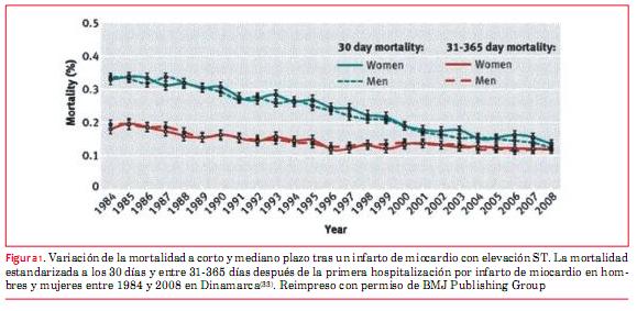
&nbsp; </span><span style="font-size: 10pt; font-family: Verdana;"><o:p></o:p></span>    <br> <span style="font-size: 10pt; font-family: Verdana;"><o:p></o:p></span>    <br> <span style="font-size: 10pt; font-family: Verdana;"><o:p></o:p></span><b> <span style="font-size: 10pt; font-family: Verdana; ">Duraci&oacute;n &oacute;ptima del seguimiento y de la internaci&oacute;n hospitalaria&nbsp;</span><o:p></o:p></b> </p>         <p style="margin: 0cm 0cm 0.0001pt;"> <span style="font-size: 10pt; font-family: Verdana; ">&nbsp;</span><span style="font-size: 10pt; font-family: Verdana;"><o:p></o:p></span></p>          <p style="margin: 0cm 0cm 0.0001pt;"> <span style="font-size: 10pt; font-family: Verdana; ">La duraci&oacute;n de la estad&iacute;a hospitalaria ha disminuido dr&aacute;sticamente con los a&ntilde;os, lo que tiene un importante impacto sobre los gastos de atenci&oacute;n m&eacute;dica y en la calidad de vida de los pacientes. La pr&aacute;ctica actual var&iacute;a ampliamente en los pa&iacute;ses y centros, y no queda claro si las altas tempranas del hospital son seguras</span><sup><span style="font-size: 10pt; font-family: Verdana; "><a name="-32"></a>(</span><span style="font-size: 10pt; font-family: Verdana; color: rgb(31, 26, 23);"><a href="#32" style="color: blue; text-decoration: underline; text-underline: single">32</a></span><span style="font-size: 10pt; font-family: Verdana; ">)</span></sup><span style="font-size: 10pt; font-family: Verdana; ">. Es muy tranquilizador que pese a la continua reducci&oacute;n de la estad&iacute;a hospitalaria, los resultados han mejorado significativamente (</span><span style="font-size: 10pt; font-family: Verdana; color: rgb(31, 26, 23);"><a href="#graf_1" style="color: blue; text-decoration: underline; text-underline: single">figura 1</a></span><span style="font-size: 10pt; font-family: Verdana; ">).</span></p>        <p style="margin: 0cm 0cm 0.0001pt;">&nbsp;</p>        <p style="margin: 0cm 0cm 0.0001pt;"> <span style="font-size: 10pt; font-family: Verdana; "><a name="graf_1"></a><img style="width: 579px; height: 283px;" alt="" src="/img/revistas/ruc/v29n3/3a14g1.JPG"></span></p>       <p style="margin: 0cm 0cm 0.0001pt;">&nbsp;</p>       ]]></body>
<body><![CDATA[<p style="margin: 0cm 0cm 0.0001pt;"><sup><a name="-33"></a>(<a href="#33" style="color: blue; text-decoration: underline; text-underline: single">33</a>)</sup></p>        <p style="margin: 0cm 0cm 0.0001pt;"> <span style="font-size: 10pt; font-family: Verdana; "> &nbsp;&nbsp;</span>    <br>    </p>            <p style="margin: 0cm 0cm 0.0001pt;"> <span style="font-size: 10pt; font-family: Verdana; ">Hay dos nuevos estudios que demuestran que es seguro y factible dar de alta a los pacientes con IAMCEST de bajo riesgo dentro de los dos primeros d&iacute;as luego de una PCI primaria</span><sup><span style="font-size: 10pt; font-family: Verdana; "><a name="-34"></a><a name="-35"></a>(</span><span style="font-size: 10pt; font-family: Verdana; color: rgb(31, 26, 23);"><a href="#34" style="color: blue; text-decoration: underline; text-underline: single">34</a></span><span style="font-size: 10pt; font-family: Verdana; ">,</span><span style="font-size: 10pt; font-family: Verdana; color: rgb(31, 26, 23);"><a href="#35" style="color: blue; text-decoration: underline; text-underline: single">35</a></span><span style="font-size: 10pt; font-family: Verdana; ">)</span></sup><span style="font-size: 10pt; font-family: Verdana; ">. M&aacute;s de 40% de los pacientes con IAMCEST en uno de los estudios cumplieron con los criterios de alta temprana</span><sup><span style="font-size: 10pt; font-family: Verdana; ">(</span><span style="font-size: 10pt; font-family: Verdana; color: rgb(31, 26, 23);"><a href="#34" style="color: blue; text-decoration: underline; text-underline: single">34</a></span><span style="font-size: 10pt; font-family: Verdana; ">)</span></sup><span style="font-size: 10pt; font-family: Verdana; ">.<sup> </sup>Un alta temprana podr&iacute;a bajar considerablemente los costos sanitarios.</span><o:p></o:p></p>          <p style="margin: 0cm 0cm 0.0001pt;"> <span style="font-size: 10pt; font-family: Verdana; ">&nbsp; </span><span style="font-size: 10pt;"><o:p></o:p></span></p>          <p style="margin: 0cm 0cm 0.0001pt;"> <span style="font-size: 10pt; font-family: Verdana; ">Con base en la literatura, propone los siguientes criterios para definir los pacientes de bajo riesgo para un alta temprana:</span><o:p></o:p></p>          <p style="margin: 0cm 0cm 0.0001pt;"> <span style="font-size: 10pt; font-family: Verdana; ">&nbsp; </span><span style="font-size: 10pt;"><o:p></o:p></span></p>          <p style="margin: 0cm 0cm 0.0001pt;"> <span style="font-size: 10pt; font-family: Verdana; ">1.&nbsp;&nbsp;&nbsp;&nbsp;Edad &lt;70 a&ntilde;os.&nbsp; </span><span style="font-size: 10pt;"><o:p></o:p></span></p>          <p style="margin: 0cm 0cm 0.0001pt;"> <span style="font-size: 10pt; font-family: Verdana; ">2.&nbsp;&nbsp;&nbsp;&nbsp;Dolor breve hasta el intervalo de reperfusi&oacute;n (&lt;4 horas).&nbsp; </span><span style="font-size: 10pt;"><o:p></o:p></span></p>          <p style="margin: 0cm 0cm 0.0001pt;"> <span style="font-size: 10pt; font-family: Verdana; ">3.&nbsp;&nbsp;&nbsp;&nbsp;PCI primaria no complicada con buen resultado (flujo TIMI [trombolisis en el infarto de miocardio] 3 y resoluci&oacute;n r&aacute;pida y completa de la elevaci&oacute;n ST).&nbsp; </span><span style="font-size: 10pt;"><o:p></o:p></span></p>          ]]></body>
<body><![CDATA[<p style="margin: 0cm 0cm 0.0001pt;"> <span style="font-size: 10pt; font-family: Verdana; ">4.&nbsp;&nbsp;&nbsp;&nbsp;Fracci&oacute;n de eyecci&oacute;n del ventr&iacute;culo izquierdo &gt;45% sin s&iacute;ntomas de insuficiencia card&iacute;aca.&nbsp; </span><span style="font-size: 10pt;"><o:p></o:p></span></p>          <p style="margin: 0cm 0cm 0.0001pt;"> <span style="font-size: 10pt; font-family: Verdana; ">5.&nbsp;&nbsp;&nbsp;&nbsp;Ausencia de arritmias significativas durante las primeras 24 horas.&nbsp; </span><span style="font-size: 10pt;"><o:p></o:p></span></p>          <p style="margin: 0cm 0cm 0.0001pt;"> <span style="font-size: 10pt; font-family: Verdana; ">6.&nbsp;&nbsp;&nbsp;&nbsp;Paciente que colabora, adhiere al tratamiento y cuenta con apoyo social.&nbsp; </span><span style="font-size: 10pt;"><o:p></o:p></span></p>          <p class="MsoNormal"><span style="font-size: 10pt;"><o:p>&nbsp;</o:p></span></p>          <p style="margin: 0cm 0cm 0.0001pt;"><b> <span style="font-size: 10pt; font-family: Verdana; ">SCA sin elevaci&oacute;n de ST</span><o:p></o:p></b></p>          <p style="margin: 0cm 0cm 0.0001pt;"><b> <span style="font-size: 10pt; font-family: Verdana; ">&nbsp;</span></b><span style="font-size: 10pt; font-family: Verdana; "> </span><span style="font-size: 10pt;"><o:p></o:p></span></p>          <p style="margin: 0cm 0cm 0.0001pt;"><b> <span style="font-size: 10pt; font-family: Verdana; ">Predicci&oacute;n del riesgo&nbsp;</span></b><span style="font-size: 10pt; font-family: Verdana; "> </span><span style="font-size: 10pt;"><o:p></o:p></span></p>          <p style="margin: 0cm 0cm 0.0001pt;"> <span style="font-size: 10pt; font-family: Verdana; ">Es importante encontrar la manera de predecir correctamente el riesgo en los pacientes con SCA para la toma de decisiones cl&iacute;nicas, especialmente con respecto a la angiograf&iacute;a coronaria. Se est&aacute;n utilizando varios modelos de predicci&oacute;n de riesgo. GRACE (Global Registry of Acute Coronary Events) es uno de los scores utilizados m&aacute;s com&uacute;nmente. Recientemente se desarroll&oacute; mini GRACE (MG), un score de riesgo que excluye la creatinina, y la clasificaci&oacute;n Killip del modelo de riesgo original GRACE de ocho factores. El score de riesgo mini GRACE ajustado (AMG, por su sigla en ingl&eacute;s) incluye la &lsquo;prescripci&oacute;n de un diur&eacute;tico de asa durante la internaci&oacute;n&rsquo; en lugar de la clase Killip y concentraci&oacute;n de creatinina. Ambas puntuaciones de riesgo mostraron una buena exactitud en el Proyecto de Auditor&iacute;a Nacional en la Isquemia Mioc&aacute;rdica (Myocardial Ischaemia National Audit Project (MINAP), donde el score de riesgo AMG funciona un poco mejor que el score de riesgo MG</span><sup><span style="font-size: 10pt; font-family: Verdana; "><a name="-36"></a>(</span><span style="font-size: 10pt; font-family: Verdana; color: rgb(31, 26, 23);"><a href="#36" style="color: blue; text-decoration: underline; text-underline: single">36</a></span><span style="font-size: 10pt; font-family: Verdana; ">)</span></sup><span style="font-size: 10pt; font-family: Verdana; ">.&nbsp; </span><span style="font-size: 10pt;"><o:p></o:p></span></p>          <p style="margin: 0cm 0cm 0.0001pt;"> <span style="font-size: 10pt; font-family: Verdana; ">Los marcadores de laboratorio pueden ayudar a estratificar el riesgo. El valor m&aacute;ximo de troponina en pacientes que se presentan con SCASEST ha mostrado ser un predictor independiente de morbimortalidad hospitalaria</span><sup><span style="font-size: 10pt; font-family: Verdana; "><a name="-37"></a>(</span><span style="font-size: 10pt; font-family: Verdana; color: rgb(31, 26, 23);"><a href="#37" style="color: blue; text-decoration: underline; text-underline: single">37</a></span><span style="font-size: 10pt; font-family: Verdana; ">)</span></sup><span style="font-size: 10pt; font-family: Verdana; ">. Entre los marcadores predictivos se incluyen la interleuquina 10, la mieloperoxidasa y el factor de crecimiento placentario</span><sup><span style="font-size: 10pt; font-family: Verdana; "><a name="-38"></a>(</span><span style="font-size: 10pt; font-family: Verdana; color: rgb(31, 26, 23);"><a href="#38" style="color: blue; text-decoration: underline; text-underline: single">38</a></span><span style="font-size: 10pt; font-family: Verdana; ">)</span></sup><span style="font-size: 10pt; font-family: Verdana; ">.<sup>&nbsp;</sup> </span> <o:p></o:p></p>          <p style="margin: 0cm 0cm 0.0001pt;"><span style="font-size: 10pt;"><o:p>&nbsp;</o:p></span></p>          ]]></body>
<body><![CDATA[<p style="margin: 0cm 0cm 0.0001pt;"><b> <span style="font-size: 10pt; font-family: Verdana; ">Papel de la PCI en SCASEST y mejor momento para realizarla&nbsp;</span></b><span style="font-size: 10pt; font-family: Verdana; "> </span></p>          <p style="margin: 0cm 0cm 0.0001pt;"> <span style="font-size: 10pt; font-family: Verdana; ">Para los pacientes de riesgo de intermedio a alto hay una fuerte evidencia que apoya la angiograf&iacute;a de rutina m&aacute;s que el manejo conservador. Sin embargo, no queda claro cu&aacute;l es el momento &oacute;ptimo para realizar la angiograf&iacute;a coronaria. Si bien un abordaje temprano invasivo parece favorable, los estudios que investigan el efecto de la oportunidad utilizaron diferentes puntos de tiempo para la angiograf&iacute;a &lsquo;temprana&rsquo; y diferida. En los pacientes de muy alto riesgo, tales como aquellos con angina refractaria, insuficiencia card&iacute;aca severa, arritmias ventriculares potencialmente fatales o inestabilidad hemodin&aacute;mica o un infarto de miocardio (IM) en evoluci&oacute;n, est&aacute; indicado un abordaje invasivo urgente. Para los pacientes que no pertenezcan a esta categor&iacute;a de alto riesgo, no queda claro cu&aacute;l ser&iacute;a el momento &oacute;ptimo. No existe ning&uacute;n beneficio claro para una estrategia invasiva temprana dentro de las primeras 24 horas con respecto a las variables evaluables cl&iacute;nicas &lsquo;duras&rsquo;, pero hay un n&uacute;mero creciente de centros que adoptan una estrategia invasiva temprana dentro de las primeras 24 horas para los pacientes con riesgo de intermedio a alto. Es probable que un abordaje as&iacute; sea razonable, ya que el actuar m&aacute;s temprano ciertamente ayuda a reducir la internaci&oacute;n. Se deben considerar factores como la presencia de diabetes, funci&oacute;n renal, funci&oacute;n del ventr&iacute;culo izquierdo, s&iacute;ntomas recurrentes, y revascularizaci&oacute;n previa, junto con el score TIMI o GRACE.</span><o:p></o:p></p>               <p style="margin: 0cm 0cm 0.0001pt;"><b>  <multicol gutter="18" cols="2"></multicol> <span style="font-size: 10pt; font-family: Verdana; ">  Imagenolog&iacute;a intravascular&nbsp;</span></b><span style="font-size: 10pt; font-family: Verdana; "> </span></p>          <p style="margin: 0cm 0cm 0.0001pt;"> <span style="font-size: 10pt; font-family: Verdana; ">El concepto de la PCI guiada por imagenolog&iacute;a intravascular ha evolucionado con el advenimiento de equipos como el ultrasonido intravascular (IVUS) y m&aacute;s recientemente la tomograf&iacute;a de coherencia &oacute;ptica (OCT, por su sigla en ingl&eacute;s). Se puede usar de dos modos diferentes, ya sea para evaluaci&oacute;n previa a la PCI, para comprender mejor las caracter&iacute;sticas de la placa coronaria (placa estable o inestable, di&aacute;metro y largo, carga del trombo, etc&eacute;tera), o para valorar la expansi&oacute;n y aposici&oacute;n del stent despu&eacute;s de una PCI. Las ventajas son obvias; a diferencia de la angiograf&iacute;a, que a veces se usa como herramienta a ojo, y que permite medir los di&aacute;metros luminales en unas pocas tomas ortogonales, el IVUS coronario ofrece una vista tomogr&aacute;fica. Asimismo, la resoluci&oacute;n es mucho mejor que la de la angiograf&iacute;a.&nbsp; </span> <o:p></o:p></p>          <p style="margin: 0cm 0cm 0.0001pt;"><span style="font-size: 10pt;"><o:p>&nbsp;</o:p></span></p>          <p style="margin: 0cm 0cm 0.0001pt;"> <span style="font-size: 10pt; font-family: Verdana; ">El primer concepto, la valoraci&oacute;n de las lesiones previo a una PCI, ha sido analizado en el estudio multic&eacute;ntrico PROSPECT (Providing Regional Observations to Study Predictors of Events in the Coronary Tree)</span><sup><span style="font-size: 10pt; font-family: Verdana; "><a name="-39"></a>(</span><span style="font-size: 10pt; font-family: Verdana; color: rgb(31, 26, 23);"><a href="#39" style="color: blue; text-decoration: underline; text-underline: single">39</a></span><span style="font-size: 10pt; font-family: Verdana; ">)</span></sup><span style="font-size: 10pt; font-family: Verdana; ">.<sup> </sup>Este estudio mostr&oacute; que el IVUS es &uacute;til para definir las caracter&iacute;sticas de las placas vulnerables. Los fenotipos de m&aacute;s alto riesgo asociados con eventos card&iacute;acos adversos mayores (MACE) de vasos no responsables incluyeron la presencia de fibroateromas de capa fina, carga de la placa &gt;70%, y &aacute;rea m&iacute;nima de la luz &lt;4,0 mm. Sin embargo, estos datos no son suficientes para recomendar el uso de las caracter&iacute;sticas de las placas que surgen del IVUS para decidir si una lesi&oacute;n necesita ser tratada<a name="-40"></a></span><sup><span style="font-size: 10pt; font-family: Verdana; ">(</span><span style="font-size: 10pt; font-family: Verdana; color: rgb(31, 26, 23);"><a href="#40" style="color: blue; text-decoration: underline; text-underline: single">40</a></span><span style="font-size: 10pt; font-family: Verdana; ">)</span></sup><span style="font-size: 10pt; font-family: Verdana; ">.&nbsp;</span><o:p></o:p></p>          <p style="margin: 0cm 0cm 0.0001pt;"> <span style="font-size: 10pt; font-family: Verdana; ">&nbsp;</span><span style="font-size: 10pt;"><o:p></o:p></span></p>          <p style="margin: 0cm 0cm 0.0001pt;"> <span style="font-size: 10pt; font-family: Verdana; ">Mientras que IVUS se basa en el ultrasonido, la OCT se basa en la luz, que tiene una longitud de onda mucho m&aacute;s corta, y, por lo tanto, logra una resoluci&oacute;n espacial diez veces mejor comparado con IVUS</span><sup><span style="font-size: 10pt; font-family: Verdana; "><a name="-41"></a>(</span><span style="font-size: 10pt; font-family: Verdana; color: rgb(31, 26, 23);"><a href="#41" style="color: blue; text-decoration: underline; text-underline: single">41</a></span><span style="font-size: 10pt; font-family: Verdana; ">)</span></sup><span style="font-size: 10pt; font-family: Verdana; ">.<sup> </sup>Esto permite definir mejor las finas capas fibrosas y la extensi&oacute;n circunferencial de los n&uacute;cleos necr&oacute;ticos. Ayuda a detectar otras caracter&iacute;sticas microestructurales como los cristales de colesterol, trombos, dep&oacute;sitos de calcio, placas fibrosas y placas ricas en l&iacute;pidos</span><sup><span style="font-size: 10pt; font-family: Verdana; "><a name="-42"></a>(</span><span style="font-size: 10pt; font-family: Verdana; color: rgb(31, 26, 23);"><a href="#42" style="color: blue; text-decoration: underline; text-underline: single">42</a></span><span style="font-size: 10pt; font-family: Verdana; ">)</span></sup><span style="font-size: 10pt; font-family: Verdana; ">. La OCT permite visualizar caracter&iacute;sticas no observables con IVUS, tales como los colgajos de la &iacute;ntima y defectos de la &iacute;ntima, distorsiones de la media, y aposici&oacute;n de los <i>struts</i> del stent.</span><o:p></o:p></p>          <p style="margin: 0cm 0cm 0.0001pt;"> <span style="font-size: 10pt; font-family: Verdana; ">&nbsp; </span><span style="font-size: 10pt;"><o:p></o:p></span></p>          <p style="margin: 0cm 0cm 0.0001pt;"> <span style="font-size: 10pt; font-family: Verdana; ">Un estudio japon&eacute;s que analiz&oacute; la lesi&oacute;n responsable en los pacientes con IAM hall&oacute; que la incidencia de las rupturas de la placa observadas por OCT fue significativamente m&aacute;s alta que la observada tanto por la angioscop&iacute;a como por IVUS</span><sup><span style="font-size: 10pt; font-family: Verdana; "><a name="-43"></a>(</span><span style="font-size: 10pt; font-family: Verdana; color: rgb(31, 26, 23);"><a href="#43" style="color: blue; text-decoration: underline; text-underline: single">43</a></span><span style="font-size: 10pt; font-family: Verdana; ">)</span></sup><span style="font-size: 10pt; font-family: Verdana; ">.<sup> </sup>La OCT tambi&eacute;n fue superior para detectar la erosi&oacute;n de la capa fibrosa y el fibroateroma con capa delgada; asimismo, podr&iacute;a estimar el espesor de la capa fibrosa.&nbsp;</span><o:p></o:p></p>          ]]></body>
<body><![CDATA[<p style="margin: 0cm 0cm 0.0001pt;"> <span style="font-size: 10pt; font-family: Verdana; ">&nbsp;</span><span style="font-size: 10pt;"><o:p></o:p></span></p>          <p style="margin: 0cm 0cm 0.0001pt;"> <span style="font-size: 10pt; font-family: Verdana; ">Sin embargo, la profundidad de la penetraci&oacute;n de la imagen se limita solo a unos pocos mil&iacute;metros con esta nueva t&eacute;cnica<a name="-44"></a></span><sup><span style="font-size: 10pt; font-family: Verdana; ">(</span><span style="font-size: 10pt; font-family: Verdana; color: rgb(31, 26, 23);"><a href="#44" style="color: blue; text-decoration: underline; text-underline: single">44</a></span><span style="font-size: 10pt; font-family: Verdana; ">)</span></sup><span style="font-size: 10pt; font-family: Verdana; ">.<sup> </sup>Por lo tanto, no logra dar una imagen de la adventicia y valorar la carga de la placa. Alfonso y colaboradores</span><sup><span style="font-size: 10pt; font-family: Verdana; "><a name="-45"></a>(</span><span style="font-size: 10pt; font-family: Verdana; color: rgb(31, 26, 23);"><a href="#45" style="color: blue; text-decoration: underline; text-underline: single">45</a></span><span style="font-size: 10pt; font-family: Verdana; ">)</span></sup><span style="font-size: 10pt; font-family: Verdana; "> propusieron el uso combinado de OCT e IVUS en pacientes con trombosis del stent. Como la longitud de la imagen fue m&aacute;s corta con OCT, suger&iacute;an corridas de OCT superpuestas para sortear el problema. El desaf&iacute;o de la OCT es que requiere tener un campo sin sangre para obtener las im&aacute;genes.&nbsp;</span><o:p></o:p></p>          <p style="margin: 0cm 0cm 0.0001pt;"> <span style="font-size: 10pt; font-family: Verdana; ">&nbsp;</span><span style="font-size: 10pt;"><o:p></o:p></span></p>          <p style="margin: 0cm 0cm 0.0001pt;"> <span style="font-size: 10pt; font-family: Verdana; ">Como la OCT tiene una resoluci&oacute;n superior al IVUS, discrimina claramente los <i>struts</i> del stent en &aacute;reas muy calcificadas que son dif&iacute;ciles de identificar con IVUS. La OCT posintervenci&oacute;n tambi&eacute;n produce una imagen m&aacute;s n&iacute;tida del l&iacute;mite entre la neo&iacute;ntima y el trombo y ofrece un diagn&oacute;stico confiable de la reestenosis intrastent o neoaterosclerosis. En la pr&aacute;ctica actual parecer&iacute;a que la OCT y el IVUS se complementan con sus respectivas ventajas y desventajas. Sin embargo, tenemos que ser conscientes de que los datos sobre los resultados cl&iacute;nicos son limitados y que estas t&eacute;cnicas aumentan los costos de los procedimientos.&nbsp; </span><span style="font-size: 10pt;"><o:p></o:p></span></p>          <p class="MsoNormal"><span style="font-size: 10pt;"><o:p>&nbsp;</o:p></span></p>          <p style="margin: 0cm 0cm 0.0001pt;"><b> <span style="font-size: 10pt; font-family: Verdana; ">Terapia antiplaquetaria&nbsp;</span></b><span style="font-size: 10pt; font-family: Verdana; "> </span> <o:p></o:p></p>          <p style="margin: 0cm 0cm 0.0001pt;"><span style="font-size: 10pt;"><o:p>&nbsp;</o:p></span></p>          <p style="margin: 0cm 0cm 0.0001pt;"> <span style="font-size: 10pt; font-family: Verdana; ">La aspirina sigue siendo la base de toda terapia antiplaquetaria. Sin embargo, la terapia dual antiplaquetaria con aspirina y un bloqueante del receptor P2Y<sub>12</sub> es claramente m&aacute;s efectiva, siendo el clopidogrel el agente usado m&aacute;s com&uacute;nmente para este prop&oacute;sito en la actualidad. Sin embargo, los problemas con este tratamiento son la demora bastante larga hasta alcanzar la inhibici&oacute;n m&aacute;xima plaquetaria y la alta tasa de malos respondedores</span><sup><span style="font-size: 10pt; font-family: Verdana; "><a name="-46"></a>(</span><span style="font-size: 10pt; font-family: Verdana; color: rgb(31, 26, 23);"><a href="#46" style="color: blue; text-decoration: underline; text-underline: single">46</a></span><span style="font-size: 10pt; font-family: Verdana; ">)</span></sup><span style="font-size: 10pt; font-family: Verdana; ">.<sup> </sup>Un enfoque que ha sido probado repetidamente es la triple terapia antiplaquetaria usando cilostazol. Aunque los resultados de esta conducta han indicado un cierto beneficio, se usa raramente</span><sup><span style="font-size: 10pt; font-family: Verdana; "><a name="-47"></a><a name="-48"></a>(</span><span style="font-size: 10pt; font-family: Verdana; color: rgb(31, 26, 23);"><a href="#47" style="color: blue; text-decoration: underline; text-underline: single">47</a></span><span style="font-size: 10pt; font-family: Verdana; ">,</span><span style="font-size: 10pt; font-family: Verdana; color: rgb(31, 26, 23);"><a href="#48" style="color: blue; text-decoration: underline; text-underline: single">48</a></span><span style="font-size: 10pt; font-family: Verdana; ">)</span></sup><span style="font-size: 10pt; font-family: Verdana; ">.<sup> </sup>Una raz&oacute;n para esto probablemente sea la aparici&oacute;n de la generaci&oacute;n m&aacute;s nueva de los bloqueadores de los receptores P2Y<sub>12</sub> como prasugrel, ticagrelor y cangrelor. Bloquean la uni&oacute;n de ADP al receptor plaquetario P2Y<sub>12</sub>, inhibiendo as&iacute; la agregaci&oacute;n plaquetaria.&nbsp;</span><o:p></o:p></p>            <p style="margin: 0cm 0cm 0.0001pt;">  <multicol gutter="18" cols="2"></multicol>  <span style="font-size: 10pt; font-family: Verdana; ">  Naturalmente ser&iacute;a de esperar que una mayor inhibici&oacute;n antiplaquetaria entra&ntilde;e un mayor riesgo de sangrado, por lo que muchos pacientes reciben inhibidores de la bomba de protones (IBP). Sin embargo, los datos no siguen completamente esta l&oacute;gica.&nbsp;</span><o:p></o:p></p>          <p style="margin: 0cm 0cm 0.0001pt;"> <span style="font-size: 10pt; font-family: Verdana; ">&nbsp;</span><span style="font-size: 10pt; font-family: Verdana;"><o:p></o:p></span></p>          ]]></body>
<body><![CDATA[<p style="margin: 0cm 0cm 0.0001pt;"> <span style="font-size: 10pt; font-family: Verdana; ">Prasugrel: el ensayo TRITON-TIMI 38 fue una comparaci&oacute;n cabeza a cabeza entre la aspirina y el prasugrel versus aspirina m&aacute;s clopidogrel en 13.608 pacientes con SCA con riesgo de moderado a alto sometidos a PCI. En la mayor&iacute;a de los casos, el f&aacute;rmaco en estudio se administr&oacute; luego de la angiograf&iacute;a coronaria. En el seguimiento a los 15 meses se redujeron los MACE (muerte cardiovascular, IM no fatal, o accidente cerebrovascular no fatal) con prasugrel (9,9% vs 12,1%; HR 0,81, IC 95% 0,73 a 0,90). Esta variable evaluable compuesta fue lograda fundamentalmente gracias a una reducci&oacute;n de los IM no fatales. Los sangrados mayores aumentaron algo con prasugrel (2,4% vs 1,8%; HR 1,32, IC 95% 1,3 a 1,68). El sangrado aument&oacute; fundamentalmente en aquellos pacientes con antecedentes de accidente cerebrovascular o ataque isqu&eacute;mico transitorio, edad &sup3;75 a&ntilde;os o con un peso corporal &pound;60 kg. El ensayo TRILOGY SCA analiz&oacute; la acci&oacute;n de prasugrel comparado con clopidogrel en pacientes con SCASEST no sometidos a PCI. No hubo diferencias estad&iacute;sticamente significativas en las tasas de MACE (13,9% vs 16,0%; HR 0,91, IC 95% 0,79 a 1,05).&nbsp;</span><o:p></o:p></p>          <p style="margin: 0cm 0cm 0.0001pt;"> <span style="font-size: 10pt; font-family: Verdana; ">&nbsp;</span><span style="font-size: 10pt; font-family: Verdana;"><o:p></o:p></span></p>          <p style="margin: 0cm 0cm 0.0001pt;"> <span style="font-size: 10pt; font-family: Verdana; ">Ticagrelor: a diferencia de clopidogrel y prasugrel, ticagrelor se une al receptor plaquetario P2Y<sub>12</sub> de manera reversible. Este agente fue estudiado en el ensayo PLATO (18.624 pacientes) en pacientes con SCA, y tambi&eacute;n en aquellos no sometidos a PCI pero que recib&iacute;an tratamiento m&eacute;dico. El tratamiento fue iniciado temprano, a una mediana de 5 horas despu&eacute;s del ingreso al hospital. Este estudio mostr&oacute; un riesgo reducido para MACE (definido como muerte cardiovascular, IM, o accidente cerebrovascular) en el brazo de ticagrelor (9,8% vs 11,7%, HR 0,84, IC 95% 0,77 a 0,92), y tambi&eacute;n hubo un riesgo reducido de mortalidad cardiovascular como una &uacute;nica variable evaluada. En t&eacute;rminos generales no hubo una diferencia significativa en las tasas de sangrado mayor entre los grupos de ticagrelor y clopidogrel (11,6% vs 11,2%, respectivamente). Sin embargo, hubo un riesgo m&aacute;s alto de sangrado mayor relacionado con la cirug&iacute;a no coronaria (4,5% vs 3,8%).&nbsp; </span> <o:p></o:p></p>          <p style="margin: 0cm 0cm 0.0001pt;"><span style="font-size: 10pt; font-family: Verdana;"><o:p>&nbsp;</o:p></span></p>          <p style="margin: 0cm 0cm 0.0001pt;"> <span style="font-size: 10pt; font-family: Verdana; ">Cangrelor: a diferencia de los medicamentos arriba mencionados, cangrelor se administra por v&iacute;a intravenosa. Se lo analiz&oacute; contra placebo y contra clopidogrel. El ensayo CHAMPION- PLATFORM (control con placebo) fue interrumpido precozmente porque un an&aacute;lisis inicial mostr&oacute; resultados decepcionantes. El ensayo CHAMPION- ICP (clopidogrel como comparador) tampoco logr&oacute; mostrar un beneficio importante. El estudio m&aacute;s reciente y de mayor tama&ntilde;o, el ensayo CHAMPION-PHOENIX, compar&oacute; cangrelor contra la precarga con 300-600 mg de clopidogrel. Este estudio no solo incluy&oacute; SCA sino tambi&eacute;n pacientes con CAD estable. Encontr&oacute; una reducci&oacute;n del riesgo de eventos isqu&eacute;micos (muerte, IM, revascularizaci&oacute;n por isquemia o trombosis del stent) en las primeras 48 horas sin un aumento del riesgo de sangrado mayor</span><sup><span style="font-size: 10pt; font-family: Verdana; "><a name="-49"></a>(</span><span style="font-size: 10pt; font-family: Verdana; color: rgb(31, 26, 23);"><a href="#49" style="color: blue; text-decoration: underline; text-underline: single">49</a></span><span style="font-size: 10pt; font-family: Verdana; ">)</span></sup><span style="font-size: 10pt; font-family: Verdana; ">.<sup> </sup>Todav&iacute;a no est&aacute; claro cu&aacute;l ser&iacute;a su papel en la pr&aacute;ctica cl&iacute;nica en el contexto de contar con ticagrelor y prasugrel, y nunca ha sido comparado contra estos agentes.&nbsp;</span><o:p></o:p></p>          <p style="margin: 0cm 0cm 0.0001pt;"> <span style="font-size: 10pt; font-family: Verdana; ">&nbsp;</span><span style="font-size: 10pt; font-family: Verdana;"><o:p></o:p></span></p>          <p style="margin: 0cm 0cm 0.0001pt;"> <span style="font-size: 10pt; font-family: Verdana; ">Cont&aacute;ndose ahora con m&aacute;s terapias antiplaquetarias y m&aacute;s potentes, puede ser dif&iacute;cil decidir qu&eacute; agente usar y cu&aacute;ndo. Hoy d&iacute;a, la decisi&oacute;n habitualmente se basa en los factores cl&iacute;nicos y de riesgo; la farmacogen&eacute;tica tambi&eacute;n puede jugar un papel en orientar las terapias en el futuro</span><sup><span style="font-size: 10pt; font-family: Verdana; "><a name="-50"></a>(</span><span style="font-size: 10pt; font-family: Verdana; color: rgb(31, 26, 23);"><a href="#50" style="color: blue; text-decoration: underline; text-underline: single">50</a></span><span style="font-size: 10pt; font-family: Verdana; ">)</span></sup><span style="font-size: 10pt; font-family: Verdana; ">.<sup>&nbsp;</sup> </span> <o:p></o:p></p>          <p style="margin: 0cm 0cm 0.0001pt;"><span style="font-size: 10pt; font-family: Verdana;">El sangrado gastrointestinal (GI) es uno de los riesgos m&aacute;s comunes de una fuerte terapia antiplaquetaria. Por lo tanto, a menudo tambi&eacute;n se indican IBP. Es interesante ver que un estudio reciente hall&oacute; que el sangrado GI bajo es m&aacute;s com&uacute;n que el sangrado GI alto en pacientes que est&aacute;n recibiendo IBP<sup><a name="-51"></a>(<a href="#51" style="color: blue; text-decoration: underline; text-underline: single">51</a>)</sup>.<sup> </sup>Asimismo, el impacto de los IBP sobre el efecto del clopidogrel ha sido un tema de controversia durante un tiempo. Los estudios de laboratorio han sugerido que si se utilizan IBP, se podr&iacute;a estar reduciendo el efecto antiplaquetario. Sin embargo, los estudios que investigaron las variables evaluables cl&iacute;nicas han mostrado resultados contradictorios. Un examen sistem&aacute;tico reciente ofrece una muy buena rese&ntilde;a general, incluyendo 33 estudios, y concluye que los datos cl&iacute;nicos son sumamente contrastantes, pero que aun los estudios nuevos y mejor dise&ntilde;ados no muestran evidencias de un efecto adverso relevante del efecto de los IBP en los pacientes que reciben clopidogrel en cuanto a los resultados cl&iacute;nicos<sup><a name="-52"></a>(<a href="#52" style="color: blue; text-decoration: underline; text-underline: single">52</a>)</sup>.&nbsp; <br style="">     <o:p></o:p></span></p>          <p style="margin: 0cm 0cm 0.0001pt;"><b>  <multicol gutter="18" cols="2"></multicol> <span style="font-size: 10pt; font-family: Verdana; ">  Muerte s&uacute;bita</span><o:p></o:p></b></p>          <p style="margin: 0cm 0cm 0.0001pt;"><b> <span style="font-size: 10pt; font-family: Verdana; ">&nbsp;</span></b><span style="font-size: 10pt; font-family: Verdana; "> </span><span style="font-size: 10pt; font-family: Verdana;"><o:p></o:p></span></p>          ]]></body>
<body><![CDATA[<p style="margin: 0cm 0cm 0.0001pt;"> <span style="font-size: 10pt; font-family: Verdana; ">La muerte s&uacute;bita (MS) es una presentaci&oacute;n poco com&uacute;n, pero a menudo fatal de los SCA</span><sup><span style="font-size: 10pt; font-family: Verdana; "><a name="-53"></a>(</span><span style="font-size: 10pt; font-family: Verdana; color: rgb(31, 26, 23);"><a href="#53" style="color: blue; text-decoration: underline; text-underline: single">53</a></span><span style="font-size: 10pt; font-family: Verdana; ">)</span></sup><span style="font-size: 10pt; font-family: Verdana; ">.<sup> </sup>Si bien hay otras razones para una MS, especialmente en pacientes m&aacute;s j&oacute;venes, la causa m&aacute;s com&uacute;n de paros card&iacute;acos por taquiarritmia en pacientes mayores de 40 a&ntilde;os es la isquemia mioc&aacute;rdica</span><sup><span style="font-size: 10pt; font-family: Verdana; ">(</span><span style="font-size: 10pt; font-family: Verdana; color: rgb(31, 26, 23);"><a href="#4" style="color: blue; text-decoration: underline; text-underline: single">4</a></span><span style="font-size: 10pt; font-family: Verdana; ">,</span><span style="font-size: 10pt; font-family: Verdana; color: rgb(31, 26, 23);"><a href="#37" style="color: blue; text-decoration: underline; text-underline: single">37</a></span><span style="font-size: 10pt; font-family: Verdana; ">) </span> </sup><span style="font-size: 10pt; font-family: Verdana; ">.<sup> </sup>La mayor&iacute;a de estas muertes s&uacute;bitas ocurren fuera del hospital (paro card&iacute;aco extrahospitalario [OHCA, por su sigla en ingl&eacute;s]). La supervivencia de los pacientes con OHCA ha sido mala durante varias d&eacute;cadas, con un promedio &lt;10% al alta hospitalaria, y puede ser incluso m&aacute;s baja, particularmente en &aacute;reas distantes. No obstante ello, en los &uacute;ltimos a&ntilde;os la supervivencia ha aumentado, especialmente en las &aacute;reas metropolitanas. El servicio de ambulancias de Londres constat&oacute; un aumento de las tasas de supervivencia, pasando de 12% a 32% entre 2007 y 2012</span><sup><span style="font-size: 10pt; font-family: Verdana; ">(</span><span style="font-size: 10pt; font-family: Verdana; color: rgb(31, 26, 23);"><a href="#5" style="color: blue; text-decoration: underline; text-underline: single">5</a></span><span style="font-size: 10pt; font-family: Verdana; ">)</span></sup><span style="font-size: 10pt; font-family: Verdana; ">.<sup>&nbsp;</sup> </span><span style="font-size: 10pt;"><o:p></o:p></span></p>          <p><span style="font-size: 10pt; font-family: Verdana; ">No se puede m&aacute;s que especular sobre las razones de esta mejora, dado que son pocas las intervenciones aisladas que han realmente demostrado ser eficaces</span><sup><span style="font-size: 10pt; font-family: Verdana; "><a name="-54"></a>(</span><span style="font-size: 10pt; font-family: Verdana; color: rgb(31, 26, 23);"><a href="#54" style="color: blue; text-decoration: underline; text-underline: single">54</a></span><span style="font-size: 10pt; font-family: Verdana; ">)</span></sup><span style="font-size: 10pt; font-family: Verdana; ">.<sup> </sup>Por consiguiente, es m&aacute;s probable que las mejoras observadas en la supervivencia sean debidas a la combinaci&oacute;n de m&uacute;ltiples tratamientos eficaces. Las compresiones de pecho realizadas precozmente y la desfibrilaci&oacute;n temprana son dos elementos de cambio indiscutidos</span><sup><span style="font-size: 10pt; font-family: Verdana; "><a name="-55"></a>(</span><span style="font-size: 10pt; font-family: Verdana; color: rgb(31, 26, 23);"><a href="#55" style="color: blue; text-decoration: underline; text-underline: single">55</a></span><span style="font-size: 10pt; font-family: Verdana; ">)</span></sup><span style="font-size: 10pt; font-family: Verdana; ">.<sup> </sup>Es probable que la disponibilidad de desfibriladores autom&aacute;ticos p&uacute;blicos, los desfibriladores de los EMS y la sensibilizaci&oacute;n del p&uacute;blico, y un aumento del n&uacute;mero de legos entrenados en compresi&oacute;n tor&aacute;cica hayan jugado un papel importante</span><sup><span style="font-size: 10pt; font-family: Verdana; "><a name="-56"></a>(</span><span style="font-size: 10pt; font-family: Verdana; color: rgb(31, 26, 23);"><a href="#56" style="color: blue; text-decoration: underline; text-underline: single">56</a></span><span style="font-size: 10pt; font-family: Verdana; ">)</span></sup><span style="font-size: 10pt; font-family: Verdana; ">.<sup>&nbsp;</sup> </span><span style="font-size: 10pt;"><o:p></o:p></span></p>          <p><span style="font-size: 10pt; font-family: Verdana; ">Sin embargo, tambi&eacute;n resultan importantes elementos como la hipotermia terap&eacute;utica y una angiograf&iacute;a inmediata para definir y potencialmente tratar la causa de base</span><sup><span style="font-size: 10pt; font-family: Verdana; "><a name="-57"></a><a name="-58"></a>(</span><span style="font-size: 10pt; font-family: Verdana; color: rgb(31, 26, 23);"><a href="#57" style="color: blue; text-decoration: underline; text-underline: single">57</a></span><span style="font-size: 10pt; font-family: Verdana; ">,</span><span style="font-size: 10pt; font-family: Verdana; color: rgb(31, 26, 23);"><a href="#58" style="color: blue; text-decoration: underline; text-underline: single">58</a></span><span style="font-size: 10pt; font-family: Verdana; ">)</span></sup><span style="font-size: 10pt; font-family: Verdana; ">. En un estudio observacional se evalu&oacute; a 9.971 pacientes con OHCA y sospecha de causa card&iacute;aca en relaci&oacute;n con el hospital al cual fueron referidos. Los pacientes tratados en hospitales con servicios de cardiolog&iacute;a intervencionista las 24 horas tuvieron una mejor supervivencia (OR 1,40, IC 95% 1,12 a 1,74; p=0,003).&nbsp; </span><span style="font-size: 10pt;"><o:p></o:p></span></p>          <p style="margin: 0cm 0cm 0.0001pt;"> <span style="font-size: 10pt; font-family: Verdana; ">Las gu&iacute;as actuales recomiendan la realizaci&oacute;n de una angiograf&iacute;a en los pacientes inmediatamente despu&eacute;s de una resucitaci&oacute;n exitosa de un OHCA (recuperaci&oacute;n de la circulaci&oacute;n espont&aacute;nea) en caso de elevaciones ST en el ECG posresucitaci&oacute;n. Sin embargo, la exactitud de los ECG posteriores a una resucitaci&oacute;n no quedan claros y no hay fundamentos como para recomendar una angiograf&iacute;a temprana en todos los pacientes mayores de 35-40 a&ntilde;os, independientemente del ECG, si no hay una causa no card&iacute;aca obvia.&nbsp; </span><span style="font-size: 10pt;"><o:p></o:p></span></p>          <p class="MsoNormal"><span style="font-size: 10pt;"><o:p>&nbsp;</o:p></span></p>          <p style="margin: 0cm 0cm 0.0001pt;"><b> <span style="font-size: 10pt; font-family: Verdana; ">Rehabilitaci&oacute;n card&iacute;aca tras un SCA</span><o:p></o:p></b></p>          <p style="margin: 0cm 0cm 0.0001pt;"><b> <span style="font-size: 10pt; font-family: Verdana; ">&nbsp;</span></b><span style="font-size: 10pt; font-family: Verdana; "> </span><span style="font-size: 10pt;"><o:p></o:p></span></p>          <p style="margin: 0cm 0cm 0.0001pt;"> <span style="font-size: 10pt; font-family: Verdana; ">Si bien parece intuitivo que los programas de rehabilitaci&oacute;n card&iacute;aca son beneficiosos porque ofrecen un seguimiento cuidadoso, actividad f&iacute;sica supervisada y orientaciones sobre cambios en el estilo de vida, la informaci&oacute;n cl&iacute;nica sobre sus efectos es controvertida. Muy recientemente, la rehabilitaci&oacute;n card&iacute;aca por SCA ha sido cuestionada nuevamente por el ECA multic&eacute;ntrico de rehabilitaci&oacute;n card&iacute;aca integral en pacientes que han presentado un IM agudo (RAMIT: Rehabilitation After Myocardial Infarction Trial)</span><sup><span style="font-size: 10pt; font-family: Verdana; "> <a name="-59"></a>(</span><span style="font-size: 10pt; font-family: Verdana; color: rgb(31, 26, 23);"><a href="#59" style="color: blue; text-decoration: underline; text-underline: single">59</a></span><span style="font-size: 10pt; font-family: Verdana; ">)</span></sup><span style="font-size: 10pt; font-family: Verdana; ">.<sup> </sup>En este estudio, la rehabilitaci&oacute;n card&iacute;aca en los pacientes que han sufrido un IAM no incidi&oacute; sobre la mortalidad o la morbilidad, la medicaci&oacute;n card&iacute;aca, los factores de riesgo o los cambios en el estilo de vida. Sin embargo, debemos ser conscientes de que el ensayo RAMIT era peque&ntilde;o y si observamos la evidencia de una forma m&aacute;s completa, reuniendo todos los ECA disponibles, como en una revisi&oacute;n Cochrane (combinando 47 estudios), hay un efecto significativo aunque modesto sobre la mortalidad</span><sup><span style="font-size: 10pt; font-family: Verdana; "><a name="-60"></a>(</span><span style="font-size: 10pt; font-family: Verdana; color: rgb(31, 26, 23);"><a href="#60" style="color: blue; text-decoration: underline; text-underline: single">60</a></span><span style="font-size: 10pt; font-family: Verdana; ">)</span></sup><span style="font-size: 10pt; font-family: Verdana; ">.<sup> </sup>Este metaan&aacute;lisis no incluy&oacute; los hallazgos del ensayo RAMIT, que habr&iacute;an reducido aun m&aacute;s el efecto estimado sobre la mortalidad por todas las causas de 13% a 11%</span><sup><span style="font-size: 10pt; font-family: Verdana; "><a name="-61"></a>(</span><span style="font-size: 10pt; font-family: Verdana; color: rgb(31, 26, 23);"><a href="#61" style="color: blue; text-decoration: underline; text-underline: single">61</a></span><span style="font-size: 10pt; font-family: Verdana; ">)</span></sup><span style="font-size: 10pt; font-family: Verdana; ">. Es importante destacar que la revisi&oacute;n Cochrane se concentr&oacute; en la rehabilitaci&oacute;n basada en el ejercicio f&iacute;sico, siendo probable que la rehabilitaci&oacute;n no basada en el ejercicio (educaci&oacute;n del paciente) tenga poco efecto sobre la mortalidad despu&eacute;s de un IM</span><sup><span style="font-size: 10pt; font-family: Verdana; "><a name="-62"></a>(</span><span style="font-size: 10pt; font-family: Verdana; color: rgb(31, 26, 23);"><a href="#62" style="color: blue; text-decoration: underline; text-underline: single">62</a></span><span style="font-size: 10pt; font-family: Verdana; ">)</span></sup><span style="font-size: 10pt; font-family: Verdana; ">.<sup>&nbsp;</sup> </span> <o:p></o:p></p>          <p style="margin: 0cm 0cm 0.0001pt;"><span style="font-size: 10pt;"><o:p>&nbsp;</o:p></span></p>            <p style="margin: 0cm 0cm 0.0001pt;"> <span style="font-size: 10pt; font-family: Verdana; ">El problema de combinar resultados de muchos ensayos, por supuesto, es que esto no tiene en cuenta la &lsquo;evoluci&oacute;n&rsquo; de dichas intervenciones</span><sup><span style="font-size: 10pt; font-family: Verdana; "><a name="-63"></a>(</span><span style="font-size: 10pt; font-family: Verdana; color: rgb(31, 26, 23);"><a href="#63" style="color: blue; text-decoration: underline; text-underline: single">63</a></span><span style="font-size: 10pt; font-family: Verdana; ">)</span></sup><span style="font-size: 10pt; font-family: Verdana; ">.<sup> </sup>Los resultados del estudio reciente OMEGA, un estudio de cohorte no aleatorizado, han demostrado que un programa de rehabilitaci&oacute;n card&iacute;aca completo a corto plazo tras un IAM agudo mejoraba el pron&oacute;stico de forma significativa al a&ntilde;o</span><sup><span style="font-size: 10pt; font-family: Verdana; "><a name="-64"></a>(</span><span style="font-size: 10pt; font-family: Verdana; color: rgb(31, 26, 23);"><a href="#64" style="color: blue; text-decoration: underline; text-underline: single">64</a></span><span style="font-size: 10pt; font-family: Verdana; ">)</span></sup><span style="font-size: 10pt; font-family: Verdana; ">.<sup> </sup>Los que asistieron a programas de rehabilitaci&oacute;n tuvieron una mortalidad por todas las causas m&aacute;s baja que la de los que no asistieron, pero es dif&iacute;cil interpretar los datos sin una asignaci&oacute;n de tratamiento aleatorizada. Hubo una relaci&oacute;n dosis-respuesta importante; cuantas m&aacute;s sesiones cumplieran, menor la mortalidad por todas las causas. Sin embargo, quienes asist&iacute;an a pocas sesiones tend&iacute;an a ser fumadores, y al hacer los ajustes para las diferencias basales en la condici&oacute;n de tabaquismo esa asociaci&oacute;n dosis-respuesta desapareci&oacute;.</span><span style="font-size: 10pt; font-family: Verdana; color: rgb(31, 26, 23);"><o:p></o:p></span><span style="font-size: 10pt; font-family: Verdana; ">&nbsp; </span><span style="font-size: 10pt;"><o:p></o:p></span></p>          ]]></body>
<body><![CDATA[<p style="margin: 0cm 0cm 0.0001pt;">  <multicol gutter="18" cols="2"></multicol>  <span style="font-size: 10pt; font-family: Verdana; ">  Si bien la rehabilitaci&oacute;n card&iacute;aca tal como se la brinda en muchos pa&iacute;ses puede no ser eficaz para reducir las variables cl&iacute;nicas duras, sigue siendo &uacute;til dar informaci&oacute;n, consejos y transmitir tranquilidad y ayuda en la prevenci&oacute;n secundaria a largo plazo</span><sup><span style="font-size: 10pt; font-family: Verdana; "><a name="-65"></a>(</span><span style="font-size: 10pt; font-family: Verdana; color: rgb(31, 26, 23);"><a href="#65" style="color: blue; text-decoration: underline; text-underline: single">65</a></span><span style="font-size: 10pt; font-family: Verdana; ">)</span></sup><span style="font-size: 10pt; font-family: Verdana; ">.<sup>&nbsp;</sup> </span><span style="font-size: 10pt;"><o:p></o:p></span></p>          <p class="MsoNormal"><span style="font-size: 10pt;"><o:p>&nbsp;</o:p></span></p>          <p style="margin: 0cm 0cm 0.0001pt;"><b> <span style="font-size: 10pt; font-family: Verdana; ">Conclusiones&nbsp;</span><o:p></o:p></b></p>          <p style="margin: 0cm 0cm 0.0001pt;"> <span style="font-size: 10pt; font-family: Verdana; ">&nbsp;</span><span style="font-size: 10pt;"><o:p></o:p></span></p>          <p style="margin: 0cm 0cm 0.0001pt;"> <span style="font-size: 10pt; font-family: Verdana; ">
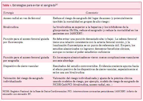
Las opciones de tratamiento para los SCA han mejorado de manera importante en los &uacute;ltimos a&ntilde;os, contribuyendo a mejoras notables de los resultados. Esto se aplica especialmente a los casos de IAMCEST, mientras que la mortalidad a largo plazo despu&eacute;s de SCASEST sigue siendo considerable. Es probable que la muy reciente introducci&oacute;n de las terapias antiplaquetarias de tercera generaci&oacute;n (prasugrel, ticagrelor) y la forma intravenosa m&aacute;s reciente, cangrelor, sigan mejorando los resultados cl&iacute;nicos despu&eacute;s de un SCA. Estos agentes m&aacute;s potentes pueden aumentar los riesgos de sangrado, y considerando la asociaci&oacute;n entre el sangrado y los resultados, las estrategias para evitar el sangrado periprocedimiento son importantes. Pueden incluir la angiograf&iacute;a por v&iacute;a radial, el acceso femoral guiado por ultrasonido, y el uso de bivalirudina.</span><o:p></o:p></p>          <p style="margin: 0cm 0cm 0.0001pt;"> <span style="font-size: 10pt; font-family: Verdana; ">&nbsp; </span><span style="font-size: 10pt;"><o:p></o:p></span></p>          <p style="margin: 0cm 0cm 0.0001pt;"><span style="font-size: 10pt;"><o:p>&nbsp;</o:p></span></p>          <p style="margin: 0cm 0cm 0.0001pt;"><b> <span style="font-size: 10pt; font-family: Verdana; ">Colaboradores. </span></b> <span style="font-size: 10pt; font-family: Verdana; ">PM redact&oacute; el manuscrito. AB, AJL y AB revisaron el manuscrito cr&iacute;ticamente para el contenido verificando su contenido intelectual. Los tres autores hicieron grandes contribuciones a este trabajo y aprobaron la versi&oacute;n final.&nbsp; </span> <o:p></o:p></p>          <p style="margin: 0cm 0cm 0.0001pt;"><b> <span style="font-size: 10pt; font-family: Verdana; ">Conflicto de intereses</span></b><span style="font-size: 10pt; font-family: Verdana; ">: ninguno.</span><o:p></o:p></p>          <p style="margin: 0cm 0cm 0.0001pt;"> <span style="font-size: 10pt; font-family: Verdana; ">Origen y revisi&oacute;n por pares: encargado, revisi&oacute;n interna por colegas.</span><span style="font-size: 10pt;"><o:p></o:p></span></p>          ]]></body>
<body><![CDATA[<p style="margin: 0cm 0cm 0.0001pt;"> <span style="font-size: 10pt; font-family: Verdana; ">&nbsp; </span><span style="font-size: 10pt;"><o:p></o:p></span></p>          <p class="MsoNormal"><span style="font-size: 10pt;"><o:p>&nbsp;</o:p></span></p>          <p style="margin: 0cm 0cm 0.0001pt;"><b> <span style="font-size: 10pt; font-family: Verdana; ">Referencias</span><o:p></o:p></b></p>          <p style="margin: 0cm 0cm 0.0001pt;"><b> <span style="font-size: 10pt; font-family: Verdana; ">&nbsp;</span></b><span style="font-size: 10pt; font-family: Verdana; "> </span><span style="font-size: 10pt;"><o:p></o:p></span></p>          <p style="margin: 0cm 0cm 0.0001pt;"> <span style="font-size: 10pt; font-family: Verdana; "><a name="1"></a> </span><span style="font-size: 10pt; font-family: Verdana; color: rgb(31, 26, 23);"> <a href="#-1" style="color: blue; text-decoration: underline; text-underline: single">1</a></span><span style="font-size: 10pt; font-family: Verdana; ">.&nbsp;&nbsp;&nbsp;&nbsp;<b>Members WG, Roger VL, Go AS, et al.</b> Heart disease and stroke statistics.2012 update: a report from the American Heart Association. Circulation 2012; 125: e2&ndash;220.&nbsp; </span><span style="font-size: 10pt; font-family: Verdana;"><o:p></o:p></span></p>          <p style="margin: 0cm 0cm 0.0001pt;"> <span style="font-size: 10pt; font-family: Verdana; "><a name="2"></a> </span><span style="font-size: 10pt; font-family: Verdana; color: rgb(31, 26, 23);"> <a href="#-2" style="color: blue; text-decoration: underline; text-underline: single">2</a></span><span style="font-size: 10pt; font-family: Verdana; ">.&nbsp;&nbsp;&nbsp;&nbsp;<b>Knight CJ, Timmis AD.</b> Almanac 2011: acute coronary syndromes. The national society journals present selected research that has driven recent advances in clinical cardiology. Heart 2011;97:1820&ndash;7.&nbsp; </span><span style="font-size: 10pt; font-family: Verdana;"><o:p></o:p></span></p>          <p style="margin: 0cm 0cm 0.0001pt;"> <span style="font-size: 10pt; font-family: Verdana; "><a name="3"></a> </span><span style="font-size: 10pt; font-family: Verdana; color: rgb(31, 26, 23);"> <a href="#-3" style="color: blue; text-decoration: underline; text-underline: single">3</a></span><span style="font-size: 10pt; font-family: Verdana; ">.&nbsp;&nbsp;&nbsp;&nbsp;<b>Yeh RW, Sidney S, Chandra M, et al.</b> Population trends in the incidence and outcomes of acute myocardial infarction. N Engl J Med 2010;362:2155&ndash;65.&nbsp; </span><span style="font-size: 10pt; font-family: Verdana;"><o:p></o:p></span></p>          <p style="margin: 0cm 0cm 0.0001pt;"> <span style="font-size: 10pt; font-family: Verdana; "><a name="4"></a> </span><span style="font-size: 10pt; font-family: Verdana; color: rgb(31, 26, 23);"> <a href="#-4" style="color: blue; text-decoration: underline; text-underline: single">4</a></span><span style="font-size: 10pt; font-family: Verdana; ">.&nbsp;&nbsp;&nbsp;&nbsp;<b>Nolan JP, Lyon RM, Sasson C, et al.</b> Advances in the hospital management of patients following an out of hospital cardiac arrest. Heart 2012;98:1201&ndash;6.&nbsp; </span><span style="font-size: 10pt; font-family: Verdana;"><o:p></o:p></span></p>          <p style="margin: 0cm 0cm 0.0001pt;"> <span style="font-size: 10pt; font-family: Verdana; "><a name="5"></a> </span><span style="font-size: 10pt; font-family: Verdana; color: rgb(31, 26, 23);"> <a href="#-5" style="color: blue; text-decoration: underline; text-underline: single">5</a></span><span style="font-size: 10pt; font-family: Verdana; ">.&nbsp;&nbsp;&nbsp;&nbsp;<b>Fothergill RT, Watson LR, Chamberlain D, et al.</b> Increases in survival from out-of-hospital cardiac arrest: a five year study. Resuscitation 2013;84: 1089&ndash;92.&nbsp; </span><span style="font-size: 10pt; font-family: Verdana;"><o:p></o:p></span></p>          <p style="margin: 0cm 0cm 0.0001pt;"> <span style="font-size: 10pt; font-family: Verdana; "><a name="6"></a> </span><span style="font-size: 10pt; font-family: Verdana; color: rgb(31, 26, 23);"> <a href="#-6" style="color: blue; text-decoration: underline; text-underline: single">6</a></span><span style="font-size: 10pt; font-family: Verdana; ">.&nbsp;&nbsp;&nbsp;&nbsp;<b>Stone GW, Maehara A, Witzenbichler B, et al. </b>Intracoronary abciximab and aspiration thrombectomy in patients with large anterior myocardial infarction: the INFUSE-AMI randomized trial. JAMA 2012;307:1817&ndash;26.&nbsp; </span><span style="font-size: 10pt; font-family: Verdana;"><o:p></o:p></span></p>          ]]></body>
<body><![CDATA[<p style="margin: 0cm 0cm 0.0001pt;"> <span style="font-size: 10pt; font-family: Verdana; "><a name="7"></a> </span><span style="font-size: 10pt; font-family: Verdana; color: rgb(31, 26, 23);"> <a href="#-7" style="color: blue; text-decoration: underline; text-underline: single">7</a></span><span style="font-size: 10pt; font-family: Verdana; ">.&nbsp;&nbsp;&nbsp;&nbsp;<b>Stone GW, Witzenbichler B, Guagliumi G, et al.</b> Heparin plus a glycoprotein IIb/IIIa inhibitor versus bivalirudin monotherapy and paclitaxel-eluting stents versus bare-metal stents in acute myocardial infarction (HORIZONS-AMI): final 3-year results from a multicentre, randomised controlled trial. Lancet 2011;377:2193&ndash;204.&nbsp; </span><span style="font-size: 10pt; font-family: Verdana;"><o:p></o:p></span></p>          <p style="margin: 0cm 0cm 0.0001pt;"> <span style="font-size: 10pt; font-family: Verdana; "><a name="8"></a> </span><span style="font-size: 10pt; font-family: Verdana; color: rgb(31, 26, 23);"> <a href="#-8" style="color: blue; text-decoration: underline; text-underline: single">8</a></span><span style="font-size: 10pt; font-family: Verdana; ">.&nbsp;&nbsp;&nbsp;&nbsp;<b>Kimmelstiel C, Zhang P, Kapur NK, et al.</b> Bivalirudin is a dual inhibitor of thrombin and collagen-dependent platelet activation in patients undergoing percutaneous coronary intervention. Circ Cardiovasc Interv 2011;4:171&ndash;9.&nbsp; </span><span style="font-size: 10pt; font-family: Verdana;"><o:p></o:p></span></p>          <p style="margin: 0cm 0cm 0.0001pt;"> <span style="font-size: 10pt; font-family: Verdana; "><a name="9"></a> </span><span style="font-size: 10pt; font-family: Verdana; color: rgb(31, 26, 23);"> <a href="#-9" style="color: blue; text-decoration: underline; text-underline: single">9</a></span><span style="font-size: 10pt; font-family: Verdana; ">.&nbsp;&nbsp;&nbsp;&nbsp;<b>Meier P, Frohlich GM, Lansky AJ. </b>Bleeding complications in percutaneous coronary interventions. Cardiology 2013;125:213&ndash;16.&nbsp; </span><span style="font-size: 10pt; font-family: Verdana;"><o:p></o:p></span></p>          <p style="margin: 0cm 0cm 0.0001pt;"> <span style="font-size: 10pt; font-family: Verdana; "><a name="10"></a> </span><span style="font-size: 10pt; font-family: Verdana; color: rgb(31, 26, 23);"> <a href="#-10" style="color: blue; text-decoration: underline; text-underline: single">10</a></span><span style="font-size: 10pt; font-family: Verdana; ">.&nbsp;&nbsp;&nbsp;&nbsp;<b>Mamas MA, Ratib K, Routledge H, et al.</b> In?uence of access site selection on PCI-related adverse events in patients with STEMI: meta-analysis of randomised controlled trials. Heart 2012;98:303&ndash;11.&nbsp; </span><span style="font-size: 10pt; font-family: Verdana;"><o:p></o:p></span></p>          <p style="margin: 0cm 0cm 0.0001pt;"> <span style="font-size: 10pt; font-family: Verdana; "><a name="11"></a> </span><span style="font-size: 10pt; font-family: Verdana; color: rgb(31, 26, 23);"> <a href="#-11" style="color: blue; text-decoration: underline; text-underline: single">11</a></span><span style="font-size: 10pt; font-family: Verdana; ">.&nbsp;&nbsp;&nbsp;&nbsp;<b>Meier P, Windecker S, Lansky AJ.</b> Radial versus femoral access for primary percutaneous coronary intervention: is there a preferred route to the heart? Heart 2012;98:269&ndash;70.&nbsp; </span><span style="font-size: 10pt; font-family: Verdana;"><o:p></o:p></span></p>          <p style="margin: 0cm 0cm 0.0001pt;"> <span style="font-size: 10pt; font-family: Verdana; "><a name="12"></a> </span><span style="font-size: 10pt; font-family: Verdana; color: rgb(31, 26, 23);"> <a href="#-12" style="color: blue; text-decoration: underline; text-underline: single">12</a></span><span style="font-size: 10pt; font-family: Verdana; ">.&nbsp;&nbsp;&nbsp;&nbsp;<b>Baklanov DV, Kaltenbach LA, Marso SP, et al.</b> The prevalence and outcomes of transradial percutaneous coronary intervention for ST-segment elevation myocardial infarction: analysis from the National Cardiovascular Data Registry (2007 to 2011). J Am Coll Cardiol 2013;61:420&ndash;6.&nbsp; </span><span style="font-size: 10pt; font-family: Verdana;"><o:p></o:p></span></p>          <p style="margin: 0cm 0cm 0.0001pt;"> <span style="font-size: 10pt; font-family: Verdana; "><a name="13"></a> </span><span style="font-size: 10pt; font-family: Verdana; color: rgb(31, 26, 23);"> <a href="#-13" style="color: blue; text-decoration: underline; text-underline: single">13</a></span><span style="font-size: 10pt; font-family: Verdana; ">.&nbsp;&nbsp;&nbsp;&nbsp;<b>Cafri C, Zahger D, Merkin M, et al.</b> Efficacy of the radial approach for the performance of primary PCI for STEMI. J Invasive Cardiol 2013;25:150&ndash;3.&nbsp; </span><span style="font-size: 10pt; font-family: Verdana;"><o:p></o:p></span></p>          <p style="margin: 0cm 0cm 0.0001pt;"> <span style="font-size: 10pt; font-family: Verdana; "><a name="14"></a> </span><span style="font-size: 10pt; font-family: Verdana; color: rgb(31, 26, 23);"> <a href="#-14" style="color: blue; text-decoration: underline; text-underline: single">14</a></span><span style="font-size: 10pt; font-family: Verdana; ">.&nbsp;&nbsp;&nbsp;&nbsp;<b>Secco GG, Marinucci L, Uguccioni L, et al.</b> Transradial versus transfemoral approach for primary percutaneous coronary interventions in elderly patients. J Invasive Cardiol 2013;25:254&ndash;6.&nbsp; </span><span style="font-size: 10pt; font-family: Verdana;"><o:p></o:p></span></p>          <p style="margin: 0cm 0cm 0.0001pt;"> <span style="font-size: 10pt; font-family: Verdana; "><a name="15"></a>  </span><span style="font-size: 10pt; font-family: Verdana; color: rgb(31, 26, 23);">  <a href="#-15" style="color: blue; text-decoration: underline; text-underline: single">15</a></span><span style="font-size: 10pt; font-family: Verdana; ">.&nbsp;&nbsp;&nbsp;&nbsp;<b>Ratib K, Mamas MA, Routledge HC, et al. </b>In?uence of access site choice on incidence of neurologic complications after percutaneous coronary intervention. Am Heart J 2013;165:317&ndash;24.&nbsp; </span><span style="font-size: 10pt; font-family: Verdana;"><o:p></o:p></span></p>          <p style="margin: 0cm 0cm 0.0001pt;"> <span style="font-size: 10pt; font-family: Verdana; "><a name="16"></a> </span><span style="font-size: 10pt; font-family: Verdana; color: rgb(31, 26, 23);"> <a href="#-16" style="color: blue; text-decoration: underline; text-underline: single">16</a></span><span style="font-size: 10pt; font-family: Verdana; ">.&nbsp;&nbsp;&nbsp;&nbsp;<b>Ratib K, Routledge H, Mamas MA, et al.</b> Trends in access site choice and PCI outcomes: insights from the UK national PCI dataset. Heart 2012;98: A28&ndash;A9.&nbsp; </span><span style="font-size: 10pt; font-family: Verdana;"><o:p></o:p></span></p>          ]]></body>
<body><![CDATA[<p style="margin: 0cm 0cm 0.0001pt;"> <span style="font-size: 10pt; font-family: Verdana; "><a name="17"></a> </span><span style="font-size: 10pt; font-family: Verdana; color: rgb(31, 26, 23);"> <a href="#-17" style="color: blue; text-decoration: underline; text-underline: single">17</a></span><span style="font-size: 10pt; font-family: Verdana; ">.&nbsp;&nbsp;&nbsp;&nbsp;<b>Feldman DN, Swaminathan RV, Kaltenbach LA, et al. </b>Adoption of radial access and comparison of outcomes to femoral access in percutaneous coronary intervention: an updated report from the National Cardiovascular Data Registry (2007&ndash;2012). Circulation 2013;127:2295&ndash;306.&nbsp; </span><span style="font-size: 10pt; font-family: Verdana;"><o:p></o:p></span></p>          <!-- ref --><p style="margin: 0cm 0cm 0.0001pt;"> <span style="font-size: 10pt; font-family: Verdana; "><a name="18"></a> </span><span style="font-size: 10pt; font-family: Verdana; color: rgb(31, 26, 23);"> <a href="#-18" style="color: blue; text-decoration: underline; text-underline: single">18</a></span><span style="font-size: 10pt; font-family: Verdana; ">.&nbsp;&nbsp;&nbsp;&nbsp;<b>De Luca G, Schaffer A, Wirianta J, et al. </b>Comprehensive meta-analysis of radial vs femoral approach in primary angioplasty for STEMI. Int J Cardiol 2013 (Epub ahead of print).    &nbsp; </span><span style="font-size: 10pt; font-family: Verdana;"><o:p></o:p></span></p>          <p style="margin: 0cm 0cm 0.0001pt;"> <span style="font-size: 10pt; font-family: Verdana; "><a name="19"></a> </span><span style="font-size: 10pt; font-family: Verdana; color: rgb(31, 26, 23);"> <a href="#-19" style="color: blue; text-decoration: underline; text-underline: single">19</a></span><span style="font-size: 10pt; font-family: Verdana; ">.&nbsp;&nbsp;&nbsp;&nbsp;<b>Mehta SR, Jolly SS, Cairns J, et al.</b> Effects of radial versus femoral artery access in patients with acute coronary syndromes with or without ST-segment elevation. J Am Coll Cardiol 2012;60:2490&ndash;9.&nbsp; </span><span style="font-size: 10pt; font-family: Verdana;"><o:p></o:p></span></p>          <p style="margin: 0cm 0cm 0.0001pt;"> <span style="font-size: 10pt; font-family: Verdana; "><a name="20"></a> </span><span style="font-size: 10pt; font-family: Verdana; color: rgb(31, 26, 23);"> <a href="#-20" style="color: blue; text-decoration: underline; text-underline: single">20</a></span><span style="font-size: 10pt; font-family: Verdana; ">.&nbsp;&nbsp;&nbsp;&nbsp;<b>Klutstein MW, Westerhout CM, Armstrong PW, et al. </b>Radial versus femoral access, bleeding and ischemic events in patients with non-ST-segment elevation acute coronary syndrome managed with an invasive strategy. Am Heart J 2013;165:583&ndash;90 e1.&nbsp; </span><span style="font-size: 10pt; font-family: Verdana;"><o:p></o:p></span></p>          <p style="margin: 0cm 0cm 0.0001pt;"> <span style="font-size: 10pt; font-family: Verdana; "><a name="21"></a> </span><span style="font-size: 10pt; font-family: Verdana; color: rgb(31, 26, 23);"> <a href="#-21" style="color: blue; text-decoration: underline; text-underline: single">21</a></span><span style="font-size: 10pt; font-family: Verdana; ">.&nbsp;&nbsp;&nbsp;&nbsp;<b>Hamon M, Pristipino C, Di Mario C, et al. </b>Consensus document on the radial approach in percutaneous cardiovascular interventions: position paper by the European Association of Percutaneous Cardiovascular Interventions and Working Groups on Acute Cardiac Care and Thrombosis of the European Society of Cardiology. EuroIntervention 2013;8: 1242&ndash;51.&nbsp; </span><span style="font-size: 10pt; font-family: Verdana;"><o:p></o:p></span></p>          <p style="margin: 0cm 0cm 0.0001pt;"> <span style="font-size: 10pt; font-family: Verdana; "><a name="22"></a> </span><span style="font-size: 10pt; font-family: Verdana; color: rgb(31, 26, 23);"> <a href="#-22" style="color: blue; text-decoration: underline; text-underline: single">22</a></span><span style="font-size: 10pt; font-family: Verdana; ">.&nbsp;&nbsp;&nbsp;&nbsp;<b>Politi L, Sgura F, Rossi R, et al.</b> A randomised trial of target-vessel versus multi-vessel revascularisation in ST-elevation myocardial infarction: major adverse cardiac events during long-term follow-up. Heart 2010;96:662&ndash;7.&nbsp; </span><span style="font-size: 10pt; font-family: Verdana;"><o:p></o:p></span></p>          <!-- ref --><p style="margin: 0cm 0cm 0.0001pt;"> <span style="font-size: 10pt; font-family: Verdana; "><a name="23"></a> </span><span style="font-size: 10pt; font-family: Verdana; color: rgb(31, 26, 23);"> <a href="#-23" style="color: blue; text-decoration: underline; text-underline: single">23</a></span><span style="font-size: 10pt; font-family: Verdana; ">.&nbsp;&nbsp;&nbsp;&nbsp;<b>Abe D, Sato A, Hoshi T, et al.</b> Initial culprit-only versus initial multivessel percutaneous coronary intervention in patients with ST-segment elevation myocardial infarction: results from the Ibaraki Cardiovascular Assessment Study registry. Heart Vessels 2013 March 26th (Epub ahead of print).    &nbsp; </span><span style="font-size: 10pt; font-family: Verdana;"><o:p></o:p></span></p>          <p style="margin: 0cm 0cm 0.0001pt;"> <span style="font-size: 10pt; font-family: Verdana; "><a name="24"></a> </span><span style="font-size: 10pt; font-family: Verdana; color: rgb(31, 26, 23);"> <a href="#-24" style="color: blue; text-decoration: underline; text-underline: single">24</a></span><span style="font-size: 10pt; font-family: Verdana; ">.&nbsp;&nbsp;&nbsp;&nbsp;<b>Brener SJ, Milford-Beland S, Roe MT, et al.</b> Culprit-only or multivessel revascularization in patients with acute coronary syndromes: an American College of Cardiology National Cardiovascular Database Registry report. Am Heart J 2008;155:140&ndash;6.&nbsp; </span><span style="font-size: 10pt; font-family: Verdana;"><o:p></o:p></span></p>          ]]></body>
<body><![CDATA[<p style="margin: 0cm 0cm 0.0001pt;"> <span style="font-size: 10pt; font-family: Verdana; "><a name="25"></a> </span><span style="font-size: 10pt; font-family: Verdana; color: rgb(31, 26, 23);"> <a href="#-25" style="color: blue; text-decoration: underline; text-underline: single">25</a></span><span style="font-size: 10pt; font-family: Verdana; ">.&nbsp;&nbsp;&nbsp;&nbsp;<b>Vlaar PJ, Mahmoud KD, Holmes DR Jr, et al. </b>Culprit vessel only versus multivessel and staged percutaneous coronary intervention for multivessel disease in patients presenting with ST-segment elevation myocardial infarction: a pairwise and network meta-analysis. J Am Coll Cardiol 2011;58:692&ndash;703.&nbsp; </span><span style="font-size: 10pt; font-family: Verdana;"><o:p></o:p></span></p>          <p style="margin: 0cm 0cm 0.0001pt;"> <span style="font-size: 10pt; font-family: Verdana; "><a name="26"></a> </span><span style="font-size: 10pt; font-family: Verdana; color: rgb(31, 26, 23);"> <a href="#-26" style="color: blue; text-decoration: underline; text-underline: single">26</a></span><span style="font-size: 10pt; font-family: Verdana; ">.&nbsp;&nbsp;&nbsp;&nbsp;<b>Wijns W, Kolh P, Danchin N, et al.</b> Guidelines on myocardial revascularization. Eur Heart J 2010;31: 2501&ndash;55.&nbsp; </span><span style="font-size: 10pt; font-family: Verdana;"><o:p></o:p></span></p>          <p style="margin: 0cm 0cm 0.0001pt;"> <span style="font-size: 10pt; font-family: Verdana; "><a name="27"></a> </span><span style="font-size: 10pt; font-family: Verdana; color: rgb(31, 26, 23);"> <a href="#-27" style="color: blue; text-decoration: underline; text-underline: single">27</a></span><span style="font-size: 10pt; font-family: Verdana; ">.&nbsp;&nbsp;&nbsp;&nbsp;<b>Kushner FG, Hand M, Smith SC Jr, et al.</b> 2009 focused updates: ACC/AHA guidelines for the management of patients with ST-elevation myocardial infarction (updating the 2004 guideline and 2007 focused update) and ACC/AHA/SCAI guidelines on percutaneous coronary intervention (updating the 2005 guideline and 2007 focused update): a report of the American College of Cardiology Foundation/ American Heart Association Task Force on Practice Guidelines. J Am Coll Cardiol 2009;54:2205&ndash;41.&nbsp; </span><span style="font-size: 10pt; font-family: Verdana;"><o:p></o:p></span></p>          <p style="margin: 0cm 0cm 0.0001pt;"> <span style="font-size: 10pt; font-family: Verdana; "><a name="28"></a> </span><span style="font-size: 10pt; font-family: Verdana; color: rgb(31, 26, 23);"> <a href="#-28" style="color: blue; text-decoration: underline; text-underline: single">28</a></span><span style="font-size: 10pt; font-family: Verdana; ">.&nbsp;&nbsp;&nbsp;&nbsp;<b>Wijns W, Kolh P, Danchin N, et al. </b>Guidelines on myocardial revascularization: the Task Force on Myocardial Revascularization of the European Society of Cardiology (ESC) and the European Association for Cardio-Thoracic Surgery (EACTS). Eur Heart J 2010;31:2501&ndash;55.&nbsp; </span><span style="font-size: 10pt; font-family: Verdana;"><o:p></o:p></span></p>          <p style="margin: 0cm 0cm 0.0001pt;"> <span style="font-size: 10pt; font-family: Verdana; "><a name="29"></a> </span><span style="font-size: 10pt; font-family: Verdana; color: rgb(31, 26, 23);"> <a href="#-29" style="color: blue; text-decoration: underline; text-underline: single">29</a></span><span style="font-size: 10pt; font-family: Verdana; ">.&nbsp;&nbsp;&nbsp;&nbsp;<b>Eagle KA, Nallamothu BK, Mehta RH, et al.</b> Trends in acute reperfusion therapy for ST-segment elevation myocardial infarction from 1999 to 2006: we are getting better but we have got a long way to go. Eur Heart J 2008;29:609&ndash;17.&nbsp; </span><span style="font-size: 10pt; font-family: Verdana;"><o:p></o:p></span></p>          <p style="margin: 0cm 0cm 0.0001pt;"> <span style="font-size: 10pt; font-family: Verdana; "><a name="30"></a> </span><span style="font-size: 10pt; font-family: Verdana; color: rgb(31, 26, 23);"> <a href="#-30" style="color: blue; text-decoration: underline; text-underline: single">30</a></span><span style="font-size: 10pt; font-family: Verdana; ">.&nbsp;&nbsp;&nbsp;&nbsp;<b>Terkelsen CJ, Sorensen JT, Maeng M, et al.</b> System delay and mortality among patients with STEMI treated with primary percutaneous coronary intervention. JAMA 2010;304:763&ndash;71.&nbsp; </span><span style="font-size: 10pt; font-family: Verdana;"><o:p></o:p></span></p>          <p style="margin: 0cm 0cm 0.0001pt;"> <span style="font-size: 10pt; font-family: Verdana; "><a name="31"></a> </span><span style="font-size: 10pt; font-family: Verdana; color: rgb(31, 26, 23);"> <a href="#-31" style="color: blue; text-decoration: underline; text-underline: single">31</a></span><span style="font-size: 10pt; font-family: Verdana; ">.&nbsp;&nbsp;&nbsp;&nbsp;<b>Rollando D, Puggioni E, Robotti S, et al.</b> Symptom onset-to-balloon time and mortality in the first seven years after STEMI treated with primary percutaneous coronary intervention. Heart 2012;98:1738&ndash;42.&nbsp; </span><span style="font-size: 10pt; font-family: Verdana;"><o:p></o:p></span></p>          <p style="margin: 0cm 0cm 0.0001pt;"> <span style="font-size: 10pt; font-family: Verdana; "><a name="32"></a> </span><span style="font-size: 10pt; font-family: Verdana; color: rgb(31, 26, 23);"> <a href="#-32" style="color: blue; text-decoration: underline; text-underline: single">32</a></span><span style="font-size: 10pt; font-family: Verdana; ">.&nbsp;&nbsp;&nbsp;&nbsp;<b>Khavandi A, Freeman P, Meier P. </b>Discharge after primary angioplasty at 24 h: feasible and safe or a step too far? Cardiology 2013;125:176&ndash;9.&nbsp; </span><span style="font-size: 10pt; font-family: Verdana;"><o:p></o:p></span></p>          <!-- ref --><p style="margin: 0cm 0cm 0.0001pt;"> <span style="font-size: 10pt; font-family: Verdana; "><a name="33"></a> </span><span style="font-size: 10pt; font-family: Verdana; color: rgb(31, 26, 23);"> <a href="#-33" style="color: blue; text-decoration: underline; text-underline: single">33</a></span><span style="font-size: 10pt; font-family: Verdana; ">.&nbsp;&nbsp;&nbsp;&nbsp;<b>Schmidt M, Jacobsen JB, Lash TL, et al.</b> 25 year trends in first time hospitalization for acute myocardial infarction, subsequent short and long term mortality, and the prognostic impact of sex and comorbidity: a Danish nationwide cohort study. BMJ 2012;344:e356.    &nbsp; </span><span style="font-size: 10pt; font-family: Verdana;"><o:p></o:p></span></p>          ]]></body>
<body><![CDATA[<p style="margin: 0cm 0cm 0.0001pt;"> <span style="font-size: 10pt; font-family: Verdana; "><a name="34"></a> </span><span style="font-size: 10pt; font-family: Verdana; color: rgb(31, 26, 23);"> <a href="#-34" style="color: blue; text-decoration: underline; text-underline: single">34</a></span><span style="font-size: 10pt; font-family: Verdana; ">.&nbsp;&nbsp;&nbsp;&nbsp;<b>Jones DA, Rathod KS, Howard JP, et al. </b>Safety and feasibility of hospital discharge 2 days following primary percutaneous intervention for ST-segment elevation myocardial infarction. Heart 2012;98: 1722&ndash;7.&nbsp; </span><span style="font-size: 10pt; font-family: Verdana;"><o:p></o:p></span></p>          <!-- ref --><p style="margin: 0cm 0cm 0.0001pt;"> <span style="font-size: 10pt; font-family: Verdana; "><a name="35"></a> </span><span style="font-size: 10pt; font-family: Verdana; color: rgb(31, 26, 23);"> <a href="#-35" style="color: blue; text-decoration: underline; text-underline: single">35</a></span><span style="font-size: 10pt; font-family: Verdana; ">.&nbsp;&nbsp;&nbsp;&nbsp;<b>Noman A, Zaman AG, Schechter C, et al. </b>Early discharge after primary percutaneous coronary intervention for ST-elevation myocardial infarction. Eur Heart J Acute Cardiovasc Care 2013 February 14th (epub ahead of print).    &nbsp; </span><span style="font-size: 10pt; font-family: Verdana;"><o:p></o:p></span></p>          <p style="margin: 0cm 0cm 0.0001pt;"> <span style="font-size: 10pt; font-family: Verdana; "><a name="36"></a> </span><span style="font-size: 10pt; font-family: Verdana; color: rgb(31, 26, 23);"> <a href="#-36" style="color: blue; text-decoration: underline; text-underline: single">36</a></span><span style="font-size: 10pt; font-family: Verdana; ">.&nbsp;&nbsp;&nbsp;&nbsp;<b>Simms AD, Reynolds S, Pieper K, et al.</b> Evaluation of the NICE mini-GRACE risk scores for acute myocardial infarction using the Myocardial Ischaemia National Audit Project (MINAP) 2003&ndash;2009: National Institute for Cardiovascular Outcomes Research (NICOR). Heart 2012;99:35&ndash;40.&nbsp; </span><span style="font-size: 10pt; font-family: Verdana;"><o:p></o:p></span></p>          <p style="margin: 0cm 0cm 0.0001pt;"> <span style="font-size: 10pt; font-family: Verdana; "><a name="37"></a> </span><span style="font-size: 10pt; font-family: Verdana; color: rgb(31, 26, 23);"> <a href="#-37" style="color: blue; text-decoration: underline; text-underline: single">37</a></span><span style="font-size: 10pt; font-family: Verdana; ">.&nbsp;&nbsp;&nbsp;&nbsp;<b>Jolly SS, Shenkman H, Brieger D, et al.</b> Quantitative troponin and death, cardiogenic shock, cardiac arrest and new heart failure in patients with non-ST-segment elevation acute coronary syndromes (NSTE ACS): insights from the Global Registry of Acute Coronary Events. Heart 2011;97:197&ndash;202.&nbsp; </span><span style="font-size: 10pt; font-family: Verdana;"><o:p></o:p></span></p>          <p style="margin: 0cm 0cm 0.0001pt;"> <span style="font-size: 10pt; font-family: Verdana; "><a name="38"></a> </span><span style="font-size: 10pt; font-family: Verdana; color: rgb(31, 26, 23);"> <a href="#-38" style="color: blue; text-decoration: underline; text-underline: single">38</a></span><span style="font-size: 10pt; font-family: Verdana; ">.&nbsp;&nbsp;&nbsp;&nbsp;<b>Oemrawsingh RM, Lenderink T, Akkerhuis KM, et al. </b>Multimarker risk model containing troponin-T, interleukin 10, myeloperoxidase and placental growth factor predicts long-term cardiovascular risk after non-ST-segment elevation acute coronary syndrome. Heart 2011;97:1061&ndash;6.&nbsp; </span><span style="font-size: 10pt; font-family: Verdana;"><o:p></o:p></span></p>          <p style="margin: 0cm 0cm 0.0001pt;"> <span style="font-size: 10pt; font-family: Verdana; "><a name="39"></a> </span><span style="font-size: 10pt; font-family: Verdana; color: rgb(31, 26, 23);"> <a href="#-39" style="color: blue; text-decoration: underline; text-underline: single">39</a></span><span style="font-size: 10pt; font-family: Verdana; ">.&nbsp;&nbsp;&nbsp;&nbsp;<b>Stone GW, Maehara A, Lansky AJ, et al.</b> A prospective natural-history study of coronary atherosclerosis. N Engl J Med 2011;364:226&ndash;35.&nbsp; </span><span style="font-size: 10pt; font-family: Verdana;"><o:p></o:p></span></p>          <!-- ref --><p style="margin: 0cm 0cm 0.0001pt;"> <span style="font-size: 10pt; font-family: Verdana; "><a name="40"></a> </span><span style="font-size: 10pt; font-family: Verdana; color: rgb(31, 26, 23);"> <a href="#-40" style="color: blue; text-decoration: underline; text-underline: single">40</a></span><span style="font-size: 10pt; font-family: Verdana; ">.&nbsp;&nbsp;&nbsp;&nbsp;<b>Lodi-Junqueira L, de Sousa MR, da Paixao LC, et al. </b>Does intravascular ultrasound provide clinical benefits for percutaneous coronary intervention with bare-metal stent implantation? A meta-analysis of randomized controlled trials. Syst Rev 2012;1:42.    &nbsp; </span><span style="font-size: 10pt; font-family: Verdana;"><o:p></o:p></span></p>          <p style="margin: 0cm 0cm 0.0001pt;"> <span style="font-size: 10pt; font-family: Verdana; "><a name="41"></a> </span><span style="font-size: 10pt; font-family: Verdana; color: rgb(31, 26, 23);"> <a href="#-41" style="color: blue; text-decoration: underline; text-underline: single">41</a></span><span style="font-size: 10pt; font-family: Verdana; ">.&nbsp;&nbsp;&nbsp;&nbsp;<b>Maehara A, Mintz GS, Weissman NJ.</b> Advances in intravascular imaging. Circ Cardiovasc Interv 2009;2:482&ndash;90.&nbsp; </span><span style="font-size: 10pt; font-family: Verdana;"><o:p></o:p></span></p>          ]]></body>
<body><![CDATA[<p style="margin: 0cm 0cm 0.0001pt;"> <span style="font-size: 10pt; font-family: Verdana; "><a name="42"></a> </span><span style="font-size: 10pt; font-family: Verdana; color: rgb(31, 26, 23);"> <a href="#-42" style="color: blue; text-decoration: underline; text-underline: single">42</a></span><span style="font-size: 10pt; font-family: Verdana; ">.&nbsp;&nbsp;&nbsp;&nbsp;<b>Yabushita H, Bouma BE, Houser SL, et al. </b>Characterization of human atherosclerosis by optical coherence tomography. Circulation 2002;106: 1640&ndash;5.&nbsp; </span><span style="font-size: 10pt; font-family: Verdana;"><o:p></o:p></span></p>          <p style="margin: 0cm 0cm 0.0001pt;"> <span style="font-size: 10pt; font-family: Verdana; "><a name="43"></a> </span><span style="font-size: 10pt; font-family: Verdana; color: rgb(31, 26, 23);"> <a href="#-43" style="color: blue; text-decoration: underline; text-underline: single">43</a></span><span style="font-size: 10pt; font-family: Verdana; ">.&nbsp;&nbsp;&nbsp;&nbsp;<b>Kubo T, Imanishi T, Takarada S, et al.</b> Assessment of culprit lesion morphology in acute myocardial infarction: ability of optical coherence tomography compared with intravascular ultrasound and coronary angioscopy. J Am Coll Cardiol 2007;50:933&ndash;9.&nbsp; </span><span style="font-size: 10pt; font-family: Verdana;"><o:p></o:p></span></p>          <p style="margin: 0cm 0cm 0.0001pt;"> <span style="font-size: 10pt; font-family: Verdana; "><a name="44"></a> </span><span style="font-size: 10pt; font-family: Verdana; color: rgb(31, 26, 23);"> <a href="#-44" style="color: blue; text-decoration: underline; text-underline: single">44</a></span><span style="font-size: 10pt; font-family: Verdana; ">.&nbsp;&nbsp;&nbsp;&nbsp;<b>Lindsay AC, Viceconte N, Di Mario C.</b> Optical coherence tomography: has its time come? Heart 2011;97:1361&ndash;2.&nbsp; </span><span style="font-size: 10pt; font-family: Verdana;"><o:p></o:p></span></p>          <p style="margin: 0cm 0cm 0.0001pt;"> <span style="font-size: 10pt; font-family: Verdana; "><a name="45"></a> </span><span style="font-size: 10pt; font-family: Verdana; color: rgb(31, 26, 23);"> <a href="#-45" style="color: blue; text-decoration: underline; text-underline: single">45</a></span><span style="font-size: 10pt; font-family: Verdana; ">.&nbsp;&nbsp;&nbsp;&nbsp;<b>Alfonso F, Dutary J, Paulo M, et al. </b>Combined use of optical coherence tomography and intravascular ultrasound imaging in patients undergoing coronary interventions for stent thrombosis. Heart 2012;98:1213&ndash;20.&nbsp; </span><span style="font-size: 10pt; font-family: Verdana;"><o:p></o:p></span></p>          <p style="margin: 0cm 0cm 0.0001pt;"> <span style="font-size: 10pt; font-family: Verdana; "><a name="46"></a> </span><span style="font-size: 10pt; font-family: Verdana; color: rgb(31, 26, 23);"> <a href="#-46" style="color: blue; text-decoration: underline; text-underline: single">46</a></span><span style="font-size: 10pt; font-family: Verdana; ">.&nbsp;&nbsp;&nbsp;&nbsp;<b>Sambu N, Radhakrishnan A, Dent H, et al.</b> Personalised antiplatelet therapy in stent thrombosis: observations from the Clopidogrel Resistance in Stent Thrombosis (CREST) registry. Heart 2012; 98:706&ndash;11.&nbsp; </span><span style="font-size: 10pt; font-family: Verdana;"><o:p></o:p></span></p>          <p style="margin: 0cm 0cm 0.0001pt;"> <span style="font-size: 10pt; font-family: Verdana; "><a name="47"></a> </span><span style="font-size: 10pt; font-family: Verdana; color: rgb(31, 26, 23);"> <a href="#-47" style="color: blue; text-decoration: underline; text-underline: single">47</a></span><span style="font-size: 10pt; font-family: Verdana; ">.&nbsp;&nbsp;&nbsp;&nbsp;<b>Park KW, Park JJ, Lee SP, et al.</b> Cilostazol attenuates on-treatment platelet reactivity in patients with CYP2C19 loss of function alleles receiving dual antiplatelet therapy: a genetic substudy of the CILON-T randomised controlled trial. Heart 2012;97:641&ndash;7.&nbsp; </span><span style="font-size: 10pt; font-family: Verdana;"><o:p></o:p></span></p>          <p style="margin: 0cm 0cm 0.0001pt;"> <span style="font-size: 10pt; font-family: Verdana; "><a name="48"></a> </span><span style="font-size: 10pt; font-family: Verdana; color: rgb(31, 26, 23);"> <a href="#-48" style="color: blue; text-decoration: underline; text-underline: single">48</a></span><span style="font-size: 10pt; font-family: Verdana; ">.&nbsp;&nbsp;&nbsp;&nbsp;<b>Tamhane U, Meier P, Chetcuti S, et al.</b> Efficacy of cilostazol in reducing restenosis in patients undergoing contemporary stent based PCI: a meta-analysis of randomised controlled trials. EuroIntervention 2009;5:384&ndash;93.&nbsp; </span><span style="font-size: 10pt; font-family: Verdana;"><o:p></o:p></span></p>          <p style="margin: 0cm 0cm 0.0001pt;"> <span style="font-size: 10pt; font-family: Verdana; "><a name="49"></a> </span><span style="font-size: 10pt; font-family: Verdana; color: rgb(31, 26, 23);"> <a href="#-49" style="color: blue; text-decoration: underline; text-underline: single">49</a></span><span style="font-size: 10pt; font-family: Verdana; ">.&nbsp;&nbsp;&nbsp;&nbsp;<b>Bhatt DL, Stone GW, Mahaffey KW, et al. </b>Effect of platelet inhibition with cangrelor during PCI on ischemic events. N Engl J Med 2013;368:1303&ndash;13.&nbsp; </span><span style="font-size: 10pt; font-family: Verdana;"><o:p></o:p></span></p>          <p style="margin: 0cm 0cm 0.0001pt;"> <span style="font-size: 10pt; font-family: Verdana; "><a name="50"></a> </span><span style="font-size: 10pt; font-family: Verdana; color: rgb(31, 26, 23);"> <a href="#-50" style="color: blue; text-decoration: underline; text-underline: single">50</a></span><span style="font-size: 10pt; font-family: Verdana; ">.&nbsp;&nbsp;&nbsp;&nbsp;<b>Verschuren JJ, Jukema JW. </b>Pharmacogenetics of antiplatelet therapy: ready for clinical application? Heart 2011;97:1268&ndash;76.&nbsp; </span><span style="font-size: 10pt; font-family: Verdana;"><o:p></o:p></span></p>          <p style="margin: 0cm 0cm 0.0001pt;"> <span style="font-size: 10pt; font-family: Verdana; "><a name="51"></a> </span><span style="font-size: 10pt; font-family: Verdana; color: rgb(31, 26, 23);"> <a href="#-51" style="color: blue; text-decoration: underline; text-underline: single">51</a></span><span style="font-size: 10pt; font-family: Verdana; ">.&nbsp;&nbsp;&nbsp;&nbsp;<b>Casado Arroyo R, Polo-Tomas M, Roncales MP, et al.</b> Lower GI bleeding is more common than upper among patients on dual antiplatelet therapy: long-term follow-up of a cohort of patients commonly using PPI co-therapy. Heart 2012;98:718&ndash;23.&nbsp; </span><span style="font-size: 10pt; font-family: Verdana;"><o:p></o:p></span></p>          ]]></body>
<body><![CDATA[<p style="margin: 0cm 0cm 0.0001pt;"> <span style="font-size: 10pt; font-family: Verdana; "><a name="52"></a> </span><span style="font-size: 10pt; font-family: Verdana; color: rgb(31, 26, 23);"> <a href="#-52" style="color: blue; text-decoration: underline; text-underline: single">52</a></span><span style="font-size: 10pt; font-family: Verdana; ">.&nbsp;&nbsp;&nbsp;&nbsp;<b>Focks JJ, Brouwer MA, van Oijen MG, et al.</b> Concomitant use of clopidogrel and proton pump inhibitors: impact on platelet function and clinical outcome- a systematic review. Heart 2013;99:520&ndash;7.&nbsp; </span><span style="font-size: 10pt; font-family: Verdana;"><o:p></o:p></span></p>          <p style="margin: 0cm 0cm 0.0001pt;"> <span style="font-size: 10pt; font-family: Verdana; "><a name="53"></a> </span><span style="font-size: 10pt; font-family: Verdana; color: rgb(31, 26, 23);"> <a href="#-53" style="color: blue; text-decoration: underline; text-underline: single">53</a></span><span style="font-size: 10pt; font-family: Verdana; ">.&nbsp;&nbsp;&nbsp;&nbsp;<b>Perkins GD, Brace SJ, Smythe M, et al. </b>Out-of-hospital cardiac arrest: recent advances in resuscitation and effects on outcome. Heart 2011;98: 529&ndash;35.&nbsp; </span><span style="font-size: 10pt; font-family: Verdana;"><o:p></o:p></span></p>          <!-- ref --><p style="margin: 0cm 0cm 0.0001pt;"> <span style="font-size: 10pt; font-family: Verdana; "><a name="54"></a> </span><span style="font-size: 10pt; font-family: Verdana; color: rgb(31, 26, 23);"> <a href="#-54" style="color: blue; text-decoration: underline; text-underline: single">54</a></span><span style="font-size: 10pt; font-family: Verdana; ">.&nbsp;&nbsp;&nbsp;&nbsp;<b>Brooks SC, Bigham BL, Morrison LJ.</b> Mechanical versus manual chest compressions for cardiac arrest. Cochrane Database Syst Rev 2011:CD007260, doi.10.1002/ 14651858.CD007260.pub2.    &nbsp; </span><span style="font-size: 10pt; font-family: Verdana;"><o:p></o:p></span></p>          <!-- ref --><p style="margin: 0cm 0cm 0.0001pt;"> <span style="font-size: 10pt; font-family: Verdana; "><a name="55"></a> </span><span style="font-size: 10pt; font-family: Verdana; color: rgb(31, 26, 23);"> <a href="#-55" style="color: blue; text-decoration: underline; text-underline: single">55</a></span><span style="font-size: 10pt; font-family: Verdana; ">.&nbsp;&nbsp;&nbsp;&nbsp;<b>Meier P, Baker P, Jost D, et al.</b> Chest compressions before defibrillation for out-of-hospital cardiac arrest: a meta-analysis of randomized controlled clinical trials. BMC Med 2010;8:52.    &nbsp; </span><span style="font-size: 10pt; font-family: Verdana;"><o:p></o:p></span></p>          <p style="margin: 0cm 0cm 0.0001pt;"> <span style="font-size: 10pt; font-family: Verdana; "><a name="56"></a> </span><span style="font-size: 10pt; font-family: Verdana; color: rgb(31, 26, 23);"> <a href="#-56" style="color: blue; text-decoration: underline; text-underline: single">56</a></span><span style="font-size: 10pt; font-family: Verdana; ">.&nbsp;&nbsp;&nbsp;&nbsp;<b>Adielsson A, Hollenberg J, Karlsson T, et al.</b> Increase in survival and bystander CPR in out-of-hospital shockable arrhythmia: bystander CPR and female gender are predictors of improved outcome. Experiences from Sweden in an 18-year perspective. Heart 2011;97:1391&ndash;6.&nbsp; </span><span style="font-size: 10pt; font-family: Verdana;"><o:p></o:p></span></p>          <!-- ref --><p style="margin: 0cm 0cm 0.0001pt;"> <span style="font-size: 10pt; font-family: Verdana; "><a name="57"></a> </span><span style="font-size: 10pt; font-family: Verdana; color: rgb(31, 26, 23);"> <a href="#-57" style="color: blue; text-decoration: underline; text-underline: single">57</a></span><span style="font-size: 10pt; font-family: Verdana; ">.&nbsp;&nbsp;&nbsp;&nbsp;<b>Arrich J, Holzer M, Havel C, et al. </b>Hypothermia for neuroprotection in adults after cardiopulmonary resuscitation. Cochrane Database Syst Rev 2012;(9): CD004128.    &nbsp; </span><span style="font-size: 10pt; font-family: Verdana;"><o:p></o:p></span></p>          <p style="margin: 0cm 0cm 0.0001pt;"> <span style="font-size: 10pt; font-family: Verdana; "><a name="58"></a> </span><span style="font-size: 10pt; font-family: Verdana; color: rgb(31, 26, 23);"> <a href="#-58" style="color: blue; text-decoration: underline; text-underline: single">58</a></span><span style="font-size: 10pt; font-family: Verdana; ">.&nbsp;&nbsp;&nbsp;&nbsp;<b>Stub D, Smith K, Bray JE, et al. </b>Hospital characteristics are associated with patient outcomes following out-of-hospital cardiac arrest. Heart 2011; 97:1489&ndash;94.&nbsp; </span><span style="font-size: 10pt; font-family: Verdana;"><o:p></o:p></span></p>          ]]></body>
<body><![CDATA[<p style="margin: 0cm 0cm 0.0001pt;"> <span style="font-size: 10pt; font-family: Verdana; "><a name="59"></a> </span><span style="font-size: 10pt; font-family: Verdana; color: rgb(31, 26, 23);"> <a href="#-59" style="color: blue; text-decoration: underline; text-underline: single">59</a></span><span style="font-size: 10pt; font-family: Verdana; ">.&nbsp;&nbsp;&nbsp;&nbsp;<b>West RR, Jones DA, Henderson AH. </b>Rehabilitation after myocardial infarction trial (RAMIT): multi-centre randomised controlled trial of comprehensive cardiac rehabilitation in patients following acute myocardial infarction. Heart 2012;98:637&ndash;44.&nbsp; </span><span style="font-size: 10pt; font-family: Verdana;"><o:p></o:p></span></p>          <!-- ref --><p style="margin: 0cm 0cm 0.0001pt;"> <span style="font-size: 10pt; font-family: Verdana; "><a name="60"></a> </span><span style="font-size: 10pt; font-family: Verdana; color: rgb(31, 26, 23);"> <a href="#-60" style="color: blue; text-decoration: underline; text-underline: single">60</a></span><span style="font-size: 10pt; font-family: Verdana; ">.&nbsp;&nbsp;&nbsp;&nbsp;<b>Heran BS, Chen JM, Ebrahim S, et al. </b>Exercise-based cardiac rehabilitation for coronary heart disease. Cochrane Database Syst Rev 2011:CD001800, doi.10.1002/ 14651858.CD001800.pub2.    &nbsp; </span><span style="font-size: 10pt; font-family: Verdana;"><o:p></o:p></span></p>          <p style="margin: 0cm 0cm 0.0001pt;"> <span style="font-size: 10pt; font-family: Verdana; "><a name="61"></a> </span><span style="font-size: 10pt; font-family: Verdana; color: rgb(31, 26, 23);"> <a href="#-61" style="color: blue; text-decoration: underline; text-underline: single">61</a></span><span style="font-size: 10pt; font-family: Verdana; ">.&nbsp;&nbsp;&nbsp;&nbsp;<b>Doherty P, Lewin R. </b>The RAMIT trial, a pragmatic RCT of cardiac rehabilitation versus usual care: what does it tell us? Heart 2012;98:605&ndash;6.&nbsp; </span><span style="font-size: 10pt; font-family: Verdana;"><o:p></o:p></span></p>          <!-- ref --><p style="margin: 0cm 0cm 0.0001pt;"> <span style="font-size: 10pt; font-family: Verdana; "><a name="62"></a> </span><span style="font-size: 10pt; font-family: Verdana; color: rgb(31, 26, 23);"> <a href="#-62" style="color: blue; text-decoration: underline; text-underline: single">62</a></span><span style="font-size: 10pt; font-family: Verdana; ">.&nbsp;&nbsp;&nbsp;&nbsp;<b>Brown JP, Clark AM, Dalal H, et al.</b> Patient education in the management of coronary heart disease. Cochrane Database Syst Rev 2011:CD008895.    &nbsp; </span><span style="font-size: 10pt; font-family: Verdana;"><o:p></o:p></span></p>          <p style="margin: 0cm 0cm 0.0001pt;"> <span style="font-size: 10pt; font-family: Verdana; "><a name="63"></a> </span><span style="font-size: 10pt; font-family: Verdana; color: rgb(31, 26, 23);"> <a href="#-63" style="color: blue; text-decoration: underline; text-underline: single">63</a></span><span style="font-size: 10pt; font-family: Verdana; ">.&nbsp;&nbsp;&nbsp;&nbsp;<b>Wood D.</b> Is cardiac rehabilitation fit for purpose in the NHS: maybe not. Heart 2012;98:607&ndash;8.&nbsp; </span><span style="font-size: 10pt; font-family: Verdana;"><o:p></o:p></span></p>          <!-- ref --><p style="margin: 0cm 0cm 0.0001pt;"> <span style="font-size: 10pt; font-family: Verdana; "><a name="64"></a> </span><span style="font-size: 10pt; font-family: Verdana; color: rgb(31, 26, 23);"> <a href="#-64" style="color: blue; text-decoration: underline; text-underline: single">64</a></span><span style="font-size: 10pt; font-family: Verdana; ">.&nbsp;&nbsp;&nbsp;&nbsp;<b>Rauch B, Riemer T, Schwaab B, et al.</b> Short-term comprehensive cardiac rehabilitation after AMI is associated with reduced 1-year mortality: results from the OMEGA study. Eur J Prev Cardiol 2013 doi: 10.1002/14651858.CD008895. pub2.    &nbsp; </span><span style="font-size: 10pt; font-family: Verdana;"><o:p></o:p></span></p>          <!-- ref --><p style="margin: 0cm 0cm 0.0001pt;"> <span style="font-size: 10pt; font-family: Verdana; "><a name="65"></a> </span><span style="font-size: 10pt; font-family: Verdana; color: rgb(31, 26, 23);"> <a href="#-65" style="color: blue; text-decoration: underline; text-underline: single">65</a></span><span style="font-size: 10pt; font-family: Verdana; ">.&nbsp;&nbsp;&nbsp;&nbsp;<b>West RR, Henderson AH.</b> Cardiac rehabilitation and exercise training. Heart  2013;99:753–4.    </span></p>               ]]></body><back>
<ref-list>
<ref id="B1">
<label>1</label><nlm-citation citation-type="journal">
<person-group person-group-type="author">
<name>
<surname><![CDATA[Members]]></surname>
<given-names><![CDATA[WG]]></given-names>
</name>
<name>
<surname><![CDATA[Roger]]></surname>
<given-names><![CDATA[VL]]></given-names>
</name>
<name>
<surname><![CDATA[Go]]></surname>
<given-names><![CDATA[AS]]></given-names>
</name>
</person-group>
<article-title xml:lang="en"><![CDATA[Heart disease and stroke statistics.2012 update: a report from the American Heart Association]]></article-title>
<source><![CDATA[Circulation]]></source>
<year>2012</year>
<volume>125</volume>
<page-range>e2-220</page-range></nlm-citation>
</ref>
<ref id="B2">
<label>2</label><nlm-citation citation-type="journal">
<person-group person-group-type="author">
<name>
<surname><![CDATA[Knight]]></surname>
<given-names><![CDATA[CJ]]></given-names>
</name>
<name>
<surname><![CDATA[Timmis]]></surname>
<given-names><![CDATA[AD]]></given-names>
</name>
</person-group>
<article-title xml:lang="en"><![CDATA[Almanac 2011: acute coronary syndromes. The national society journals present selected research that has driven recent advances in clinical cardiology]]></article-title>
<source><![CDATA[Heart]]></source>
<year>2011</year>
<volume>97</volume>
<page-range>1820-7</page-range></nlm-citation>
</ref>
<ref id="B3">
<label>3</label><nlm-citation citation-type="journal">
<person-group person-group-type="author">
<name>
<surname><![CDATA[Yeh]]></surname>
<given-names><![CDATA[RW]]></given-names>
</name>
<name>
<surname><![CDATA[Sidney]]></surname>
<given-names><![CDATA[S]]></given-names>
</name>
<name>
<surname><![CDATA[Chandra]]></surname>
<given-names><![CDATA[M]]></given-names>
</name>
</person-group>
<article-title xml:lang="en"><![CDATA[Population trends in the incidence and outcomes of acute myocardial infarction]]></article-title>
<source><![CDATA[N Engl J Med]]></source>
<year>2010</year>
<volume>362</volume>
<page-range>2155-65</page-range></nlm-citation>
</ref>
<ref id="B4">
<label>4</label><nlm-citation citation-type="journal">
<person-group person-group-type="author">
<name>
<surname><![CDATA[Nolan]]></surname>
<given-names><![CDATA[JP]]></given-names>
</name>
<name>
<surname><![CDATA[Lyon]]></surname>
<given-names><![CDATA[RM]]></given-names>
</name>
<name>
<surname><![CDATA[Sasson]]></surname>
<given-names><![CDATA[C]]></given-names>
</name>
</person-group>
<article-title xml:lang="en"><![CDATA[Advances in the hospital management of patients following an out of hospital cardiac arrest]]></article-title>
<source><![CDATA[Heart]]></source>
<year>2012</year>
<volume>98</volume>
<page-range>1201-6</page-range></nlm-citation>
</ref>
<ref id="B5">
<label>5</label><nlm-citation citation-type="journal">
<person-group person-group-type="author">
<name>
<surname><![CDATA[Fothergill]]></surname>
<given-names><![CDATA[RT]]></given-names>
</name>
<name>
<surname><![CDATA[Watson]]></surname>
<given-names><![CDATA[LR]]></given-names>
</name>
<name>
<surname><![CDATA[Chamberlain]]></surname>
<given-names><![CDATA[D]]></given-names>
</name>
</person-group>
<article-title xml:lang="en"><![CDATA[Increases in survival from out-of-hospital cardiac arrest: a five year study]]></article-title>
<source><![CDATA[Resuscitation]]></source>
<year>2013</year>
<volume>84</volume>
<page-range>1089-92</page-range></nlm-citation>
</ref>
<ref id="B6">
<label>6</label><nlm-citation citation-type="journal">
<person-group person-group-type="author">
<name>
<surname><![CDATA[Stone]]></surname>
<given-names><![CDATA[GW]]></given-names>
</name>
<name>
<surname><![CDATA[Maehara]]></surname>
<given-names><![CDATA[A]]></given-names>
</name>
<name>
<surname><![CDATA[Witzenbichler]]></surname>
<given-names><![CDATA[B]]></given-names>
</name>
</person-group>
<article-title xml:lang="en"><![CDATA[Intracoronary abciximab and aspiration thrombectomy in patients with large anterior myocardial infarction: the INFUSE-AMI randomized trial]]></article-title>
<source><![CDATA[JAMA]]></source>
<year>2012</year>
<volume>307</volume>
<page-range>1817-26</page-range></nlm-citation>
</ref>
<ref id="B7">
<label>7</label><nlm-citation citation-type="journal">
<person-group person-group-type="author">
<name>
<surname><![CDATA[Stone]]></surname>
<given-names><![CDATA[GW]]></given-names>
</name>
<name>
<surname><![CDATA[Witzenbichler]]></surname>
<given-names><![CDATA[B]]></given-names>
</name>
<name>
<surname><![CDATA[Guagliumi]]></surname>
<given-names><![CDATA[G]]></given-names>
</name>
</person-group>
<article-title xml:lang="en"><![CDATA[Heparin plus a glycoprotein IIb/IIIa inhibitor versus bivalirudin monotherapy and paclitaxel-eluting stents versus bare-metal stents in acute myocardial infarction (HORIZONS-AMI): final 3-year results from a multicentre, randomised controlled trial]]></article-title>
<source><![CDATA[Lancet]]></source>
<year>2011</year>
<volume>377</volume>
<page-range>2193-204</page-range></nlm-citation>
</ref>
<ref id="B8">
<label>8</label><nlm-citation citation-type="journal">
<person-group person-group-type="author">
<name>
<surname><![CDATA[Kimmelstiel]]></surname>
<given-names><![CDATA[C]]></given-names>
</name>
<name>
<surname><![CDATA[Zhang]]></surname>
<given-names><![CDATA[P]]></given-names>
</name>
<name>
<surname><![CDATA[Kapur]]></surname>
<given-names><![CDATA[NK]]></given-names>
</name>
</person-group>
<article-title xml:lang="en"><![CDATA[Bivalirudin is a dual inhibitor of thrombin and collagen-dependent platelet activation in patients undergoing percutaneous coronary intervention]]></article-title>
<source><![CDATA[Circ Cardiovasc Interv]]></source>
<year>2011</year>
<volume>4</volume>
<page-range>171-9</page-range></nlm-citation>
</ref>
<ref id="B9">
<label>9</label><nlm-citation citation-type="journal">
<person-group person-group-type="author">
<name>
<surname><![CDATA[Meier]]></surname>
<given-names><![CDATA[P]]></given-names>
</name>
<name>
<surname><![CDATA[Frohlich]]></surname>
<given-names><![CDATA[GM]]></given-names>
</name>
<name>
<surname><![CDATA[Lansky]]></surname>
<given-names><![CDATA[AJ]]></given-names>
</name>
</person-group>
<article-title xml:lang="en"><![CDATA[Bleeding complications in percutaneous coronary interventions]]></article-title>
<source><![CDATA[Cardiology]]></source>
<year>2013</year>
<volume>125</volume>
<page-range>213-16</page-range></nlm-citation>
</ref>
<ref id="B10">
<label>10</label><nlm-citation citation-type="journal">
<person-group person-group-type="author">
<name>
<surname><![CDATA[Mamas]]></surname>
<given-names><![CDATA[MA]]></given-names>
</name>
<name>
<surname><![CDATA[Ratib]]></surname>
<given-names><![CDATA[K]]></given-names>
</name>
<name>
<surname><![CDATA[Routledge]]></surname>
<given-names><![CDATA[H]]></given-names>
</name>
</person-group>
<article-title xml:lang="en"><![CDATA[Influence of access site selection on PCI-related adverse events in patients with STEMI: meta-analysis of randomised controlled trials]]></article-title>
<source><![CDATA[Heart]]></source>
<year>2012</year>
<volume>98</volume>
<page-range>303-11</page-range></nlm-citation>
</ref>
<ref id="B11">
<label>11</label><nlm-citation citation-type="journal">
<person-group person-group-type="author">
<name>
<surname><![CDATA[Meier]]></surname>
<given-names><![CDATA[P]]></given-names>
</name>
<name>
<surname><![CDATA[Windecker]]></surname>
<given-names><![CDATA[S]]></given-names>
</name>
<name>
<surname><![CDATA[Lansky]]></surname>
<given-names><![CDATA[AJ]]></given-names>
</name>
</person-group>
<article-title xml:lang="en"><![CDATA[Radial versus femoral access for primary percutaneous coronary intervention: is there a preferred route to the heart?]]></article-title>
<source><![CDATA[Heart]]></source>
<year>2012</year>
<volume>98</volume>
<page-range>269-70</page-range></nlm-citation>
</ref>
<ref id="B12">
<label>12</label><nlm-citation citation-type="journal">
<person-group person-group-type="author">
<name>
<surname><![CDATA[Baklanov]]></surname>
<given-names><![CDATA[DV]]></given-names>
</name>
<name>
<surname><![CDATA[Kaltenbach]]></surname>
<given-names><![CDATA[LA]]></given-names>
</name>
<name>
<surname><![CDATA[Marso]]></surname>
<given-names><![CDATA[SP]]></given-names>
</name>
</person-group>
<article-title xml:lang="en"><![CDATA[The prevalence and outcomes of transradial percutaneous coronary intervention for ST-segment elevation myocardial infarction: analysis from the National Cardiovascular Data Registry (2007 to 2011)]]></article-title>
<source><![CDATA[J Am Coll Cardiol]]></source>
<year>2013</year>
<volume>61</volume>
<page-range>420-6</page-range></nlm-citation>
</ref>
<ref id="B13">
<label>13</label><nlm-citation citation-type="journal">
<person-group person-group-type="author">
<name>
<surname><![CDATA[Cafri]]></surname>
<given-names><![CDATA[C]]></given-names>
</name>
<name>
<surname><![CDATA[Zahger]]></surname>
<given-names><![CDATA[D]]></given-names>
</name>
<name>
<surname><![CDATA[Merkin]]></surname>
<given-names><![CDATA[M]]></given-names>
</name>
</person-group>
<article-title xml:lang="en"><![CDATA[Efficacy of the radial approach for the performance of primary PCI for STEMI]]></article-title>
<source><![CDATA[J Invasive Cardiol]]></source>
<year>2013</year>
<volume>25</volume>
<page-range>150-3</page-range></nlm-citation>
</ref>
<ref id="B14">
<label>14</label><nlm-citation citation-type="journal">
<person-group person-group-type="author">
<name>
<surname><![CDATA[Secco]]></surname>
<given-names><![CDATA[GG]]></given-names>
</name>
<name>
<surname><![CDATA[Marinucci]]></surname>
<given-names><![CDATA[L]]></given-names>
</name>
<name>
<surname><![CDATA[Uguccioni]]></surname>
<given-names><![CDATA[L]]></given-names>
</name>
</person-group>
<article-title xml:lang="en"><![CDATA[Transradial versus transfemoral approach for primary percutaneous coronary interventions in elderly patients]]></article-title>
<source><![CDATA[J Invasive Cardiol]]></source>
<year>2013</year>
<volume>25</volume>
<page-range>254-6</page-range></nlm-citation>
</ref>
<ref id="B15">
<label>15</label><nlm-citation citation-type="journal">
<person-group person-group-type="author">
<name>
<surname><![CDATA[Ratib]]></surname>
<given-names><![CDATA[K]]></given-names>
</name>
<name>
<surname><![CDATA[Mamas]]></surname>
<given-names><![CDATA[MA]]></given-names>
</name>
<name>
<surname><![CDATA[Routledge]]></surname>
<given-names><![CDATA[HC]]></given-names>
</name>
</person-group>
<article-title xml:lang="en"><![CDATA[Influence of access site choice on incidence of neurologic complications after percutaneous coronary intervention]]></article-title>
<source><![CDATA[Am Heart J]]></source>
<year>2013</year>
<volume>165</volume>
<page-range>317-24</page-range></nlm-citation>
</ref>
<ref id="B16">
<label>16</label><nlm-citation citation-type="journal">
<person-group person-group-type="author">
<name>
<surname><![CDATA[Ratib]]></surname>
<given-names><![CDATA[K]]></given-names>
</name>
<name>
<surname><![CDATA[Routledge]]></surname>
<given-names><![CDATA[H]]></given-names>
</name>
<name>
<surname><![CDATA[Mamas]]></surname>
<given-names><![CDATA[MA]]></given-names>
</name>
</person-group>
<article-title xml:lang="en"><![CDATA[Trends in access site choice and PCI outcomes: insights from the UK national PCI dataset]]></article-title>
<source><![CDATA[Heart]]></source>
<year>2012</year>
<volume>98</volume>
<page-range>A28-A9</page-range></nlm-citation>
</ref>
<ref id="B17">
<label>17</label><nlm-citation citation-type="journal">
<person-group person-group-type="author">
<name>
<surname><![CDATA[Feldman]]></surname>
<given-names><![CDATA[DN]]></given-names>
</name>
<name>
<surname><![CDATA[Swaminathan]]></surname>
<given-names><![CDATA[RV]]></given-names>
</name>
<name>
<surname><![CDATA[Kaltenbach]]></surname>
<given-names><![CDATA[LA]]></given-names>
</name>
</person-group>
<article-title xml:lang="en"><![CDATA[Adoption of radial access and comparison of outcomes to femoral access in percutaneous coronary intervention: an updated report from the National Cardiovascular Data Registry (2007-2012)]]></article-title>
<source><![CDATA[Circulation]]></source>
<year>2013</year>
<volume>127</volume>
<page-range>2295-306</page-range></nlm-citation>
</ref>
<ref id="B18">
<label>18</label><nlm-citation citation-type="journal">
<person-group person-group-type="author">
<name>
<surname><![CDATA[De Luca]]></surname>
<given-names><![CDATA[G]]></given-names>
</name>
<name>
<surname><![CDATA[Schaffer]]></surname>
<given-names><![CDATA[A]]></given-names>
</name>
<name>
<surname><![CDATA[Wirianta]]></surname>
<given-names><![CDATA[J]]></given-names>
</name>
</person-group>
<article-title xml:lang="en"><![CDATA[Comprehensive meta-analysis of radial vs femoral approach in primary angioplasty for STEMI]]></article-title>
<source><![CDATA[Int J Cardiol]]></source>
<year>2013</year>
</nlm-citation>
</ref>
<ref id="B19">
<label>19</label><nlm-citation citation-type="journal">
<person-group person-group-type="author">
<name>
<surname><![CDATA[Mehta]]></surname>
<given-names><![CDATA[SR]]></given-names>
</name>
<name>
<surname><![CDATA[Jolly]]></surname>
<given-names><![CDATA[SS]]></given-names>
</name>
<name>
<surname><![CDATA[Cairns]]></surname>
<given-names><![CDATA[J]]></given-names>
</name>
</person-group>
<article-title xml:lang="en"><![CDATA[Effects of radial versus femoral artery access in patients with acute coronary syndromes with or without ST-segment elevation]]></article-title>
<source><![CDATA[J Am Coll Cardiol]]></source>
<year>2012</year>
<volume>60</volume>
<page-range>2490-9</page-range></nlm-citation>
</ref>
<ref id="B20">
<label>20</label><nlm-citation citation-type="journal">
<person-group person-group-type="author">
<name>
<surname><![CDATA[Klutstein]]></surname>
<given-names><![CDATA[MW]]></given-names>
</name>
<name>
<surname><![CDATA[Westerhout]]></surname>
<given-names><![CDATA[CM]]></given-names>
</name>
<name>
<surname><![CDATA[Armstrong]]></surname>
<given-names><![CDATA[PW]]></given-names>
</name>
</person-group>
<article-title xml:lang="en"><![CDATA[Radial versus femoral access, bleeding and ischemic events in patients with non-ST-segment elevation acute coronary syndrome managed with an invasive strategy]]></article-title>
<source><![CDATA[Am Heart J]]></source>
<year>2013</year>
<volume>165</volume>
<page-range>583-90 e1</page-range></nlm-citation>
</ref>
<ref id="B21">
<label>21</label><nlm-citation citation-type="journal">
<person-group person-group-type="author">
<name>
<surname><![CDATA[Hamon]]></surname>
<given-names><![CDATA[M]]></given-names>
</name>
<name>
<surname><![CDATA[Pristipino]]></surname>
<given-names><![CDATA[C]]></given-names>
</name>
<name>
<surname><![CDATA[Di Mario]]></surname>
<given-names><![CDATA[C]]></given-names>
</name>
</person-group>
<article-title xml:lang="en"><![CDATA[Consensus document on the radial approach in percutaneous cardiovascular interventions: position paper by the European Association of Percutaneous Cardiovascular Interventions and Working Groups on Acute Cardiac Care and Thrombosis of the European Society of Cardiology]]></article-title>
<source><![CDATA[EuroIntervention]]></source>
<year>2013</year>
<volume>8</volume>
<page-range>1242-51</page-range></nlm-citation>
</ref>
<ref id="B22">
<label>22</label><nlm-citation citation-type="journal">
<person-group person-group-type="author">
<name>
<surname><![CDATA[Politi]]></surname>
<given-names><![CDATA[L]]></given-names>
</name>
<name>
<surname><![CDATA[Sgura]]></surname>
<given-names><![CDATA[F]]></given-names>
</name>
<name>
<surname><![CDATA[Rossi]]></surname>
<given-names><![CDATA[R]]></given-names>
</name>
</person-group>
<article-title xml:lang="en"><![CDATA[A randomised trial of target-vessel versus multi-vessel revascularisation in ST-elevation myocardial infarction: major adverse cardiac events during long-term follow-up]]></article-title>
<source><![CDATA[Heart]]></source>
<year>2010</year>
<volume>96</volume>
<page-range>662-7</page-range></nlm-citation>
</ref>
<ref id="B23">
<label>23</label><nlm-citation citation-type="journal">
<person-group person-group-type="author">
<name>
<surname><![CDATA[Abe]]></surname>
<given-names><![CDATA[D]]></given-names>
</name>
<name>
<surname><![CDATA[Sato]]></surname>
<given-names><![CDATA[A]]></given-names>
</name>
<name>
<surname><![CDATA[Hoshi]]></surname>
<given-names><![CDATA[T]]></given-names>
</name>
</person-group>
<article-title xml:lang="en"><![CDATA[Initial culprit-only versus initial multivessel percutaneous coronary intervention in patients with ST-segment elevation myocardial infarction: results from the Ibaraki Cardiovascular Assessment Study registry]]></article-title>
<source><![CDATA[Heart Vessels]]></source>
<year>2013</year>
<month> M</month>
<day>ar</day>
</nlm-citation>
</ref>
<ref id="B24">
<label>24</label><nlm-citation citation-type="journal">
<person-group person-group-type="author">
<name>
<surname><![CDATA[Brener]]></surname>
<given-names><![CDATA[SJ]]></given-names>
</name>
<name>
<surname><![CDATA[Milford-Beland]]></surname>
<given-names><![CDATA[S]]></given-names>
</name>
<name>
<surname><![CDATA[Roe]]></surname>
<given-names><![CDATA[MT]]></given-names>
</name>
</person-group>
<article-title xml:lang="en"><![CDATA[Culprit-only or multivessel revascularization in patients with acute coronary syndromes: an American College of Cardiology National Cardiovascular Database Registry report]]></article-title>
<source><![CDATA[Am Heart J]]></source>
<year>2008</year>
<volume>155</volume>
<page-range>140-6</page-range></nlm-citation>
</ref>
<ref id="B25">
<label>25</label><nlm-citation citation-type="journal">
<person-group person-group-type="author">
<name>
<surname><![CDATA[Vlaar]]></surname>
<given-names><![CDATA[PJ]]></given-names>
</name>
<name>
<surname><![CDATA[Mahmoud]]></surname>
<given-names><![CDATA[KD]]></given-names>
</name>
<name>
<surname><![CDATA[Holmes]]></surname>
<given-names><![CDATA[DR Jr]]></given-names>
</name>
</person-group>
<article-title xml:lang="en"><![CDATA[Culprit vessel only versus multivessel and staged percutaneous coronary intervention for multivessel disease in patients presenting with ST-segment elevation myocardial infarction: a pairwise and network meta-analysis]]></article-title>
<source><![CDATA[J Am Coll Cardiol]]></source>
<year>2011</year>
<volume>58</volume>
<page-range>692-703</page-range></nlm-citation>
</ref>
<ref id="B26">
<label>26</label><nlm-citation citation-type="journal">
<person-group person-group-type="author">
<name>
<surname><![CDATA[Wijns]]></surname>
<given-names><![CDATA[W]]></given-names>
</name>
<name>
<surname><![CDATA[Kolh]]></surname>
<given-names><![CDATA[P]]></given-names>
</name>
<name>
<surname><![CDATA[Danchin]]></surname>
<given-names><![CDATA[N]]></given-names>
</name>
</person-group>
<article-title xml:lang="en"><![CDATA[Guidelines on myocardial revascularization]]></article-title>
<source><![CDATA[Eur Heart J]]></source>
<year>2010</year>
<volume>31</volume>
<page-range>2501-55</page-range></nlm-citation>
</ref>
<ref id="B27">
<label>27</label><nlm-citation citation-type="journal">
<person-group person-group-type="author">
<name>
<surname><![CDATA[Kushner]]></surname>
<given-names><![CDATA[FG]]></given-names>
</name>
<name>
<surname><![CDATA[Hand]]></surname>
<given-names><![CDATA[M]]></given-names>
</name>
<name>
<surname><![CDATA[Smith]]></surname>
<given-names><![CDATA[SC Jr]]></given-names>
</name>
</person-group>
<article-title xml:lang="en"><![CDATA[2009 focused updates: ACC/AHA guidelines for the management of patients with ST-elevation myocardial infarction (updating the 2004 guideline and 2007 focused update) and ACC/AHA/SCAI guidelines on percutaneous coronary intervention (updating the 2005 guideline and 2007 focused update): a report of the American College of Cardiology Foundation/ American Heart Association Task Force on Practice Guidelines.]]></article-title>
<source><![CDATA[J Am Coll Cardiol]]></source>
<year>2009</year>
<volume>54</volume>
<page-range>2205-41</page-range></nlm-citation>
</ref>
<ref id="B28">
<label>29</label><nlm-citation citation-type="journal">
<person-group person-group-type="author">
<name>
<surname><![CDATA[Eagle]]></surname>
<given-names><![CDATA[KA]]></given-names>
</name>
<name>
<surname><![CDATA[Nallamothu]]></surname>
<given-names><![CDATA[BK]]></given-names>
</name>
<name>
<surname><![CDATA[Mehta]]></surname>
<given-names><![CDATA[RH]]></given-names>
</name>
</person-group>
<article-title xml:lang="en"><![CDATA[Trends in acute reperfusion therapy for ST-segment elevation myocardial infarction from 1999 to 2006: we are getting better but we have got a long way to go]]></article-title>
<source><![CDATA[Eur Heart J]]></source>
<year>2008</year>
<volume>29</volume>
<page-range>609-17</page-range></nlm-citation>
</ref>
<ref id="B29">
<label>30</label><nlm-citation citation-type="journal">
<person-group person-group-type="author">
<name>
<surname><![CDATA[Terkelsen]]></surname>
<given-names><![CDATA[CJ]]></given-names>
</name>
<name>
<surname><![CDATA[Sorensen]]></surname>
<given-names><![CDATA[JT]]></given-names>
</name>
<name>
<surname><![CDATA[Maeng]]></surname>
<given-names><![CDATA[M]]></given-names>
</name>
</person-group>
<article-title xml:lang="en"><![CDATA[System delay and mortality among patients with STEMI treated with primary percutaneous coronary intervention]]></article-title>
<source><![CDATA[JAMA]]></source>
<year>2010</year>
<volume>304</volume>
<page-range>763-71</page-range></nlm-citation>
</ref>
<ref id="B30">
<label>31</label><nlm-citation citation-type="journal">
<person-group person-group-type="author">
<name>
<surname><![CDATA[Rollando]]></surname>
<given-names><![CDATA[D]]></given-names>
</name>
<name>
<surname><![CDATA[Puggioni]]></surname>
<given-names><![CDATA[E]]></given-names>
</name>
<name>
<surname><![CDATA[Robotti]]></surname>
<given-names><![CDATA[S]]></given-names>
</name>
</person-group>
<article-title xml:lang="en"><![CDATA[Symptom onset-to-balloon time and mortality in the first seven years after STEMI treated with primary percutaneous coronary intervention]]></article-title>
<source><![CDATA[Heart]]></source>
<year>2012</year>
<volume>98</volume>
<page-range>1738-42</page-range></nlm-citation>
</ref>
<ref id="B31">
<label>32</label><nlm-citation citation-type="journal">
<person-group person-group-type="author">
<name>
<surname><![CDATA[Khavandi]]></surname>
<given-names><![CDATA[A]]></given-names>
</name>
<name>
<surname><![CDATA[Freeman]]></surname>
<given-names><![CDATA[P]]></given-names>
</name>
<name>
<surname><![CDATA[Meier]]></surname>
<given-names><![CDATA[P]]></given-names>
</name>
</person-group>
<article-title xml:lang="en"><![CDATA[Discharge after primary angioplasty at 24 h: feasible and safe or a step too far?]]></article-title>
<source><![CDATA[Cardiology]]></source>
<year>2013</year>
<volume>125</volume>
<page-range>176-9</page-range></nlm-citation>
</ref>
<ref id="B32">
<label>33</label><nlm-citation citation-type="journal">
<person-group person-group-type="author">
<name>
<surname><![CDATA[Schmidt]]></surname>
<given-names><![CDATA[M]]></given-names>
</name>
<name>
<surname><![CDATA[Jacobsen]]></surname>
<given-names><![CDATA[JB]]></given-names>
</name>
<name>
<surname><![CDATA[Lash]]></surname>
<given-names><![CDATA[TL]]></given-names>
</name>
</person-group>
<article-title xml:lang="en"><![CDATA[25 year trends in first time hospitalization for acute myocardial infarction, subsequent short and long term mortality, and the prognostic impact of sex and comorbidity: a Danish nationwide cohort study]]></article-title>
<source><![CDATA[BMJ]]></source>
<year>2012</year>
<volume>344</volume>
<page-range>e356</page-range></nlm-citation>
</ref>
<ref id="B33">
<label>34</label><nlm-citation citation-type="journal">
<person-group person-group-type="author">
<name>
<surname><![CDATA[Jones]]></surname>
<given-names><![CDATA[DA]]></given-names>
</name>
<name>
<surname><![CDATA[Rathod]]></surname>
<given-names><![CDATA[KS]]></given-names>
</name>
<name>
<surname><![CDATA[Howard]]></surname>
<given-names><![CDATA[JP]]></given-names>
</name>
</person-group>
<article-title xml:lang="en"><![CDATA[Safety and feasibility of hospital discharge 2 days following primary percutaneous intervention for ST-segment elevation myocardial infarction]]></article-title>
<source><![CDATA[Heart]]></source>
<year>2012</year>
<volume>98</volume>
<page-range>1722-7</page-range></nlm-citation>
</ref>
<ref id="B34">
<label>35</label><nlm-citation citation-type="journal">
<person-group person-group-type="author">
<name>
<surname><![CDATA[Noman]]></surname>
<given-names><![CDATA[A]]></given-names>
</name>
<name>
<surname><![CDATA[Zaman]]></surname>
<given-names><![CDATA[AG]]></given-names>
</name>
<name>
<surname><![CDATA[Schechter]]></surname>
<given-names><![CDATA[C]]></given-names>
</name>
</person-group>
<article-title xml:lang="en"><![CDATA[Early discharge after primary percutaneous coronary intervention for ST-elevation myocardial infarction]]></article-title>
<source><![CDATA[Eur Heart J Acute Cardiovasc Care]]></source>
<year>2013</year>
<month> F</month>
<day>eb</day>
</nlm-citation>
</ref>
<ref id="B35">
<label>36</label><nlm-citation citation-type="journal">
<person-group person-group-type="author">
<name>
<surname><![CDATA[Simms]]></surname>
<given-names><![CDATA[AD]]></given-names>
</name>
<name>
<surname><![CDATA[Reynolds]]></surname>
<given-names><![CDATA[S]]></given-names>
</name>
<name>
<surname><![CDATA[Pieper]]></surname>
<given-names><![CDATA[K]]></given-names>
</name>
</person-group>
<article-title xml:lang="en"><![CDATA[Evaluation of the NICE mini-GRACE risk scores for acute myocardial infarction using the Myocardial Ischaemia National Audit Project (MINAP) 2003-2009: National Institute for Cardiovascular Outcomes Research (NICOR)]]></article-title>
<source><![CDATA[Heart]]></source>
<year>2012</year>
<volume>99</volume>
<page-range>35-40</page-range></nlm-citation>
</ref>
<ref id="B36">
<label>37</label><nlm-citation citation-type="journal">
<person-group person-group-type="author">
<name>
<surname><![CDATA[Jolly]]></surname>
<given-names><![CDATA[SS]]></given-names>
</name>
<name>
<surname><![CDATA[Shenkman]]></surname>
<given-names><![CDATA[H]]></given-names>
</name>
<name>
<surname><![CDATA[Brieger]]></surname>
<given-names><![CDATA[D]]></given-names>
</name>
</person-group>
<article-title xml:lang="en"><![CDATA[Quantitative troponin and death, cardiogenic shock, cardiac arrest and new heart failure in patients with non-ST-segment elevation acute coronary syndromes (NSTE ACS): insights from the Global Registry of Acute Coronary Events]]></article-title>
<source><![CDATA[Heart]]></source>
<year>2011</year>
<volume>97</volume>
<page-range>197-202</page-range></nlm-citation>
</ref>
<ref id="B37">
<label>38</label><nlm-citation citation-type="journal">
<person-group person-group-type="author">
<name>
<surname><![CDATA[Oemrawsingh]]></surname>
<given-names><![CDATA[RM]]></given-names>
</name>
<name>
<surname><![CDATA[Lenderink]]></surname>
<given-names><![CDATA[T]]></given-names>
</name>
<name>
<surname><![CDATA[Akkerhuis]]></surname>
<given-names><![CDATA[KM]]></given-names>
</name>
</person-group>
<article-title xml:lang="en"><![CDATA[Multimarker risk model containing troponin-T, interleukin 10, myeloperoxidase and placental growth factor predicts long-term cardiovascular risk after non-ST-segment elevation acute coronary syndrome]]></article-title>
<source><![CDATA[Heart]]></source>
<year>2011</year>
<volume>97</volume>
<page-range>1061-6</page-range></nlm-citation>
</ref>
<ref id="B38">
<label>39</label><nlm-citation citation-type="journal">
<person-group person-group-type="author">
<name>
<surname><![CDATA[Stone]]></surname>
<given-names><![CDATA[GW]]></given-names>
</name>
<name>
<surname><![CDATA[Maehara]]></surname>
<given-names><![CDATA[A]]></given-names>
</name>
<name>
<surname><![CDATA[Lansky]]></surname>
<given-names><![CDATA[AJ]]></given-names>
</name>
</person-group>
<article-title xml:lang="en"><![CDATA[A prospective natural-history study of coronary atherosclerosis]]></article-title>
<source><![CDATA[N Engl J Med]]></source>
<year>2011</year>
<volume>364</volume>
<page-range>226-35</page-range></nlm-citation>
</ref>
<ref id="B39">
<label>40</label><nlm-citation citation-type="journal">
<person-group person-group-type="author">
<name>
<surname><![CDATA[Lodi-Junqueira]]></surname>
<given-names><![CDATA[L]]></given-names>
</name>
<name>
<surname><![CDATA[de Sousa]]></surname>
<given-names><![CDATA[MR]]></given-names>
</name>
<name>
<surname><![CDATA[da Paixao]]></surname>
<given-names><![CDATA[LC]]></given-names>
</name>
</person-group>
<article-title xml:lang="en"><![CDATA[Does intravascular ultrasound provide clinical benefits for percutaneous coronary intervention with bare-metal stent implantation?: A meta-analysis of randomized controlled trials]]></article-title>
<source><![CDATA[Syst Rev]]></source>
<year>2012</year>
<volume>1</volume>
<page-range>42</page-range></nlm-citation>
</ref>
<ref id="B40">
<label>41</label><nlm-citation citation-type="journal">
<person-group person-group-type="author">
<name>
<surname><![CDATA[Maehara]]></surname>
<given-names><![CDATA[A]]></given-names>
</name>
<name>
<surname><![CDATA[Mintz]]></surname>
<given-names><![CDATA[GS]]></given-names>
</name>
<name>
<surname><![CDATA[Weissman]]></surname>
<given-names><![CDATA[NJ]]></given-names>
</name>
</person-group>
<article-title xml:lang="en"><![CDATA[Advances in intravascular imaging]]></article-title>
<source><![CDATA[Circ Cardiovasc Interv]]></source>
<year>2009</year>
<volume>2</volume>
<page-range>482-90</page-range></nlm-citation>
</ref>
<ref id="B41">
<label>42</label><nlm-citation citation-type="journal">
<person-group person-group-type="author">
<name>
<surname><![CDATA[Yabushita]]></surname>
<given-names><![CDATA[H]]></given-names>
</name>
<name>
<surname><![CDATA[Bouma]]></surname>
<given-names><![CDATA[BE]]></given-names>
</name>
<name>
<surname><![CDATA[Houser]]></surname>
<given-names><![CDATA[SL]]></given-names>
</name>
</person-group>
<article-title xml:lang="en"><![CDATA[Characterization of human atherosclerosis by optical coherence tomography]]></article-title>
<source><![CDATA[Circulation]]></source>
<year>2002</year>
<volume>106</volume>
<page-range>1640-5</page-range></nlm-citation>
</ref>
<ref id="B42">
<label>43</label><nlm-citation citation-type="journal">
<person-group person-group-type="author">
<name>
<surname><![CDATA[Kubo]]></surname>
<given-names><![CDATA[T]]></given-names>
</name>
<name>
<surname><![CDATA[Imanishi]]></surname>
<given-names><![CDATA[T]]></given-names>
</name>
<name>
<surname><![CDATA[Takarada]]></surname>
<given-names><![CDATA[S]]></given-names>
</name>
</person-group>
<article-title xml:lang="en"><![CDATA[Assessment of culprit lesion morphology in acute myocardial infarction: ability of optical coherence tomography compared with intravascular ultrasound and coronary angioscopy]]></article-title>
<source><![CDATA[J Am Coll Cardiol]]></source>
<year>2007</year>
<volume>50</volume>
<page-range>933-9</page-range></nlm-citation>
</ref>
<ref id="B43">
<label>44</label><nlm-citation citation-type="journal">
<person-group person-group-type="author">
<name>
<surname><![CDATA[Lindsay]]></surname>
<given-names><![CDATA[AC]]></given-names>
</name>
<name>
<surname><![CDATA[Viceconte]]></surname>
<given-names><![CDATA[N]]></given-names>
</name>
<name>
<surname><![CDATA[Di Mario]]></surname>
<given-names><![CDATA[C]]></given-names>
</name>
</person-group>
<article-title xml:lang="en"><![CDATA[Optical coherence tomography: has its time come?]]></article-title>
<source><![CDATA[Heart]]></source>
<year>2011</year>
<volume>97</volume>
<page-range>1361-2</page-range></nlm-citation>
</ref>
<ref id="B44">
<label>45</label><nlm-citation citation-type="journal">
<person-group person-group-type="author">
<name>
<surname><![CDATA[Alfonso]]></surname>
<given-names><![CDATA[F]]></given-names>
</name>
<name>
<surname><![CDATA[Dutary]]></surname>
<given-names><![CDATA[J]]></given-names>
</name>
<name>
<surname><![CDATA[Paulo]]></surname>
<given-names><![CDATA[M]]></given-names>
</name>
</person-group>
<article-title xml:lang="en"><![CDATA[Combined use of optical coherence tomography and intravascular ultrasound imaging in patients undergoing coronary interventions for stent thrombosis]]></article-title>
<source><![CDATA[Heart]]></source>
<year>2012</year>
<volume>98</volume>
<page-range>1213-20</page-range></nlm-citation>
</ref>
<ref id="B45">
<label>46</label><nlm-citation citation-type="journal">
<person-group person-group-type="author">
<name>
<surname><![CDATA[Sambu]]></surname>
<given-names><![CDATA[N]]></given-names>
</name>
<name>
<surname><![CDATA[Radhakrishnan]]></surname>
<given-names><![CDATA[A]]></given-names>
</name>
<name>
<surname><![CDATA[Dent]]></surname>
<given-names><![CDATA[H]]></given-names>
</name>
</person-group>
<article-title xml:lang="en"><![CDATA[Personalised antiplatelet therapy in stent thrombosis: observations from the Clopidogrel Resistance in Stent Thrombosis (CREST) registry]]></article-title>
<source><![CDATA[Heart]]></source>
<year>2012</year>
<volume>98</volume>
<page-range>706-11</page-range></nlm-citation>
</ref>
<ref id="B46">
<label>47</label><nlm-citation citation-type="journal">
<person-group person-group-type="author">
<name>
<surname><![CDATA[Park]]></surname>
<given-names><![CDATA[KW]]></given-names>
</name>
<name>
<surname><![CDATA[Park]]></surname>
<given-names><![CDATA[JJ]]></given-names>
</name>
<name>
<surname><![CDATA[Lee]]></surname>
<given-names><![CDATA[SP]]></given-names>
</name>
</person-group>
<article-title xml:lang="en"><![CDATA[Cilostazol attenuates on-treatment platelet reactivity in patients with CYP2C19 loss of function alleles receiving dual antiplatelet therapy: a genetic substudy of the CILON-T randomised controlled trial]]></article-title>
<source><![CDATA[Heart]]></source>
<year>2012</year>
<volume>97</volume>
<page-range>641-7</page-range></nlm-citation>
</ref>
<ref id="B47">
<label>48</label><nlm-citation citation-type="journal">
<person-group person-group-type="author">
<name>
<surname><![CDATA[Tamhane]]></surname>
<given-names><![CDATA[U]]></given-names>
</name>
<name>
<surname><![CDATA[Meier]]></surname>
<given-names><![CDATA[P]]></given-names>
</name>
<name>
<surname><![CDATA[Chetcuti]]></surname>
<given-names><![CDATA[S]]></given-names>
</name>
</person-group>
<article-title xml:lang="en"><![CDATA[Efficacy of cilostazol in reducing restenosis in patients undergoing contemporary stent based PCI: a meta-analysis of randomised controlled trials]]></article-title>
<source><![CDATA[EuroIntervention]]></source>
<year>2009</year>
<volume>5</volume>
<page-range>384-93</page-range></nlm-citation>
</ref>
<ref id="B48">
<label>49</label><nlm-citation citation-type="journal">
<person-group person-group-type="author">
<name>
<surname><![CDATA[Bhatt]]></surname>
<given-names><![CDATA[DL]]></given-names>
</name>
<name>
<surname><![CDATA[Stone]]></surname>
<given-names><![CDATA[GW]]></given-names>
</name>
<name>
<surname><![CDATA[Mahaffey]]></surname>
<given-names><![CDATA[KW]]></given-names>
</name>
</person-group>
<article-title xml:lang="en"><![CDATA[Effect of platelet inhibition with cangrelor during PCI on ischemic events]]></article-title>
<source><![CDATA[N Engl J Med]]></source>
<year>2013</year>
<volume>368</volume>
<page-range>1303-13</page-range></nlm-citation>
</ref>
<ref id="B49">
<label>50</label><nlm-citation citation-type="journal">
<person-group person-group-type="author">
<name>
<surname><![CDATA[Verschuren]]></surname>
<given-names><![CDATA[JJ]]></given-names>
</name>
<name>
<surname><![CDATA[Jukema]]></surname>
<given-names><![CDATA[JW]]></given-names>
</name>
</person-group>
<article-title xml:lang="en"><![CDATA[Pharmacogenetics of antiplatelet therapy: ready for clinical application?]]></article-title>
<source><![CDATA[Heart]]></source>
<year>2011</year>
<volume>97</volume>
<page-range>1268-76</page-range></nlm-citation>
</ref>
<ref id="B50">
<label>51</label><nlm-citation citation-type="journal">
<person-group person-group-type="author">
<name>
<surname><![CDATA[Casado Arroyo]]></surname>
<given-names><![CDATA[R]]></given-names>
</name>
<name>
<surname><![CDATA[Polo-Tomas]]></surname>
<given-names><![CDATA[M]]></given-names>
</name>
<name>
<surname><![CDATA[Roncales]]></surname>
<given-names><![CDATA[MP]]></given-names>
</name>
</person-group>
<article-title xml:lang="en"><![CDATA[Lower GI bleeding is more common than upper among patients on dual antiplatelet therapy: long-term follow-up of a cohort of patients commonly using PPI co-therapy]]></article-title>
<source><![CDATA[Heart]]></source>
<year>2012</year>
<volume>98</volume>
<page-range>718-23</page-range></nlm-citation>
</ref>
<ref id="B51">
<label>52</label><nlm-citation citation-type="journal">
<person-group person-group-type="author">
<name>
<surname><![CDATA[Focks]]></surname>
<given-names><![CDATA[JJ]]></given-names>
</name>
<name>
<surname><![CDATA[Brouwer]]></surname>
<given-names><![CDATA[MA]]></given-names>
</name>
<name>
<surname><![CDATA[van Oijen]]></surname>
<given-names><![CDATA[MG]]></given-names>
</name>
</person-group>
<article-title xml:lang="en"><![CDATA[Concomitant use of clopidogrel and proton pump inhibitors: impact on platelet function and clinical outcome- a systematic review]]></article-title>
<source><![CDATA[Heart]]></source>
<year>2013</year>
<volume>99</volume>
<page-range>520-7</page-range></nlm-citation>
</ref>
<ref id="B52">
<label>53</label><nlm-citation citation-type="journal">
<person-group person-group-type="author">
<name>
<surname><![CDATA[Perkins]]></surname>
<given-names><![CDATA[GD]]></given-names>
</name>
<name>
<surname><![CDATA[Brace]]></surname>
<given-names><![CDATA[SJ]]></given-names>
</name>
<name>
<surname><![CDATA[Smythe]]></surname>
<given-names><![CDATA[M]]></given-names>
</name>
</person-group>
<article-title xml:lang="en"><![CDATA[Out-of-hospital cardiac arrest: recent advances in resuscitation and effects on outcome]]></article-title>
<source><![CDATA[Heart]]></source>
<year>2011</year>
<volume>98</volume>
<page-range>529-35</page-range></nlm-citation>
</ref>
<ref id="B53">
<label>54</label><nlm-citation citation-type="journal">
<person-group person-group-type="author">
<name>
<surname><![CDATA[Brooks]]></surname>
<given-names><![CDATA[SC]]></given-names>
</name>
<name>
<surname><![CDATA[Bigham]]></surname>
<given-names><![CDATA[BL]]></given-names>
</name>
<name>
<surname><![CDATA[Morrison]]></surname>
<given-names><![CDATA[LJ]]></given-names>
</name>
</person-group>
<article-title xml:lang="en"><![CDATA[Mechanical versus manual chest compressions for cardiac arrest]]></article-title>
<source><![CDATA[Cochrane Database Syst Rev]]></source>
<year>2011</year>
<page-range>CD007260.pub2</page-range></nlm-citation>
</ref>
<ref id="B54">
<label>55</label><nlm-citation citation-type="journal">
<person-group person-group-type="author">
<name>
<surname><![CDATA[Meier]]></surname>
<given-names><![CDATA[P]]></given-names>
</name>
<name>
<surname><![CDATA[Baker]]></surname>
<given-names><![CDATA[P]]></given-names>
</name>
<name>
<surname><![CDATA[Jost]]></surname>
<given-names><![CDATA[D]]></given-names>
</name>
</person-group>
<article-title xml:lang="en"><![CDATA[Chest compressions before defibrillation for out-of-hospital cardiac arrest: a meta-analysis of randomized controlled clinical trials]]></article-title>
<source><![CDATA[BMC Med]]></source>
<year>2010</year>
<volume>8</volume>
<page-range>52</page-range></nlm-citation>
</ref>
<ref id="B55">
<label>56</label><nlm-citation citation-type="journal">
<person-group person-group-type="author">
<name>
<surname><![CDATA[Adielsson]]></surname>
<given-names><![CDATA[A]]></given-names>
</name>
<name>
<surname><![CDATA[Hollenberg]]></surname>
<given-names><![CDATA[J]]></given-names>
</name>
<name>
<surname><![CDATA[Karlsson]]></surname>
<given-names><![CDATA[T]]></given-names>
</name>
</person-group>
<article-title xml:lang="en"><![CDATA[Increase in survival and bystander CPR in out-of-hospital shockable arrhythmia: bystander CPR and female gender are predictors of improved outcome. Experiences from Sweden in an 18-year perspective]]></article-title>
<source><![CDATA[Heart]]></source>
<year>2011</year>
<volume>97</volume>
<page-range>1391-6</page-range></nlm-citation>
</ref>
<ref id="B56">
<label>57</label><nlm-citation citation-type="journal">
<person-group person-group-type="author">
<name>
<surname><![CDATA[Arrich]]></surname>
<given-names><![CDATA[J]]></given-names>
</name>
<name>
<surname><![CDATA[Holzer]]></surname>
<given-names><![CDATA[M]]></given-names>
</name>
<name>
<surname><![CDATA[Havel]]></surname>
<given-names><![CDATA[C]]></given-names>
</name>
</person-group>
<article-title xml:lang="en"><![CDATA[Hypothermia for neuroprotection in adults after cardiopulmonary resuscitation]]></article-title>
<source><![CDATA[Cochrane Database Syst Rev]]></source>
<year>2012</year>
<numero>9</numero>
<issue>9</issue>
<page-range>CD004128</page-range></nlm-citation>
</ref>
<ref id="B57">
<label>58</label><nlm-citation citation-type="journal">
<person-group person-group-type="author">
<name>
<surname><![CDATA[Stub]]></surname>
<given-names><![CDATA[D]]></given-names>
</name>
<name>
<surname><![CDATA[Smith]]></surname>
<given-names><![CDATA[K]]></given-names>
</name>
<name>
<surname><![CDATA[Bray]]></surname>
<given-names><![CDATA[JE]]></given-names>
</name>
</person-group>
<article-title xml:lang="en"><![CDATA[Hospital characteristics are associated with patient outcomes following out-of-hospital cardiac arrest]]></article-title>
<source><![CDATA[Heart]]></source>
<year>2011</year>
<volume>97</volume>
<page-range>1489-94</page-range></nlm-citation>
</ref>
<ref id="B58">
<label>59</label><nlm-citation citation-type="journal">
<person-group person-group-type="author">
<name>
<surname><![CDATA[West]]></surname>
<given-names><![CDATA[RR]]></given-names>
</name>
<name>
<surname><![CDATA[Jones]]></surname>
<given-names><![CDATA[DA]]></given-names>
</name>
<name>
<surname><![CDATA[Henderson]]></surname>
<given-names><![CDATA[AH]]></given-names>
</name>
</person-group>
<article-title xml:lang="en"><![CDATA[Rehabilitation after myocardial infarction trial (RAMIT): multi-centre randomised controlled trial of comprehensive cardiac rehabilitation in patients following acute myocardial infarction]]></article-title>
<source><![CDATA[Heart]]></source>
<year>2012</year>
<volume>98</volume>
<page-range>637-44</page-range></nlm-citation>
</ref>
<ref id="B59">
<label>60</label><nlm-citation citation-type="journal">
<person-group person-group-type="author">
<name>
<surname><![CDATA[Heran]]></surname>
<given-names><![CDATA[BS]]></given-names>
</name>
<name>
<surname><![CDATA[Chen]]></surname>
<given-names><![CDATA[JM]]></given-names>
</name>
<name>
<surname><![CDATA[Ebrahim]]></surname>
<given-names><![CDATA[S]]></given-names>
</name>
</person-group>
<article-title xml:lang="en"><![CDATA[Exercise-based cardiac rehabilitation for coronary heart disease]]></article-title>
<source><![CDATA[Cochrane Database Syst Rev]]></source>
<year>2011</year>
<page-range>CD001800</page-range></nlm-citation>
</ref>
<ref id="B60">
<label>61</label><nlm-citation citation-type="journal">
<person-group person-group-type="author">
<name>
<surname><![CDATA[Doherty]]></surname>
<given-names><![CDATA[P]]></given-names>
</name>
<name>
<surname><![CDATA[Lewin]]></surname>
<given-names><![CDATA[R]]></given-names>
</name>
</person-group>
<article-title xml:lang="en"><![CDATA[The RAMIT trial, a pragmatic RCT of cardiac rehabilitation versus usual care: what does it tell us?]]></article-title>
<source><![CDATA[Heart]]></source>
<year>2012</year>
<volume>98</volume>
<page-range>605-6</page-range></nlm-citation>
</ref>
<ref id="B61">
<label>62</label><nlm-citation citation-type="journal">
<person-group person-group-type="author">
<name>
<surname><![CDATA[Brown]]></surname>
<given-names><![CDATA[JP]]></given-names>
</name>
<name>
<surname><![CDATA[Clark]]></surname>
<given-names><![CDATA[AM]]></given-names>
</name>
<name>
<surname><![CDATA[Dalal]]></surname>
<given-names><![CDATA[H]]></given-names>
</name>
</person-group>
<article-title xml:lang="en"><![CDATA[Patient education in the management of coronary heart disease]]></article-title>
<source><![CDATA[Cochrane Database Syst Rev]]></source>
<year>2011</year>
<page-range>CD008895</page-range></nlm-citation>
</ref>
<ref id="B62">
<label>63</label><nlm-citation citation-type="journal">
<person-group person-group-type="author">
<name>
<surname><![CDATA[Wood]]></surname>
<given-names><![CDATA[D]]></given-names>
</name>
</person-group>
<article-title xml:lang="en"><![CDATA[Is cardiac rehabilitation fit for purpose in the NHS: maybe not]]></article-title>
<source><![CDATA[Heart]]></source>
<year>2012</year>
<volume>98</volume>
<page-range>607-8</page-range></nlm-citation>
</ref>
<ref id="B63">
<label>64</label><nlm-citation citation-type="journal">
<person-group person-group-type="author">
<name>
<surname><![CDATA[Rauch]]></surname>
<given-names><![CDATA[B]]></given-names>
</name>
<name>
<surname><![CDATA[Riemer]]></surname>
<given-names><![CDATA[T]]></given-names>
</name>
<name>
<surname><![CDATA[Schwaab]]></surname>
<given-names><![CDATA[B]]></given-names>
</name>
</person-group>
<article-title xml:lang="en"><![CDATA[Short-term comprehensive cardiac rehabilitation after AMI is associated with reduced 1-year mortality: results from the OMEGA study]]></article-title>
<source><![CDATA[Eur J Prev Cardiol]]></source>
<year>2013</year>
<page-range>CD008895. pub2</page-range></nlm-citation>
</ref>
<ref id="B64">
<label>65</label><nlm-citation citation-type="journal">
<person-group person-group-type="author">
<name>
<surname><![CDATA[West]]></surname>
<given-names><![CDATA[RR]]></given-names>
</name>
<name>
<surname><![CDATA[Henderson]]></surname>
<given-names><![CDATA[AH]]></given-names>
</name>
</person-group>
<article-title xml:lang="en"><![CDATA[Cardiac rehabilitation and exercise training]]></article-title>
<source><![CDATA[Heart]]></source>
<year>2013</year>
<volume>99</volume>
<page-range>753-4</page-range></nlm-citation>
</ref>
</ref-list>
</back>
</article>
