<?xml version="1.0" encoding="ISO-8859-1"?><article xmlns:mml="http://www.w3.org/1998/Math/MathML" xmlns:xlink="http://www.w3.org/1999/xlink" xmlns:xsi="http://www.w3.org/2001/XMLSchema-instance">
<front>
<journal-meta>
<journal-id>1688-0420</journal-id>
<journal-title><![CDATA[Revista Uruguaya de Cardiología]]></journal-title>
<abbrev-journal-title><![CDATA[Rev.Urug.Cardiol.]]></abbrev-journal-title>
<issn>1688-0420</issn>
<publisher>
<publisher-name><![CDATA[Sociedad Uruguaya de Cardiología]]></publisher-name>
</publisher>
</journal-meta>
<article-meta>
<article-id>S1688-04202011000100008</article-id>
<title-group>
<article-title xml:lang="en"><![CDATA[Syncope: an overview of diagnosis and treatment]]></article-title>
</title-group>
<contrib-group>
<contrib contrib-type="author">
<name>
<surname><![CDATA[G Benditt]]></surname>
<given-names><![CDATA[David]]></given-names>
</name>
</contrib>
</contrib-group>
<aff id="A01">
<institution><![CDATA[,University of Minnesota Medical School Department of Medicine]]></institution>
<addr-line><![CDATA[Minneapolis MN]]></addr-line>
</aff>
<pub-date pub-type="pub">
<day>00</day>
<month>03</month>
<year>2011</year>
</pub-date>
<pub-date pub-type="epub">
<day>00</day>
<month>03</month>
<year>2011</year>
</pub-date>
<volume>26</volume>
<numero>1</numero>
<fpage>55</fpage>
<lpage>70</lpage>
<copyright-statement/>
<copyright-year/>
<self-uri xlink:href="http://www.scielo.edu.uy/scielo.php?script=sci_arttext&amp;pid=S1688-04202011000100008&amp;lng=en&amp;nrm=iso"></self-uri><self-uri xlink:href="http://www.scielo.edu.uy/scielo.php?script=sci_abstract&amp;pid=S1688-04202011000100008&amp;lng=en&amp;nrm=iso"></self-uri><self-uri xlink:href="http://www.scielo.edu.uy/scielo.php?script=sci_pdf&amp;pid=S1688-04202011000100008&amp;lng=en&amp;nrm=iso"></self-uri></article-meta>
</front><body><![CDATA[         <basefont size="3"> <multicol gutter="18" cols="2"></multicol>     <p align="left"><font color="#1f1a17" face="Verdana" size="2">ART&Iacute;CULO DE REVISI&Oacute;N </font> <font face="Verdana" size="2">    <br>          &nbsp;</font></p>            <p align="left"><b><font color="#1f1a17" face="Verdana" size="4"> Syncope: an overview of diagnosis        and treatment </font></b></p>            <p align="left"><font color="#1f1a17" face="Verdana" size="2"> David G Benditt. MD, FACC, FHRS, FESC, FRCPC<sup> <a href="#a1">1</a></sup> </font></p>        <basefont size="3">     <p align="left"><font color="#1f1a17" face="Verdana" size="2"><a name="a1"></a> 1. Professor of Medicine, Co-Director Cardiac Arrhythmia Center, University   of Minnesota Medical School. From the Cardiac Arrhythmia Center, Cardiovascular   Division, Department of Medicine, University of Minnesota Medical School,   MMC 508, 420 Delaware Street SE, Minneapolis, MN 55455, USA.    <br>          Correspondence:   David G Benditt MD. Mail Code 508, 420 Delaware St SE. Minneapolis, MN,   55455    <br>          Email <a href="bendi001@umn.edu">bendi001@umn.edu</a> </font></p>        <font face="Verdana" size="2">            <br>        </font>            <p align="left"><font color="#1f1a17" face="Verdana" size="2"> INTRODUCTION </font></p>            ]]></body>
<body><![CDATA[<p align="left"><font color="#1f1a17" face="Verdana" size="2"> Syncope is a syndrome in which a relatively sudden-onset, brief loss of  consciousness results from a temporary self-terminating period of total  cerebral hypoperfusion </font><font color="#1f1a17" face="Century Schoolbook" size="2"> <font color="#1f1a17" face="Verdana"><sup>(<a href="#1">1</a>-<a href="#4">4</a>)</sup></font><font color="#1f1a17" face="Verdana" size="2"><a name=".1"></a><a name=".2"></a><a name=".3"></a><a name=".4"></a>. In this regard, it is important to note that  other conditions (e.g., epilepsy, concussions, metabolic disturbances and  intoxications) may also cause a temporary loss of consciousness (T-LOC)  but nonetheless are not &lsquo;syncope&rsquo; </font> <font color="#1f1a17" face="Verdana"><sup>(<a href="#3">3</a>-<a href="#5">5</a>)</sup></font><font color="#1f1a17" face="Verdana" size="2"><a name=".5"></a>. Each of these differ from syncope  either by the need for medical intervention to reverse the process (e.g.,  hypoglycemia) or by the underlying mechanism of the loss of consciousness  (e.g., electrical disturbance in epilepsy, trauma in head injury, etc.)  or both. Other conditions may also mimic syncope. These are often termed  &lsquo;syncope mimics&rsquo; or &lsquo;pseudosyncope&rsquo;, but differ from syncope inasmuch as  they do not cause true loss of consciousness (e.g., conversion reactions,  malingering, and cataplexy).</font></font><font color="#1f1a17" face="Verdana" size="2"> </font></p>            <p align="left"><font color="#1f1a17" face="Verdana" size="2"> In itself, &lsquo;syncope&rsquo; is not a complete diagnosis. Identifying the cause  is important, since syncope may be a marker of increased mortality risk  in some cases, but even more often may lead to physical injury resulting  from falls or accidents, diminished quality-of-life, and possible restriction  from employment or avocation. The goal should be to determine the cause  of syncope with sufficient confidence to provide a reasonable assessment  of prognosis, recurrence risk, and treatment options. The initial step  is always the documentation of a comprehensive and detailed medical history </font><font color="#1f1a17" face="Century Schoolbook" size="2">  <font color="#1f1a17" face="Verdana"><sup>(<a href="#3">3</a>-<a href="#7">7</a>)</sup></font><font color="#1f1a17" face="Verdana" size="2"><a name=".6"></a><a name=".7"></a>.</font></font><font color="#1f1a17" face="Verdana" size="2"> </font></p>       <font face="Verdana" size="2">           <br>        </font>            <p align="left"><font color="#1f1a17" face="Verdana" size="2"> LOSS OF CONSCIOUSNESS </font></p>            <p align="left"><font color="#1f1a17" face="Verdana" size="2"> Consciousness and &lsquo;loss of consciousness&rsquo; (LOC) are complex concepts, but  most physicians have a working understanding of what is meant </font><font color="#1f1a17" face="Century Schoolbook" size="2"> <font color="#1f1a17" face="Verdana"><sup>(<a href="#8">8</a>)</sup></font><font color="#1f1a17" face="Verdana" size="2"><a name=".8"></a>. Essentially,  LOC implies not only loss of awareness and appropriate responsiveness to  external stimuli, but also loss of postural tone. Occasionally, however,  symptoms may suggest that &lsquo;syncope&rsquo; is imminent, but the full clinical  picture does not evolve at that time; such cases are often termed &lsquo;near-syncope&rsquo;.  In such instances, the patient may experience near loss of vision (&lsquo;grey-out&rsquo;  due principally to transient loss of blood supply to the retina), diminution  of hearing, and feeling &lsquo;out-of-touch&rsquo; with their surroundings and/or confused.  On the other hand, many times patients complain of less well defined symptoms  such as &ldquo;dizziness&rdquo; or &ldquo;lightheadedness&rdquo;. These latter complaints (especially  in the elderly) may be due to an ill-defined functional cerebral dysfunction  triggered by transient hypotension (perhaps not severe enough to cause  TLOC), but in most cases it is believed that such symptoms are not related  to either &lsquo;syncope&rsquo; or &lsquo;near-syncope&rsquo;, and should not be reported as such.</font></font><font color="#1f1a17" face="Verdana" size="2"> </font></p>       <font face="Verdana" size="2">           <br>        </font>            <p align="left"><font color="#1f1a17" face="Verdana" size="2"> <b>EPIDEMIOLOGY AND SOCIAL COST OF SYNCOPE</b> </font></p>            <p align="left"><font color="#1f1a17" face="Verdana" size="2"> Syncope is known to be a relatively common cause of emergency department  evaluation and hospital admission, but precise estimates of frequency are  hard to establish, since in many reports the precision with which syncope  has been differentiated from TLOC is unclear. Given this limitation, various  reports estimate that syncope accounts for 1% to 3% of emergency department  visits and 1% to 6% of hospital admissions </font><font color="#1f1a17" face="Century Schoolbook" size="2"> <font color="#1f1a17" face="Verdana"><sup>(<a href="#1">1</a>,<a href="#2">2</a>)</sup></font><font color="#1f1a17" face="Verdana" size="2">.</font></font><font color="#1f1a17" face="Verdana" size="2"> </font></p>         <multicol gutter="18" cols="2"></multicol>     <p align="left"><font color="#1f1a17" face="Verdana" size="2"> An early report from the Framingham follow-up study </font><font color="#1f1a17" face="Century Schoolbook" size="2"> <font color="#1f1a17" face="Verdana"><sup>(<a href="#9">9</a>)</sup></font><font color="#1f1a17" face="Verdana" size="2"><a name=".9"></a> found that only  3,2% of adults admitted to one or more syncope spells. By contrast, in  a more recent report from the same study </font> <font color="#1f1a17" face="Verdana"><sup>(<a href="#10">10</a>)</sup></font><font color="#1f1a17" face="Verdana" size="2"><a name=".10"></a> noted that 10% of 7814 subjects  admitted to at least one syncope spell over a 17-year sampling time. In  another extensive community-based study of American adults aged 45 years  and older, Chen et al (2006) </font><font color="#1f1a17" face="Verdana"><sup>(<a href="#11">11</a>)</sup></font><font color="#1f1a17" face="Verdana" size="2"><a name=".11"></a> reported that 19% of adults admitted  to at least one syncope spell. Studies from Calgary (Canada), and Amsterdam  (The Netherlands), reported similar results for estimates of community  lifetime cumulative incidence. Ganzeboom et al </font> <font color="#1f1a17" face="Verdana"><sup>(<a href="#12">12</a>)</sup></font><font color="#1f1a17" face="Verdana" size="2"><a name=".12"></a> surveyed medical students  and found that 39% had fainted at least once. The Calgary group </font> <font color="#1f1a17" face="Verdana"><sup>(<a href="#13">13</a>)</sup></font><font color="#1f1a17" face="Verdana" size="2"><a name=".13"></a> reported  that by age 60 years 31% of males and 42% of females had fainted, very  similar to the proportions reported by Amsterdam study </font> <font color="#1f1a17" face="Verdana"><sup>(<a href="#12">12</a>,<a href="#13">13</a>)</sup></font><font color="#1f1a17" face="Verdana" size="2">. Thus, females  were more likely to faint than males, or are at least more likely to volunteer  the information. Taken together, the studies consistently suggest that  40% of people faint at least once in their lives with females perhaps being  somewhat more susceptible. Further, within three years of the initial episode,  about 35% of patients experience recurrences.</font></font><font color="#1f1a17" face="Verdana" size="2"> </font></p>            <p align="left"><font color="#1f1a17" face="Verdana" size="2"> Recent estimates place the proportion of emergency room visits due to syncope  at about 1% in Italy, France, and the United States </font><font color="#1f1a17" face="Century Schoolbook" size="2"> <font color="#1f1a17" face="Verdana"><sup>(<a href="#1">1</a>,<a href="#2">2</a>)</sup></font><font color="#1f1a17" face="Verdana" size="2">. In the US, this  translated into &gt;1.127 million visits in 2006 based on &lsquo;primary diagnoses&rsquo;  recorded in the 2006 National Hospital Ambulatory Care survey, and &gt;411,000  hospital admissions when syncope and collapse were listed among discharge  diagnoses.</font></font><font color="#1f1a17" face="Verdana" size="2"> </font></p>            ]]></body>
<body><![CDATA[<p align="left"><font color="#1f1a17" face="Verdana" size="2"> The direct cost of diagnosing and treating syncope is substantial. In the  USA, an estimate of direct cost may be obtained from the Medicare database.  In the year 2000 </font><font color="#1f1a17" face="Century Schoolbook" size="2"> <font color="#1f1a17" face="Verdana"><sup>(<a href="#20">20</a>)</sup></font><font color="#1f1a17" face="Verdana" size="2"> the estimated total annual charges for syncope-related  admissions were $5.4 billion, with a mean charge of $12,000 per hospitalization.  Data in 2005-2006 from the United Kingdom provide estimates of &pound;70 million  per annum </font><font color="#1f1a17" face="Verdana"><sup>(<a href="#14">14</a>,<a href="#15">15</a>)</sup></font><font color="#1f1a17" face="Verdana" size="2"><a name=".14"></a><a name=".15"></a>. On the other hand, determining &lsquo;total cost&rsquo; is difficult  since the indirect costs related to loss of earning by patients or family  members are much harder to measure.</font></font><font color="#1f1a17" face="Verdana" size="2"> </font></p>       <font face="Verdana" size="2">           <br>        </font>            <p align="left"><font color="#1f1a17" face="Verdana" size="2"> <b>CLASSIFICATION OF THE CAUSES OF SYNCOPE</b> </font></p>            <p align="left"><font color="#1f1a17" face="Verdana" size="2"> Syncope has many possible causes, and it is essential to approach the diagnostic  possibilities in an organized manner (<a href="#Figura1">Figure 1</a>). However, even after a  thorough assessment, it may not be possible to assign a single cause for  fainting. Patients often have multiple co-morbidities and as a consequence  they may have several equally probable causes of fainting. </font></p>             <p>  </p>        <font face="Verdana" size="2">&nbsp;<a name="Figura1"></a><img style="width: 566px; height: 343px;" alt="" src="/img/revistas/ruc/v26n1/1a08f01.JPG"></font><multicol gutter="18" cols="2"></multicol><font face="Verdana" size="2"> </font>     <p><font face="Verdana" size="2">Figure 1. Classification of the principal causes of syncope. See text for details.</font></p>            <p></p>            <p><font face="Verdana" size="2">    <br>        </font>        </p>            <p align="left"><font color="#1f1a17" face="Verdana" size="2"> NEURALLY-MEDIATED REFLEX SYNCOPE SYNDROMES </font></p>            ]]></body>
<body><![CDATA[<p align="left"><font color="#1f1a17" face="Verdana" size="2"> Neurally-mediated reflex syncope refers to a group of related conditions  in which symptomatic hypotension occurs as a result of neural reflex vasodilatation  and/or bradycardia </font><font color="#1f1a17" face="Century Schoolbook" size="2"> <font color="#1f1a17" face="Verdana"><sup>(<a href="#1">1</a>,<a href="#2">2</a>)</sup></font><font color="#1f1a17" face="Verdana" size="2">. The term &lsquo;vasovagal syncope&rsquo; refers to a particular  type of neurally-mediated reflex syncope also known as the &lsquo;common faint&rsquo;  </font><font color="#1f1a17" face="Verdana"><sup>(<a href="#16">16</a>)</sup></font><font color="#1f1a17" face="Verdana" size="2"><a name=".16"></a>. Vasovagal syncope has many manifestations and is generally considered  to encompass faints triggered by emotional upset, fear, and pain, as well  as those occurring in less well-defined circumstances. &lsquo;Situational&rsquo; faints  are essentially identical to &lsquo;vasovagal faints&rsquo;, but occur as the result  of readily identified triggers (e.g., micturition, swallowing, etc). Carotid  sinus syndrome (CSS) also falls into this category.</font></font><font color="#1f1a17" face="Verdana" size="2"> </font></p>            <p align="left"><font color="#1f1a17" face="Verdana" size="2"> The neurally-mediated reflex faints, especially the vasovagal faint, are  the most common causes of syncope overall, and are particularly prevalent  in individuals without evidence of underlying heart or vascular disease  (<a href="#Tabla1">Table 1</a>). The principal pathophysiological mechanism is the triggering  of a neural reflex resulting in both hypotension due to vasodilation and  an inappropriate chronotropic response (occasionally marked bradycardia  or asystole) </font><font color="#1f1a17" face="Century Schoolbook" size="2"> <font color="#1f1a17" face="Verdana"><sup>(<a href="#17">17</a>,<a href="#18">18</a>)</sup></font><font color="#1f1a17" face="Verdana" size="2">.<a name=".17"></a><a name=".18"></a></font></font><font color="#1f1a17" face="Verdana" size="2"> </font></p>      <font face="Verdana" size="2">      <span style="font-family: Swis721 BlkCn BT;"><a name="Tabla1"></a><img style="width: 401px; height: 329px;" alt="" src="/img/revistas/ruc/v26n1/1a08t01.JPG"></span>    <br>      </font>          <p>&nbsp;</p>            <p align="left"><font color="#1f1a17" face="Verdana" size="2"> VASOVAGAL SYNCOPE (COMMON FAINT) </font></p>            <p align="left"><font color="#1f1a17" face="Verdana" size="2"> In susceptible individuals vasovagal syncope may be triggered by prolonged  periods of upright posture, relative dehydration, excessively warm closed-in  environments, or extreme emotions. Common places for these events are churches,  restaurants and long queues. Warning symptoms may occur, and include feeling:  hot or cold, sweaty, tachycardic, &lsquo;short of air&rsquo;, loss of hearing, nausea  and change in breathing pattern. Physical findings often reported by bystanders  (if the physician asks) in these cases include marked pallor, damp and  cold (&ldquo;clammy&rdquo;) skin, and confusion. After the faint, if the patient is  permitted to remain recumbent, recovery typically is very rapid, but a  subsequent period of fatigue of variable duration is quite common. </font></p>            <p align="left"><font color="#1f1a17" face="Verdana" size="2"> Typically, the diagnosis is made from the medical history alone and no  testing is needed. However, if the medical history does not provide sufficient  basis to make the diagnosis, head-up tilt-table testing (HUT) may be helpful  to support a diagnosis of vasovagal syncope </font><font color="#1f1a17" face="Century Schoolbook" size="2"> <font color="#1f1a17" face="Verdana"><sup>(<a href="#19">19</a>,<a href="#20">20</a>)</sup></font><font color="#1f1a17" face="Verdana" size="2"><a name=".19"></a><a name=".20"></a>. Such testing, in the  absence of pharmacological provocation, has a specificity of approximately  90%. HUT is not known to be useful in the other neurally-mediated reflex  faints.</font></font></p>         <p align="left"><font color="#1f1a17" face="Verdana" size="2"> CAROTID SINUS SYNDROME </font></p>            <p align="left"><font color="#1f1a17" face="Verdana" size="2"> Carotid sinus syndrome (CSS) and carotid sinus hypersensitivity (CSH) are  two distinctly different entities. The first is a clinical syndrome resulting  in syncope or near-syncope due to bradycardia and/or vasodilatation secondary  to hypersensitivity of the carotid sinus barororeceptor. CSH, on the other  hand, is the physiologic observation that may or may not have any clinical  sequelae. If it is responsible for syncope, then the patient is diagnosed  with CSS. </font></p>            <p align="left"><font color="#1f1a17" face="Verdana" size="2"> CSS is believed to be due to accidental manipulation of neck that results  in external pressure on the carotid sinus baroreceptors. The susceptible  individual (usually older male patients &gt;65 years of age or individuals  with previous neck surgery or irradiation) can often be demonstrated to  exhibit carotid sinus hypersensitivity (CSH) during deliberate diagnostic  carotid sinus massage applied by a suitably experienced physician </font><font color="#1f1a17" face="Century Schoolbook" size="2"> <font color="#1f1a17" face="Verdana"><sup>(<a href="#21">21</a>-<a href="#23">23</a>)<a name=".21"></a><a name=".22"></a><a name=".23"></a></sup></font><font color="#1f1a17" face="Verdana" size="2">.</font></font><font color="#1f1a17" face="Verdana" size="2"> </font></p>            ]]></body>
<body><![CDATA[<p align="left"><font color="#1f1a17" face="Verdana" size="2"> The incidence of spontaneous carotid sinus syndrome (CSS) as a cause of  faints has been thought to be relatively rare but recent studies from Newcastle  UK, suggest that it may be responsible for falls in the elderly far more  often than previously believed. Whether pacing can be an effective deterrent  to &lsquo;falls&rsquo; in the elderly is currently the subject of clinical trials such  as PERF-CSH (Pacing in Elderly Recurrent Fallers with Carotid Sinus Hypersensitivity) </font><font color="#1f1a17" face="Century Schoolbook" size="2">  <font color="#1f1a17" face="Verdana"><sup>(<a href="#23">23</a>)</sup></font><font color="#1f1a17" face="Verdana" size="2">.</font></font><font color="#1f1a17" face="Verdana" size="2"> </font></p>         <multicol gutter="18" cols="2"></multicol>     <p align="left"><font color="#1f1a17" face="Verdana" size="2"> SITUATIONAL SYNCOPE </font></p>            <p align="left"><font color="#1f1a17" face="Verdana" size="2"> Situational faints are diagnosed by their distinctive history, and it is  usually unnecessary to evaluate these fainters in the clinical laboratory.  As noted above, the pathophysiology of situational faints is similar to  that of the conventional vasovagal faint except that the afferent trigger  is identifiable. Thus, these faints include micturition syncope, deglutition  or swallowing syncope, etc. Cough may also trigger reflex hypotension </font><font color="#1f1a17" face="Century Schoolbook" size="2"> <font color="#1f1a17" face="Verdana"><sup>(<a href="#24">24</a>)</sup></font><font color="#1f1a17" face="Verdana" size="2"><a name=".24"></a>,  although other non-reflex mechanisms for cough syncope have been proposed  as well.</font></font><font color="#1f1a17" face="Verdana" size="2"> </font></p>            <p align="left"><font color="#1f1a17" face="Verdana" size="2"> ORTHOSTATIC SYNCOPE </font></p>            <p align="left"><font color="#1f1a17" face="Verdana" size="2"> Orthostatic hypotension (OH) leading to syncope (orthostatic syncope),  as its name implies, occurs as a result of a transient excessive cerebral  hypotension that may occur when susceptible individuals arise from a lying  or sitting to a standing position </font><font color="#1f1a17" face="Century Schoolbook" size="2"> <font color="#1f1a17" face="Verdana"><sup>(<a href="#25">25</a>-<a href="#27">27</a>)</sup></font><font color="#1f1a17" face="Verdana" size="2">.<a name=".25"></a><a name=".26"></a><a name=".27"></a> Two basic forms are recognized.  The first is so-called &lsquo;immediate or initial hypotension&rsquo; and occurs almost  immediately upon &lsquo;active&rsquo; standing, and can be observed in young healthy  individuals as well as in older patients. In fact, many healthy individuals  experience a minor form of &lsquo;immediate hypotension&rsquo; when they need to support  themselves momentarily as they stand up. Essentially, in these instances,  &lsquo;immediate hypotension&rsquo; causes a transient self-limited &lsquo;grey-out&rsquo;. However,  immediate hypotension may not always be benign; instability and falls are  a risk in more frail individuals, and frank syncope can also occur. The  second form of orthostatic hypotension is the classical form, otherwise  termed the &lsquo;delayed&rsquo; form. Symptoms usually occur several moments after  standing up. The patient has already walked some distance, then collapses.  The cause in both cases is deemed to be the failure of autonomic nervous  system to respond to a sudden upright posture, but the delayed form tends  not to reverse until gravity intervenes (i.e., the patient has fallen).</font></font><font color="#1f1a17" face="Verdana" size="2"> </font></p>            <p align="left"><font color="#1f1a17" face="Verdana" size="2"> Either extrinsic factors or primary autonomic failure may account for orthostatic  syncope. Extrinsic factors include dehydration from prolonged exposure  to hot environments, inadequate fluid intake or excessive use of diuretics,  anti-hypertensives or vasodilators. Chronic diseases such as diabetes,  or peripheral neuropathy secondary to alcohol or other agents, may predispose  patients to orthostatic syncope. Less commonly, orthostatic hypotension  is the result of a primary autonomic diseases with inadequate reflex adaptations  to upright posture (e.g., multisystem atrophy or Parkinson&rsquo;s disease) </font><font color="#1f1a17" face="Century Schoolbook" size="2"> <font color="#1f1a17" face="Verdana"><sup>(<a href="#26">26</a>,<a href="#28">28</a>)</sup></font><font color="#1f1a17" face="Verdana" size="2"><a name=".28"></a>.</font></font><font color="#1f1a17" face="Verdana" size="2"> </font></p>            <p align="left"><font color="#1f1a17" face="Verdana" size="2"> CARDIAC ARRHYTHMIAS AS PRIMARY CAUSE OF SYNCOPE </font></p>            <p align="left"><font color="#1f1a17" face="Verdana" size="2"> Primary cardiac arrhythmias (i.e., those that are not secondary to neural-reflexes)  are less commonly the cause of syncope than is either neurally-mediated  reflex faints or orthostatic hypotension. However, given the propensity  for arrhythmias to accompany other health conditions, and especially structural  heart disease, the prognosis associated with arrhythmic syncope is of concern  (albeit not usually due to the syncope per se, but more often as a result  of the nature and severity of the underlying heart disease). </font></p>            <p align="left"><font color="#1f1a17" face="Verdana" size="2"> Cardiac tachyarrhythmias or bradyarrhythmias may be the primary cause of  syncope if the abnormal heart rate (in conjunction with vascular compensatory  responses) cannot maintain stable cerebral flow. Either or both may occur  as a result of: </font></p>            <p align="left"><font color="#1f1a17" face="Verdana" size="2"> 1)&nbsp;&nbsp;&nbsp;&nbsp;&lsquo;intrinsic&rsquo; disease of the cardiac conduction system (e.g., intrinsic  sinus node dysfunction or AV conduction system disease, or accessory conduction  pathways), or </font></p>            ]]></body>
<body><![CDATA[<p align="left"><font color="#1f1a17" face="Verdana" size="2"> 2)&nbsp;&nbsp;&nbsp;&nbsp;channelopathies (e.g., long QT syndrome, Brugada syndrome), or </font></p>            <p align="left"><font color="#1f1a17" face="Verdana" size="2"> 3)&nbsp;&nbsp;&nbsp;&nbsp;structural cardiac or cardiopulmonary disease (i.e., structural cardiac  or cardiopulmonary abnormalities, or </font></p>            <p align="left"><font color="#1f1a17" face="Verdana" size="2"> 4)&nbsp;&nbsp;&nbsp;&nbsp;extrinsic effects such as drug-induced proarrhythmia. </font></p>            <p align="left"><font color="#1f1a17" face="Verdana" size="2"> Determining which, if any of the cardiac arrhythmias are responsible for  syncope in a given individual can be difficult, especially if symptomatic  events are infrequent. Electrophysiological testing may be helpful in some  cases, but more often than not the findings are non-specific. Ambulatory  ECG monitoring offers the opportunity to obtain symptom-arrhythmia concordance.  However, 24-hour or 48-hour Holter type monitoring is not very effective  because the likelihood that syncope will occur in that relatively brief  time period is small. Longer-term monitoring (e.g., ECG &lsquo;event&rsquo; recorders,  mobile cardiac outpatient telemetry (MCOT) systems, implantable Loop recorders  [ILRs]) increases the chance of finding a correlation. In particular, MCOT  recorders and ILRs are especially valuable as they offer not only long  recording periods, but also can detect and store events automatically </font><font color="#1f1a17" face="Century Schoolbook" size="2"> <font color="#1f1a17" face="Verdana"><sup>(<a href="#29">29</a>-<a href="#32">32</a>)</sup></font><font color="#1f1a17" face="Verdana" size="2"><a name=".29"></a><a name=".30"></a><a name=".31"></a><a name=".32"></a>.</font></font><font color="#1f1a17" face="Verdana" size="2"> </font></p>         <multicol gutter="18" cols="2"></multicol>     <p align="left"><font color="#1f1a17" face="Verdana" size="2"> SINUS NODE DYSFUNCTION </font></p>            <p align="left"><font color="#1f1a17" face="Verdana" size="2"> Symptoms in patients with sinus node dysfunction may be due to either brady-  or tachyarrhythmias. Thus, while sinus/junctional bradycardia, sinus arrest  or a sinus pause are common, paroxysmal atrial tachyarrhythmias fall into  this category as well. In either case, the heart rate (HR) may be inadequate  to support cerebral blood flow. In some patients both brady- and tachycardia  may be contributory; for example, an abrupt spontaneous termination of  an atrial tachycardia may be followed by long asystolic pause prior to  recovery of a stable heart rate. </font></p>            <p align="left"><font color="#1f1a17" face="Verdana" size="2"> Electrophysiologic (EP) laboratory testing is of limited value in most  patients with suspected sinus node dysfunction </font><font color="#1f1a17" face="Century Schoolbook" size="2"> <font color="#1f1a17" face="Verdana"><sup>(<a href="#33">33</a>)</sup></font><font color="#1f1a17" face="Verdana" size="2"><a name=".33"></a>. Although measures  such as sinus node recovery time (SNRT) and sinoatrial conduction time  (SACT) exhibit a high level of specificity for sinus node disease, they  are not very sensitive and do not provide essential symptom-arrhythmia  concordance upon which to base treatment recommendations. MCOT and ILR  recorders with direct rhythm-symptom correlation are better for this application.  If, however, a correlation cannot be established despite reasonable effort,  then it may be necessary to make a clinical treatment judgment based on  other indirect evidence. Thus, severe sinus bradycardia while awake (&lt;  40 beats per minute) and repetitive sinoatrial block or sinus pauses longer  than 3 seconds may be used to justify pacemaker implantation. However,  it is essential to continue clinical surveillance using the diagnostics  within implanted devices to ascertain whether the correct course of action  has been taken.</font></font><font color="#1f1a17" face="Verdana" size="2"> </font></p>            <p align="left"><font color="#1f1a17" face="Verdana" size="2"> ATRIOVENTRICULAR CONDUCTION DISORDERS </font></p>            <p align="left"><font color="#1f1a17" face="Verdana" size="2"> Paroxysmal or persistent atrioventricular (AV) block can cause severe bradycardia  and thereby lead to syncope (<a href="#Figura2">Figure 2</a>). If a patient is found to have Mobitz  type II second degree AV block, third degree AV block or alternating left  and right bundle branch block, a causative diagnosis for the basis of syncope  can be made with reasonable certainty. In any case, these diagnoses generally  warrant pacemaker implantation. The American Heart Association/American  College of Cardiology/Heart Rhythm Society pacemaker practice guidelines  provide further details </font><font color="#1f1a17" face="Century Schoolbook" size="2"> <font color="#1f1a17" face="Verdana"><sup>(<a href="#34">34</a>)</sup></font><font color="#1f1a17" face="Verdana" size="2">.<a name=".34"></a></font></font><font color="#1f1a17" face="Verdana" size="2"> </font></p>            <p align="left"><font face="Verdana" size="2"><a name="Figura2"></a><img style="width: 566px; height: 178px;" alt="" src="/img/revistas/ruc/v26n1/1a08f02.JPG">    ]]></body>
<body><![CDATA[<br>       Figure 2. Paroxysmal AV block resulting in transient bradycardia in an older woman with recurrent syncope. A pacemaker was placed.</font></p>            <p></p>            <p align="left"><font color="#1f1a17" face="Verdana" size="2"> Other observations that may be suggestive of an AV conduction disorder  being the cause of syncope, but at best provide only indirect evidence,  include: </font></p>            <p align="left"><font color="#1f1a17" face="Verdana" size="2"> 1)&nbsp;&nbsp;&nbsp;&nbsp;bifascicular block (left bundle branch block, right bundle branch bock  with left anterior or left posterior fascicular block), and </font></p>            <p align="left"><font color="#1f1a17" face="Verdana" size="2"> 2)&nbsp;&nbsp;&nbsp;&nbsp;Mobitz type I second degree AV block in older persons (usually defined  as &gt;70 years of age). In such cases further tests (such as MCOT and/or  ILR monitoring, or EP testing) may be essential. </font></p>            <p align="left"><font color="#1f1a17" face="Verdana" size="2"> Once again, direct symptom-arrhythmia correlation by recording a spontaneous  event is the best means of directing appropriate therapeutic intervention.  However, a tentative diagnosis can be made in case of ventricular pauses  &gt; 3 seconds in duration when the patient is awake, or if Mobitz type II  second degree or third degree (i.e., complete or &lsquo;high grade&rsquo;) AV block  is discovered. In the setting of nothing more than bifascicular block or  non-specific intra-ventricular conduction delay, invasive EP study may  be helpful. </font></p>         <multicol gutter="18" cols="2"></multicol>     <p align="left"><font color="#1f1a17" face="Verdana" size="2"> In the EP laboratory, the cardiac conduction system can be &lsquo;stressed&rsquo; by  pacing and / or drug infusions (e.g., procainamide, ajamline) with the  objective of unmaking susceptibility to higher levels of conduction block  that might induce syncope. Thus, a typical study strategy would incorporate  measurement of the infra-His conduction time (H-V interval), incremental  atrial pacing to ascertain the conduction capability of the AV node-His-Purkinje  system, and if needed provocative testing with direct acting intravenously  administered antiarrhythmic drugs, such as ajmaline (not available in USA)  or procainamide. </font></p>            <p align="left"><font color="#1f1a17" face="Verdana" size="2"> EP laboratory findings can generally only provide a presumptive basis for  syncope. Thus, if the H-V interval is greater than 100ms, or if incremental  atrial pacing produces second or third degree AV block at heart rates </font><font color="#1f1a17" face="Century Schoolbook" size="2"> <font color="#1f1a17" face="Verdana">&pound;</font><font color="#1f1a17" face="Verdana" size="2">120  beats/minute, or if high degree AV block is observed at relatively slow  paced rates (typically </font><font color="#1f1a17" face="Verdana">&pound;</font><font color="#1f1a17" face="Verdana" size="2">120/minute) after drug provocation, a basis for  symptoms may be presumed </font><font color="#1f1a17" face="Verdana"><sup>(<a href="#35">35</a>,<a href="#36">36</a>)</sup></font><font color="#1f1a17" face="Verdana" size="2"><a name=".35"></a><a name=".36"></a>. However, although a pacemaker may be  justified, further monitoring to ascertain whether future syncope is prevented  is essential; consequently, it is best to use an implanted device with  comprehensive monitoring capability.</font></font></p>          <p align="left">&nbsp;</p>            <p align="left"><font color="#1f1a17" face="Verdana" size="2"> SUPRAVENTRICULAR AND VENTRICULAR TACHYARRHYTHMIAS </font></p>            ]]></body>
<body><![CDATA[<p align="left"><font color="#1f1a17" face="Verdana" size="2"> Supraventricular tachycardia (SVT) and ventricular tachyarrhythmias (VT)  account for a minority of syncope cases, but are important given their  potential for cure by ablation in the case of SVT, and concern regarding  prognosis in VT patients. </font></p>            <p align="left"><font color="#1f1a17" face="Verdana" size="2"> The diagnostic evaluation of patients with suspected SVT or VT, in the  absence of &lsquo;hard&rsquo; ECG evidence during ambulatory ECG monitoring, usually  entails EP testing. In the case of SVTs, reproducible induction of tachycardia  can be relied upon as likely unmasking the cause and the therapeutic options  in that case usually focus on ablation. Similarly, SVT or atrial fibrillation  in the setting of preexcitation syndrome (i.e., WPW syndrome) is usually  sufficient to warrant accessory connection catheter ablation. </font></p>            <p align="left"><font color="#1f1a17" face="Verdana" size="2"> In the case of VT, EP study is of value mainly in the subset of patients  with ischemic heart disease. In other scenarios (see below) EP testing  is usually not considered to be helpful, or at best its use is controversial.  Further, even in the setting of ischemic disease, the potential for induction  of non-specific tachyarrhythmias in the EP laboratory is a problem. Given  that limitation, it would be helpful to have effective non-invasive &lsquo;risk  stratification&rsquo; tools in order to improve the likelihood that an invasive  study will be productive, but these not yet available. Tests that have  at various times been advocated to risk stratify patients include signal  averaged ECG (SAECG), heart rate variability (HRV) and micro-volt T wave  alternans. Unfortunately, experience indicates that while these tests have  a reasonable &lsquo;negative predictive value&rsquo;, they exhibit only a low positive  predictive value. </font></p>            <p align="left"><font color="#1f1a17" face="Verdana" size="2"> Ventricular tachyarrhythmias (VT) tend to be more closely associated with  syncope than are SVTs, particularly if structural heart disease is present.  Thus, VT occurring in the setting of ischemic heart disease, valvular heart  disease or certain dilated cardiomyopathies may result in symptomatic hypotension.  Similarly, VT in obstructive cardiomyopathies and arrhythmogenic right  ventricular dysplasia / cardiomyopathy (ARVD/C) may cause syncope. However,  VT also occurs in some individuals with important examples are long-QT  syndrome, and Brugada syndrome. </font></p>            <p align="left"><font color="#1f1a17" face="Verdana" size="2"> In terms of presenting with syncope, torsade de pointes ventricular tachycardia  is a particularly important form of VT to keep in mind (<a href="#Figura3">Figure 3</a>). This  arrhythmia is a characteristic feature of the long-QT syndromes, may be  very fast and is often it is self-terminating. Consequently syncope may  occur </font><font color="#1f1a17" face="Century Schoolbook" size="2"> <font color="#1f1a17" face="Verdana"><sup>(<a href="#37">37</a>)</sup></font><font color="#1f1a17" face="Verdana" size="2">.<a name=".37"></a> However, on occasion progression of the arrhythmia to ventricular  fibrillation can happen; this emphasizes the importance of not overlooking  these diagnoses.</font></font><font color="#1f1a17" face="Verdana" size="2"> </font></p>            <p align="left"><font face="Verdana" size="2"><a name="Figura3"></a><img style="width: 566px; height: 114px;" alt="" src="/img/revistas/ruc/v26n1/1a08f03.JPG">    <br>        </font>        </p>            <p><font face="Verdana" size="2">Figure 3. Example of no-sustained Torsade de Pointes VT in a patient with long QT syndrome and syncope.</font></p>               <p align="left"><font color="#1f1a17" face="Verdana" size="2"> Non-sustained VT (NSVT) is a common finding during evaluation of syncope  patients (whether by Holter or by extended-duration AECG monitoring), but  is diagnostically a much less specific finding than is sustained VT. The  isolated presence of NSVT, particularly in the absence of evident structural  cardiac disease or ECG evidence of a &lsquo;channelopathy&rsquo; should not typically  be considered a &lsquo;causative&rsquo; diagnosis. On the other hand, if monomorphic  sustained VT is reproducibly inducible during EP study, a basis for syncope  seems likely. In such instances, therapy (drugs, ablation) directed at  the arrhythmia is appropriate. Implantable device prophylaxis (e.g., implantable  cardioverter-defibrillator, ICD) may also be warranted. However, while  ICDs are helpful to prevent sudden arrhythmic death, they may not prevent  episodes of dizziness or syncope, which may occur at the onset of the abnormal  rhythm before the device is activated. </font></p>         <multicol gutter="18" cols="2"></multicol>     <p align="left"><font color="#1f1a17" face="Verdana" size="2"> CARDIOVASCULAR AND CARDIOPULMONARY DISEASE </font></p>            ]]></body>
<body><![CDATA[<p align="left"><font color="#1f1a17" face="Verdana" size="2"> Syncope may occur as a direct result of severe underlying cardiac or cardiopulmonary  disease. The most common causes are acute coronary syndromes (ACS), and  acute myocardial infarction. Others, although less common, are severe aortic  stenosis or hypertrophic obstructive cardiomyopathy (HOCM), acute aortic  dissection, and severe pulmonary hypertension. In each of these, despite  the potential for important hemodynamic consequences of the conditions,  it is generally believed that the syncope is due to a neurally-mediated  reflex, rather than a direct consequence of disturbed hemodynamics. </font></p>            <p align="left"><font color="#1f1a17" face="Verdana" size="2"> CEREBROVASCULAR DISEASE AND RELATED CONDITIONS </font></p>            <p align="left"><font color="#1f1a17" face="Verdana" size="2"> Cerebrovascular disease and related conditions (with the possible exception  of syncope associated with migraine) are very rare causes of syncope. Evaluation  of these conditions (e.g., vertebrobasilar transient ischemic attacks,  subclavian steal syndrome) should be reserved to those few instances in  which there is strong medical history evidence favoring the possibility.  Otherwise, much energy and cost will be expended with low diagnostic yield. </font></p>            <p align="left"><font color="#1f1a17" face="Verdana" size="2"> Migraines are probably the most important condition in this group of causes  of syncope </font><font color="#1f1a17" face="Century Schoolbook" size="2"> <font color="#1f1a17" face="Verdana"><sup>(<a href="#58">58</a>,<a href="#59">59</a>)</sup></font><font color="#1f1a17" face="Verdana" size="2"><a name=".58"></a><a name=".59"></a>. However, as a rule, migraines do not cause syncope  directly. Evidence suggests that migraine may trigger a neurally-mediated  vasovagal reflex syncope in most cases.</font></font></p>              <p align="left"><font color="#1f1a17" face="Verdana" size="2"> <b>SYNCOPE MIMICS AND PSEUDO-SYNCOPE</b> </font></p>            <p align="left"><font color="#1f1a17" face="Verdana" size="2"> There are two important groups of conditions which may present with real  or apparent T-LOC but that should not be considered as &lsquo;syncope&rsquo;. First  of this group are conditions which cause true TLOC, but their pathophysiological  mechanism differs from true syncope. These may be considered as &lsquo;Non-syncope  TLOC&rsquo;, with epilepsy being the most important example, but trauma leading  to concussion (such as might occur after an accidental fall in an older  patient) must be kept in mind as well. The second group of conditions comprises  those in which consciousness is never really lost. These are best termed  &lsquo;Pseudosyncope&rsquo; (neurologists may use the term &lsquo;pseudoseizure&rsquo;). The most  important cause is psychiatric conversion disorders. Malingering might  also present as pseudosyncope, but this seems to be rare. Since these two  groups of conditions are not &lsquo;true&rsquo; syncope, they are not discussed further  here. However, &lsquo;drop attacks&rsquo; warrant additional mention. </font></p>            <p align="left"><font color="#1f1a17" face="Verdana" size="2"> &lsquo;Drop attacks&rsquo; belong in the &lsquo;pseudo-syncope&rsquo; group. In &lsquo;classical&rsquo; description,  the &lsquo;drop attack&rsquo; is an uncommon condition in which postural tone is lost  abruptly and patient (most often females) falls to the ground. The history-taker  can determine by careful questioning of the patient&rsquo;s recollection during  the attack whether consciousness was retained. Nevertheless, in some cases  the patient may actually have lost consciousness and simply has no recollection  of having done so. Consequently, in some individuals these episodes can  be mistaken for syncope, whereas in others they may actually have been  a &lsquo;true&rsquo; syncope. Establishing a certain diagnosis is challenging and may  be frustrating. </font></p>         <multicol gutter="18" cols="2"></multicol>     <p align="left"><font color="#1f1a17" face="Verdana" size="2"> Clinical experience suggests that many patients with pseudo-syncope often  have a history of &lsquo;true&rsquo; syncope as well. Perhaps, the rare true faints  lead to psychiatric- or stress-related responses that ultimately result  in an increasingly frequent series of &lsquo;syncope-like&rsquo; episodes (i.e., &lsquo;pseudosyncope&rsquo;). </font></p>       <font face="Verdana" size="2">           <br>        </font>            <p align="left"><font color="#1f1a17" face="Verdana" size="2"> <b>ESTABLISHING THE CAUSE OF SYNCOPE
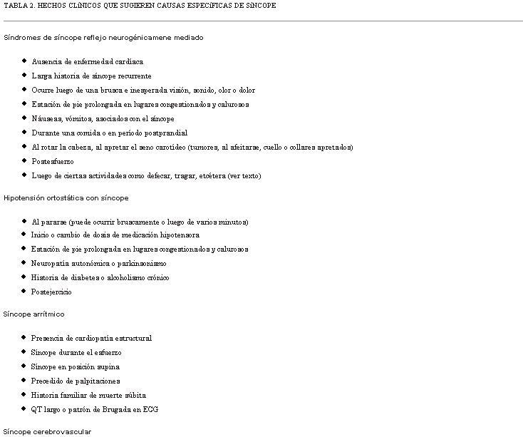
</b> </font></p>            ]]></body>
<body><![CDATA[<p align="left"><font color="#1f1a17" face="Verdana" size="2"> INITIAL EVALUATION OF THE PATIENT WITH SUSPECTED SYNCOPE </font></p>            <p align="left"><font color="#1f1a17" face="Verdana" size="2"> The evaluation of patients with suspected syncope begins with a careful  history taking (<a href="/img/revistas/ruc/v26n1/1a07t02.JPG">Table 2</a>). However, obtaining a reliable history may be  a problem. Elderly patients and those with cognitive impairment may not  be able to remember all events, while other patients may not volunteer  a complete history due to risk of losing their job, or driving privileges.  Consequently, witnesses to symptomatic events should be included in the  history-taking process, whenever possible. </font></p>            <p align="left"><font color="#1f1a17" face="Verdana" size="2"> In brief, it is essential that as many symptom events be assessed in detail.  The preceding circumstances, premonitory symptoms, and subsequent outcome  should be documented for as many episodes as possible. If a pattern emerges,  the diagnosis may become evident without the need for further testing.  Similarly, careful note should be made of patient&rsquo;s co-morbidities (for  example diabetic neuropathy, autonomic dysfunction). A pre-prepared patient  questionnaire may prove helpful to save time and still acquire the needed  details. </font></p>            <p align="left"><font color="#1f1a17" face="Verdana" size="2"> Based on the initial evaluation, it should be possible to classify patients  into one of three categories: Certain diagnosis, Suspected diagnosis or  Unknown diagnosis. Thereafter, the diagnostic flow can reasonably follow  the strategy depicted in <a href="#Figura4">Figure 4</a> which has been modified from that devised  by the European Society of Cardiology Syncope Guidelines Task Force </font><font color="#1f1a17" face="Century Schoolbook" size="2"> <font color="#1f1a17" face="Verdana"><sup>(<a href="#2">2</a>)</sup></font><font color="#1f1a17" face="Verdana" size="2">.  The goal is to choose subsequent diagnostic tests in a careful manner to  maximize cost-effectiveness and minimize the number of studies.</font></font><font color="#1f1a17" face="Verdana" size="2"> </font></p>            <p align="left"><font face="Verdana" size="2"><a name="Figura4"></a><img style="width: 566px; height: 394px;" alt="" src="/img/revistas/ruc/v26n1/1a08f04.JPG">    <br>        </font>        </p>            <p><font face="Verdana" size="2">Figure 4. Diagnostic flow of Syncope (adapted from reference 2)</font></p>            <p></p>            <p align="left"><font color="#1f1a17" face="Verdana" size="2"> SHOULD THE DIAGNOSIS BE PURSUED IN HOSPITAL OR IN THE OUTPATIENT SETTING? </font></p>            <p align="left"><font color="#1f1a17" face="Verdana" size="2"> In general, the driving force determining whether the patient with presumed  syncope should be hospitalized for diagnostic evaluation and if necessary  for treatment initiation, is most often concern regarding the individual&rsquo;s  immediate mortality risk. Secondary issues include potential for physical  injury (e.g., falls risk) and to a lesser extent whether certain treatments  inherently require hospital monitoring for safe initiation. Thus, for example,  patients with syncope accompanying complete heart block, ventricular tachycardia,  acute aortic dissection, or pulmonary embolism, should be admitted to the  hospital and preferably to an ECG monitored unit. On the other hand, most  vasovagal fainters can be sent home after careful discussion of the nature  of the problem and simple preventative maneuvers (e.g., hydration, avoidance  of hot crowded environments, etc). Clinic follow-up suffices in most of  these cases. </font></p>         <multicol gutter="18" cols="2"></multicol>     ]]></body>
<body><![CDATA[<p align="left"><font color="#1f1a17" face="Verdana" size="2"> For syncope patients in whom the etiology remains unknown after the initial  emergency department or ambulatory clinic evaluation, the need for hospitalization  is less well defined and consequently so-called &ldquo;risk stratification&rdquo; methods  have been advocated </font><font color="#1f1a17" face="Century Schoolbook" size="2"> <font color="#1f1a17" face="Verdana"><sup>(<a href="#38">38</a>-<a href="#45">45</a>)</sup></font><font color="#1f1a17" face="Verdana" size="2"><a name=".38"></a><a name=".39"></a><a name=".40"></a><a name=".41"></a><a name=".42"></a><a name=".43"></a><a name=".44"></a><a name=".45"></a>. In essence, the goal is to ascertain the relative  risk for adverse outcome using patients&rsquo; clinical features and presenting  characteristics. Based on this assessment one attempts to determine if  hospitalization is prudent.</font></font><font color="#1f1a17" face="Verdana" size="2"> </font></p>            <p align="left"><font color="#1f1a17" face="Verdana" size="2"> The risk stratification methods differ in various published studies. Nonetheless,  the Syncope Evaluation in the Emergency Department Study (SEEDS), The Osservatorio  Epidemiologico sula Sincope nel Lazio (OESIL) study, The San Francisco  Syncope Rule (SFSR) study, and the European Society of Cardiology and American  College of Emergency Physicians (ACEP) guidelines </font><font color="#1f1a17" face="Century Schoolbook" size="2"> <font color="#1f1a17" face="Verdana"><sup>(<a href="#40">40</a>-<a href="#45">45</a>)</sup></font><font color="#1f1a17" face="Verdana" size="2"> each uses clinical  data that is readily accessible to the ED physician or general practitioner.  These data include symptoms, signs, basic laboratory results and clinical  experience (&lsquo;judgment&rsquo;).</font></font><font color="#1f1a17" face="Verdana" size="2"> </font></p>            <p align="left"><font color="#1f1a17" face="Verdana" size="2"> At present, it is not possible to determine whether one or other risk stratification  scheme is superior to the others. Head-to-head testing is needed. In this  regard, the Risk Stratification Of Syncope in the Emergency Department  (ROSE) study compared the performance of OESIL score, and the SFSR recommendations  with emergency department guidelines of a single center in United Kingdom  (Royal Infirmary of Edinburgh) </font><font color="#1f1a17" face="Century Schoolbook" size="2"> <font color="#1f1a17" face="Verdana"><sup>(<a href="#44">44</a>)</sup></font><font color="#1f1a17" face="Verdana" size="2">. The latter institution used a guideline  based on ESC, American College of Physicians and ACEP guidelines. The goal  was to determine which of these Risk Stratification tools best predicted  short-term (1 week and 1 month) and medium-term (3 months) serious outcomes  for patients presenting with syncope. In this regard, each of the scores  was able to identify an increased probability of medium-term serious outcome  in patients with syncope. The SFSR showed good sensitivity at the expense  of an increased frequency of admission to the hospital. On the other hand,  the Royal Infirmary&rsquo;s center&rsquo;s own guideline, and the OESIL score, was  not sufficiently sensitive to be able to reduce admissions without missing  patients at risk of serious outcome.</font></font><font color="#1f1a17" face="Verdana" size="2"> </font></p>             <p>  </p>        <multicol gutter="18" cols="2"></multicol>     <p align="left"><font color="#1f1a17" face="Verdana" size="2"> ROLE OF SYNCOPE MANAGEMENT UNITS (SMUS) </font></p>            <p align="left"><font color="#1f1a17" face="Verdana" size="2"> An as yet incompletely answered question is whether &ldquo;SMUs&rdquo; can help solve  the problem of too many low- and intermediate-risk syncope patients being  admitted to hospital where they often are submitted to unneeded expensive  diagnostic tests as pointed out in the EGSYS (The Evaluation of Guidelines  in Syncope Study) reports </font><font color="#1f1a17" face="Century Schoolbook" size="2"> <font color="#1f1a17" face="Verdana"><sup>(69)</sup></font><font color="#1f1a17" face="Verdana" size="2">. In this regard, 2 recent prospective observational  studies demonstrated improved syncope management in the hospital by using  guideline-based decision making software and a team of specially-trained  personnel in a SMU. In the SEEDS study (<a href="#46">46</a>)<a name=".46"></a>, 103 patients were randomized  to &lsquo;standard care&rsquo; or SMU after initial assessment. The study found that  a presumptive diagnosis of the cause of syncope was significantly increased  from 10% in the &lsquo;standard care&rsquo; patients to 67% among those who underwent  SMU evaluation; hospital admission was reduced from 98% among the &lsquo;standard  care&rsquo; patients to 43% among the SMU patients. Similarly, the total length  of patient-hospital days was reduced by &gt;50% for patients in the SMU group.</font></font><font color="#1f1a17" face="Verdana" size="2"> </font></p>            <p align="left"><font color="#1f1a17" face="Verdana" size="2"> The potential for the ESC Guidelines to facilitate management of syncope  patients referred to ED&rsquo;s of 11 Italian general hospitals was investigated  in EGSYS-2 </font><font color="#1f1a17" face="Century Schoolbook" size="2"> <font color="#1f1a17" face="Verdana"><sup>(<a href="#46">46</a>)</sup></font><font color="#1f1a17" face="Verdana" size="2">, and facilitated by use of purpose-designed software in  addition to personnel training at test sites. A definite diagnosis was  established in 98% of cases, with the vast majority being either neurally-mediated  reflex or orthostatic faints. The initial evaluation (history, physical  examination, and electrocardiogram) established a diagnosis in 50% of cases.  The investigators further compared the outcomes of 745 patients managed  with this &ldquo;standardized care&rdquo; system to 929 patients managed with usual  care. In the group designated to &ldquo;standardized-care&rdquo;, hospitalizations  were fewer, in-hospital stay was shorter, fewer tests were performed per  patient, and cost per patient and mean cost per diagnosis were lower.</font></font><font color="#1f1a17" face="Verdana" size="2"> </font></p>       <font face="Verdana" size="2">           <br>        </font>            <p align="left"><font color="#1f1a17" face="Verdana" size="2"> <b>TREATMENT</b> </font></p>            <p align="left"><font color="#1f1a17" face="Verdana" size="2"> Treatment of the syncope patient may be divided into 2 parts. The first  is management of an acute syncopal event. Although physicians are only  infrequently involved in this aspect of care, treatment of the acute episode  requires protection of the patient from injury, assuring that the victim  is placed safely in a gravitationally neutral position, and documenting  adequacy of respiration and circulation. Thereafter, recovery is spontaneous.  The second part is prevention of syncope recurrences. </font></p>             ]]></body>
<body><![CDATA[<p>  </p>        <multicol gutter="18" cols="2"></multicol>     <p align="left"><font color="#1f1a17" face="Verdana" size="2"> NEURALLY-MEDIATED REFLEX FAINTS </font></p>            <p align="left"><font color="#1f1a17" face="Verdana" size="2"> In most cases, vasovagal syncope or situational faints are solitary or  at most very infrequent events. Therefore, most patients need little more  than reassurance and education about the nature of this condition and the  types of circumstances that might increase their risk of recurrence (e.g.,  prolonged standing, warm environments, etc.). However, in certain individuals,  the faints are more frequent for at least a period of time during their  lives. In these cases, should also be taught to be alert to warning symptoms  such as feeling of being hot or cold, sweaty, clammy, short of breath,  or nauseated. The goal is to educate susceptible individuals to recognize  impending events and take action in order to prevent the episodes (e.g.,  sitting or lying down with feet elevated). Similarly, if a patient knows  that sight of blood could bring about a vasovagal faint, he/she may either  avoid the situation or possibly be preconditioned to perform maneuvers  to prevent the faint (e.g., aggressive hydration, arm-tensing, leg crossing). </font></p>            <p align="left"><font color="#1f1a17" face="Verdana" size="2"> With regard to techniques for acute intervention to abort an imminent vasovagal  or situational faints, patients who have warning symptoms should be educated  about specific physical counter-maneuvers (PCM); thus, squatting, arm-tensing,  leg-crossing, and leg-crossing with lower body muscle tensing have proved  useful for averting an abrupt vasovagal reaction </font><font color="#1f1a17" face="Century Schoolbook" size="2"> <font color="#1f1a17" face="Verdana"><sup>(<a href="#47">47</a>-<a href="#50">50</a>)</sup></font><font color="#1f1a17" face="Verdana" size="2">.<a name=".47"></a><a name=".48"></a><a name=".49"></a><a name=".50"></a> In the recent  Physical Counterpressure Manoeuvres Trial (PC-Trial), van Dijk et al. demonstrated  that physical counterpressure maneuvers (PCM) can reduce the total burden  and recurrence rate of syncopal events </font> <font color="#1f1a17" face="Verdana"><sup>(<a href="#50">50</a>)</sup></font><font color="#1f1a17" face="Verdana" size="2">. Consequently, PCM education  should be part of the treatment strategy, especially in patients with warning  symptoms prior to their faints.</font></font><font color="#1f1a17" face="Verdana" size="2"> </font></p>            <p align="left"><font color="#1f1a17" face="Verdana" size="2"> Longer-term prevention of vasovagal syncope recurrences focuses on use  of fluids rich in electrolytes and increased dietary salts. Examples would  include &lsquo;sport drinks&rsquo; although those with lowest carbohydrate content  are preferred. Highly motivated patients could also be provided with instructions  regarding upright standing training (so-called tilt-training). &lsquo;Tilt training&rsquo;  involves progressively lengthening periods of enforced upright posture,  the goal of which is to improve tolerance to upright posture </font><font color="#1f1a17" face="Century Schoolbook" size="2"> <font color="#1f1a17" face="Verdana"><sup>(<a href="#51">51</a>)</sup></font><font color="#1f1a17" face="Verdana" size="2"><a name=".51"></a>. In addition,  a number of pharmacologic approaches have been proposed for vasovagal syncope.  However, most of these have not been studied in randomized, controlled  trials. Examples of such medications include fluorocortisone, beta-adrenergic  blocking drugs, and vasoconstrictor agents like midodrine and serotonin  re-uptake inhibitors. Although these may work well in individual patients,  only midodrine has shown effectiveness in controlled studies </font> <font color="#1f1a17" face="Verdana"><sup>(<a href="#52">52</a>-<a href="#54">54</a>)</sup></font><font color="#1f1a17" face="Verdana" size="2"><a name=".52"></a><a name=".53"></a><a name=".54"></a>.</font></font><font color="#1f1a17" face="Verdana" size="2"> </font></p>            <p align="left"><font color="#1f1a17" face="Verdana" size="2"> Cardiac pacing has received considerable study as a potential treatment  option in patients with very frequent vasovagal faints. However, despite  early favorable reports, pacing is not considered very effective for preventing  syncope in most patients. On the other hand, the ISSUE-2 trial indicated  that if marked bradycardia during spontaneous syncope has been documented  by ILR recording, then pacing may be warranted in recurrent fainters </font><font color="#1f1a17" face="Century Schoolbook" size="2"> <font color="#1f1a17" face="Verdana"><sup>(<a href="#30">30</a>)</sup></font><font color="#1f1a17" face="Verdana" size="2">.  ISSUE-2 was an observational study. ISSUE-3, which just concluded enrollment  in November 2010, is reassessing this issue in a randomized controlled  study.</font></font><font color="#1f1a17" face="Verdana" size="2"> </font></p>            <p align="left"><font color="#1f1a17" face="Verdana" size="2"> Carotid sinus syndrome (CSS) is a special form of neurally-mediated reflex  syncope that tends to occur in older individuals. Falls and injury is a  real concern in these patients if untreated </font><font color="#1f1a17" face="Century Schoolbook" size="2"> <font color="#1f1a17" face="Verdana"><sup>(<a href="#23">23</a>,<a href="#55">55</a>)</sup></font><font color="#1f1a17" face="Verdana" size="2"><a name=".55"></a>. In CSS, despite the  presence of both cardioinhibitory and vasodepressor features, pacing appears  to be highly effective for preventing recurrences (<a href="#figura_5">Figure 5</a>).</font></font><font color="#1f1a17" face="Verdana" size="2"> </font></p>            <p align="left"><font face="Verdana" size="2"><a name="figura_5"></a><img style="width: 566px; height: 316px;" alt="" src="/img/revistas/ruc/v26n1/1a08f05.JPG">    <br>        </font>        </p>            <p><font face="Verdana" size="2">Figure 5. Recording of ECG leads !, II, aVL, and V1, and arterial pressure during carotid sinus massage (CSM) in an older male with syncope and suspected carotid sinus syndrome. Note that despite recovery of the heart rate after the initial asystolic period, the blood pressure remains well below baseline value. This observation (i.e., the persistent hypotension) is supportive of a &lsquo;vasodepressor&rsquo; component to the CSM response in addition to the induced bradycardia (also known as the cardioinhibitory aspect of the CSM response).</font></p>            ]]></body>
<body><![CDATA[<p></p>            <p align="left"><font color="#1f1a17" face="Verdana" size="2"> ORTHOSTATIC SYNCOPE </font></p>            <p align="left"><font color="#1f1a17" face="Verdana" size="2"> Treatment of orthostatic syncope parallels in many respects the strategy  discussed earlier for vasovagal and situational faints. The principal differences  are: 1) the duration of treatment is likely to be longer, 2) affected individuals  are typically older and more frail making physical maneuvers more difficult  to employ, and 3) patients are more prone to supine hypertension thereby  complicating the overall treatment strategy. </font></p>            <p align="left"><font color="#1f1a17" face="Verdana" size="2"> In many cases, syncope associated with postural change may be caused by  low circulating plasma volume, or inadequate vascular constriction upon  moving to the upright posture (often drug-induced), or both. Consequently,  one of the basic tenets of treatment is expanding central circulating volume.  Additionally, affected individuals should be instructed to try and avoid  certain predisposing conditions (e.g., prolonged exposure to hot environments)  or medications that decrease volume status (e.g., diuretics) or that impair  vasoconstriction (e.g., vasodilators, beta-adrenergic blockers). In some  severe cases, use of vasoconstrictors (like midodrine) or volume expanders  (e.g., fludrocortisone) may be necessary to maintain adequate cerebral  perfusion. Finally, patients who exhibit poor autonomic function may benefit  from tilt-training (discussed earlier), counter-pressure clothing such  as fitted stockings and abdominal compression devices. In patients with  severe pure autonomic failure, bolus water intake, especially before arising  from bed in the morning, may result in a substantial and sustained increase  in blood pressure </font><font color="#1f1a17" face="Century Schoolbook" size="2"> <font color="#1f1a17" face="Verdana"><sup>(<a href="#56">56</a>)</sup></font><font color="#1f1a17" face="Verdana" size="2"><a name=".56"></a>.</font></font><font color="#1f1a17" face="Verdana" size="2"> </font></p>         <multicol gutter="18" cols="2"></multicol>     <p align="left"><font color="#1f1a17" face="Verdana" size="2"> PRIMARY CARDIAC ARRHYTHMIAS </font></p>            <p align="left"><font color="#1f1a17" face="Verdana" size="2"> The treatment of cardiac arrhythmias causing syncope is determined by the  specific arrhythmia that is deemed to be at fault. Given the numerous possibilities  and the many factors that go into selecting appropriate therapies for these  arrhythmias, only a very brief overview is provided here. </font></p>            <p align="left"><font color="#1f1a17" face="Verdana" size="2"> As noted earlier, sinus node disease may cause syncope either due to bradycardia  or tachycardia mechanisms as discussed earlier. When a temporal correlation  between syncope and bradycardia has been established, pacemaker implantation  is the treatment of choice. Dual-chamber or atrial pacing is preferred.  In the case of a tachyarrhythmia-induced syncope, both antiarrhytmic drugs  and ablation are reasonable treatment considerations. The final choice  depends on specific clinical circumstances and patient preferences. </font></p>            <p align="left"><font color="#1f1a17" face="Verdana" size="2"> Treatment of AV conduction system disease in syncope patients does not  differ measurably from their treatment in other patients. However, once  again it is crucial to obtain concordance between syncopal episodes and  bradycardia. If such a correlation is found and the cause of AV block is  irreversible, pacemaker implantation is a class I indication. </font></p>            <p align="left"><font color="#1f1a17" face="Verdana" size="2"> In general, first degree AV block and Type I second degree AV block (Wenkebach  type) with a narrow QRS do not cause syncope and are not indications for  a pacemaker. However, when second degree Type I block occurs in the setting  of evident infra-nodal conduction system disease (e.g., wide QRS), or in  older individuals (&gt;70 years of age) pacemaker therapy is indicated. Similarly  patients who have Mobitz type II AV block carry a high risk for intermittent  high grade AV block causing syncope, and are candidates for pacing therapy.  On the other hand, these same patients often exhibit substantial structural  heart disease and therefore, and are also prone to ventricular tachyarrhythmias.  Management in such cases may be better accomplished by ICD therapy. </font></p>            <p align="left"><font color="#1f1a17" face="Verdana" size="2"> When reentrant paroxysmal supraventricular tachyarrhythmias are deemed  to be the cause of syncope transcatheter ablation is the treatment of choice  in most cases. Drug therapy remains an option, however, given the high  success rate with ablation and its ready availability, the ablation track  seems more desirable. Similarly, most atrial tachyarrhythmias are now subject  to cure by transcatheter ablation technique. This certainly is true for  atrial flutter, most ectopic atrial tachyacrdias and increasingly for paroxysmal  atrial fibrillation. However, drug therapy remains the most commonly used  approach by most physicians. In this regard, drugs that reduce heart rate  in tachycardia may be sufficient to prevent syncope. The most common of  these agents are: beta-adrenergic blockers, and calcium channel blockers.  If the desire is also to try and suppress tachycardia recurrences, a membrane  active antiarrhythmic drug is also needed. Finally, in the case of refractory  symptomatic atrial tachyarrhythmias (particularly atrial fibrillation)  with rapid ventricular rates, treatment by His Bundle ablation and placement  of a permanent pacemaker has proved to be safe and highly effective </font><font color="#1f1a17" face="Century Schoolbook" size="2"> <font color="#1f1a17" face="Verdana"><sup>(<a href="#57">57</a>)</sup></font><font color="#1f1a17" face="Verdana" size="2">.<a name=".57"></a></font></font><font color="#1f1a17" face="Verdana" size="2"> </font></p>            ]]></body>
<body><![CDATA[<p align="left"><font color="#1f1a17" face="Verdana" size="2"> The treatment of ventricular tachyarrhythmias in syncope patients depnds  on the clinical circumstance. Patients with ischemic heart disease or dilated  cardiomyopathies and severely diminished left ventricular function (i.e.,  LV ejection fractions &lt;35%) have a high mortality rate and benefit from  ICD therapy. As alluded to earlier, whether syncope will be prevented by  ICD treatment is less certain since the devices must take time before intervening  with pacing or shock treatment </font><font color="#1f1a17" face="Century Schoolbook" size="2"> <font color="#1f1a17" face="Verdana"><sup>(<a href="#58">58</a>)</sup></font><font color="#1f1a17" face="Verdana" size="2">. Consequently, in the syncope patient  concomitant antiarrhythmic drug therapy and/or ablation may be needed.</font></font><font color="#1f1a17" face="Verdana" size="2"> </font></p>         <multicol gutter="18" cols="2"></multicol>     <p align="left"><font color="#1f1a17" face="Verdana" size="2"> Ventricular tachyarrhythmias arising from the Right Ventricular Outflow  Tract (RVOT) or Left Ventricular Outflow Tract (LVOT) or the intraventricular  fascicular system or bundle-branch system (i.e., bundle-branch reentry)  are infrequent causes of syncope. However, when they are responsible for  symptoms, transcatheter ablation is probably the treatment of choice. In  arrhythmogenic right ventricular dysplasia (ARVD), treatment strategies  are controversial. Medical treatment has not been found to be very effective.  Furthermore, since there are potentially many regions of the heart that  are affected, long-term efficacy of transcatheter ablation is limited.  Therefore, syncope patients with ARVD, in whom ventricular tachyarrhythmias  have been documented, may be best served by placement of an ICD. Once again,  however, syncope may not necessarily be completely averted and concomitant  medications may be needed. </font></p>            <p align="left"><font color="#1f1a17" face="Verdana" size="2"> Long QT syndrome, Brugada syndrome, and other &lsquo;channelopathies&rsquo; are increasingly  recognized as causes of syncope due to polymorphous VT (so-called &lsquo;torsades  de pointes&rsquo;) (<a href="#Figura4">Figure 4</a>). If there is no evidence for a reversible problem,  the use of ICD therapy may be essential to prevent further syncope and  also reduce sudden death risk. </font></p>            <p align="left"><font color="#1f1a17" face="Verdana" size="2"> STRUCTURAL CARDIOVASCULAR OR CARDIOPULMONARY DISEASE </font></p>            <p align="left"><font color="#1f1a17" face="Verdana" size="2"> In these conditions, syncope may be only one subset of symptoms being experienced  by the patient. Correction of the structural lesion or its consequence  is usually the treatment of choice. As an example, patients with valvular  aortic stenosis, pericardial disease, atrial myxoma and congenital cardiac  anomaly benefit from a direct corrective approach. In other cases, such  as primary pulmonary hypertension or restrictive cardiomyopathy, the structural  lesions are not correctable. In the case of HOCM, modification of the outflow  gradients surgically may be accompanied by substantial risk and morbidity,  and medical therapy is not very effective. If it is clear that syncope  is due to the obstruction (as opposed to arrhythmias discussed earlier)  cardiac pacing to diminish dynamic outflow gradients and transcatheter  alcohol septal ablation may prove helpful. </font></p>            <p align="left"><font color="#1f1a17" face="Verdana" size="2"> CEREBROVASCULAR </font></p>            <p align="left"><font color="#1f1a17" face="Verdana" size="2"> As noted previously, cerebrovascular disease is rarely the cause of syncope.  Migraines may be the most important consideration. The basis for the faints  in migraineurs is unclear, but there is an established relationship between  migraines and autonomic dysfunction </font><font color="#1f1a17" face="Century Schoolbook" size="2"> <font color="#1f1a17" face="Verdana"><sup>(<a href="#59">59</a>)</sup></font><font color="#1f1a17" face="Verdana" size="2">. In any case, the medical management  includes use of </font><font color="#1f1a17" face="Verdana">&acirc;</font><font color="#1f1a17" face="Verdana" size="2">-blockers and cranial/basilar artery vasoconstrictors  such as sumatriptan </font><font color="#1f1a17" face="Verdana"><sup>(<a href="#60">60</a>)</sup></font><font color="#1f1a17" face="Verdana" size="2"><a name=".60"></a>.</font></font><font color="#1f1a17" face="Verdana" size="2"> </font></p>            <p align="left"><font color="#1f1a17" face="Verdana" size="2"> Strokes and carotid system transient ischemic attacks (TIA) are almost  never the cause of syncope. On the other hand, vertebrobasilar TIAs are  a possible but extremely rare cause of faints. Treatment includes anti-platelet  agents or anti-coagulation. When diagnoses in this category are suspected,  neurological or neurosurgical consultation should be sought. </font></p>            <p align="left"><font color="#1f1a17" face="Verdana" size="2"> Subclavian steal syndrome is another, but also very rare cause of syncope  in this category. Its treatment requires intervention, either surgically  or by catheter based angioplasty </font><font color="#1f1a17" face="Century Schoolbook" size="2"> <font color="#1f1a17" face="Verdana"><sup>(<a href="#61">61</a>)</sup></font><font color="#1f1a17" face="Verdana" size="2">.<a name=".61"></a></font></font><font color="#1f1a17" face="Verdana" size="2"> </font></p>            <p align="left"><font color="#1f1a17" face="Verdana" size="2"> SYNCOPE MIMICS </font></p>            ]]></body>
<body><![CDATA[<p align="left"><font color="#1f1a17" face="Verdana" size="2"> The &lsquo;syncope mimics&rsquo; include serious medical conditions like epilepsy,  diabetic coma, intoxication, severe hypoxia or hypercapnia. While these  conditions require urgent medical attention, they are not the cause of  true syncope (see earlier discussion). Among the conditions that are defined  as syncope mimics, psychogenic pseudo-syncope often accompanied by anxiety  attacks and hyperventilation is among the most common, and is very difficult  to treat. In these patients it is very easy for health care providers to  overlook serious underlying health issues. Only when one is convinced that  there is no clinically significant cardiac or pulmonary problem should  subsequent care be directed toward psychiatric consultation, and biofeedback. </font></p>       <font face="Verdana" size="2">           <br>        </font>            <p align="left"><font color="#1f1a17" face="Verdana" size="2"> <b>CONCLUSIONS</b> </font></p>            <p align="left"><font color="#1f1a17" face="Verdana" size="2"> Syncope is a form of transient loss of consciousness (TLOC) that is self-limited  and reversible due to transient spontaneously reversible cerebral hypoperfusion.  Delineating the underlying causes and the risk of adverse outcome may be  challenging. However, careful assessment is important as syncope tends  to recur; physical injury resulting from falls or accidents, diminished  quality-of-life, and possible restriction from employment or avocation  are real concerns. Determining that certain individuals are at &lsquo;low mortality  risk&rsquo; is insufficient. The goal in every case should be to determine the  cause of syncope with sufficient confidence to provide patients and family  members with a reliable assessment of prognosis, recurrence risk, and treatment  options. </font></p>       <font face="Verdana" size="2">           <br>         </font>         <multicol gutter="18" cols="2"></multicol>     <p align="left"><font color="#1f1a17" face="Verdana" size="2"> <b>REFERENCES</b> </font></p>            <p align="left"> <font color="#000000" face="Verdana" size="2"><a name="1"></a><a href="#.1">1</a>.&nbsp;&nbsp;&nbsp;&nbsp;<b>Moya A, Sutton R, Ammirati F, Blanc JJ, Brignole M, Dahm JB, et al.</b> Guidelines  on management (diagnosis and treatment) of syncope &ndash; 2009. The Task Force  for the Diagnosis and Management of Syncope of the European Society of  Cardiology (ESC). Guidelines for the diagnosis and management of syncope  (version 2009). European Heart J 2009; 30: 2631-71.</font><font color="#1f1a17" face="Verdana" size="2"> </font></p>            <!-- ref --><p align="left"> <font color="#000000" face="Verdana" size="2"><a name="2"></a><a href="#.2">2</a>.&nbsp;&nbsp;&nbsp;&nbsp;<b>Brignole M, Alboni P, Benditt D, Bergfeldt L, Blanc JJ, Bloch Thomsen  PE, et al.</b> Guidelines on management (diagnosis and treatment) of syncope,  update 2004. Europace 2004; 6: 467-537.    </font><font color="#1f1a17" face="Verdana" size="2"> </font></p>            <!-- ref --><p align="left"> <font color="#000000" face="Verdana" size="2"><a name="3"></a><a href="#.3">3</a>.&nbsp;&nbsp;&nbsp;&nbsp;<b>Wieling W, Thijs RD, van Dijk N, Wilde AA, Benditt DG. van Dijk JG</b>. Symptoms  and signs of syncope: a review of the link between physiology and clinical  clues. Brain 2009; 132: 2630-42.    </font><font color="#1f1a17" face="Verdana" size="2"> </font></p>            <!-- ref --><p align="left"> <font color="#000000" face="Verdana" size="2"><a name="4"></a><a href="#.4">4</a>.&nbsp;&nbsp;&nbsp;&nbsp;<b>Benditt DG, Nguyen JT.</b> Syncope: Therapeutic approaches. J Am Coll Cardiol  2009; 53(19): 1741-51.    </font><font color="#1f1a17" face="Verdana" size="2"> </font></p>            <!-- ref --><p align="left"> <font color="#000000" face="Verdana" size="2"><a name="5"></a><a href="#.5">5</a>.&nbsp;&nbsp;&nbsp;&nbsp;<b>van Dijk JG, Thijs RD, Benditt DG,Wieling W.</b> A guide to disorders causing  transient loss of consciousness: focus on syncope. Nature Rev Medicine  2009; 5: 438-48.    </font><font color="#1f1a17" face="Verdana" size="2"> </font></p>            <!-- ref --><p align="left"> <font color="#000000" face="Verdana" size="2"><a name="6"></a><a href="#.6">6</a>.&nbsp;&nbsp;&nbsp;&nbsp;<b>Alboni P, Brignole M, Menozzi C, Raviele A, Del Rosso A, Dinelli M, et  al. </b>Diagnostic value of history in patients with syncope with or without  heart disease. J Am Coll Cardiol 2001; 37: 1921-8.    </font><font color="#1f1a17" face="Verdana" size="2"> </font></p>            <!-- ref --><p align="left"> <font color="#000000" face="Verdana" size="2"><a name="7"></a><a href="#.7">7</a>.&nbsp;&nbsp;&nbsp;&nbsp;<b>Colman N, Nahm K, van Dijk JG, Reitsma JB, Wieling W, Kaufmann H.</b> Diagnostic  value of history taking in reflex syncope. Clin Auton Res 2004; 14 Suppl  1: 37-44.    </font><font color="#1f1a17" face="Verdana" size="2"> </font></p>            <p align="left"> <font color="#000000" face="Verdana" size="2"><a name="8"></a><a href="#.8">8</a>.&nbsp;&nbsp;&nbsp;&nbsp;<b>Posner JB, Plum F, R2 Library.</b> Plum and posner&rsquo;s diagnosis of stupor  and coma. Oxford: Oxford University Press; 2007: 71.</font><font color="#1f1a17" face="Verdana" size="2"> </font></p>            ]]></body>
<body><![CDATA[<!-- ref --><p align="left"> <font color="#000000" face="Verdana" size="2"><a name="9"></a><a href="#.9">9</a>.&nbsp;&nbsp;&nbsp;&nbsp;<b>Savage DD, Corwin L, McGee DL, Kannel WB, Wolf PA. </b>Epidemiologic features  of isolated syncope: The Framingham study. Stroke 1985; 16: 626-9.    </font><font color="#1f1a17" face="Verdana" size="2"> </font></p>            <!-- ref --><p align="left"> <font color="#000000" face="Verdana" size="2"><a name="10"></a><a href="#.10">10</a>.&nbsp;&nbsp;&nbsp;&nbsp;<b>Soteriades ES, Evans JC, Larson MG, Chen MH, Chen L, Benjamin EJ, et  al.</b> Incidence and prognosis of syncope. N Engl J Med 2002; 347: 878-85.    </font><font color="#1f1a17" face="Verdana" size="2"> </font></p>            <!-- ref --><p align="left"> <font color="#000000" face="Verdana" size="2"><a name="11"></a><a href="#.11">11</a>.&nbsp;&nbsp;&nbsp;&nbsp;<b>Chen LY, Shen WK, Mahoney DW, Jacobsen SJ, Rodeheffer RJ.</b> Prevalence  of syncope in a population aged more than 45 years. Am J Med 2006; 119:  1088.e1-1088.e7.    </font><font color="#1f1a17" face="Verdana" size="2"> </font></p>            <!-- ref --><p align="left"> <font color="#000000" face="Verdana" size="2"><a name="12"></a><a href="#.12">12</a>.&nbsp;&nbsp;&nbsp;&nbsp;<b>Ganzeboom KS, Colman N, Reitsma JB, Shen WK, Wieling W.</b> Prevalence and  triggers of syncope in medical students. Am J Cardiol 2003; 91: 1006-8,  A8.    </font><font color="#1f1a17" face="Verdana" size="2"> </font></p>            <!-- ref --><p align="left"> <font color="#000000" face="Verdana" size="2"><a name="13"></a><a href="#.13">13</a>.&nbsp;&nbsp;&nbsp;&nbsp;<b>Serletis A, Rose S, Sheldon AG, Sheldon RS. </b>Vasovagal syncope in medical  students and their first-degree relatives. Eur Heart J 2006; 27: 1965-70.    </font><font color="#1f1a17" face="Verdana" size="2"> </font></p>            ]]></body>
<body><![CDATA[<!-- ref --><p align="left"> <font color="#000000" face="Verdana" size="2"><a name="14"></a><a href="#.14">14</a>.&nbsp;&nbsp;&nbsp;&nbsp;<b>Sun BC, Emond JA, Camargo CA Jr.</b> Characteristics and admission patterns  of patients presenting with syncope to US emergency departments, 1992-2000.  Acad Emerg Med 2004; 11: 1029-34.    </font><font color="#1f1a17" face="Verdana" size="2"> </font></p>            <!-- ref --><p align="left"> <font color="#000000" face="Verdana" size="2"><a name="15"></a><a href="#.15">15</a>.&nbsp;&nbsp;&nbsp;&nbsp;<b>Sun BC, Emond J, Comargo C Jr.</b> Direct medical costs of syncope=related  hospitalizations in the United States. Am J Cardiol 2005; 95: 668-71.    </font><font color="#1f1a17" face="Verdana" size="2"> </font></p>            <p align="left"> <font color="#000000" face="Verdana" size="2"><a name="16"></a><a href="#.16">16</a>.&nbsp;&nbsp;&nbsp;&nbsp;<b>Thijs RD, Benditt DG, Mathias CJ, Schondorf R, Sutton R, Wieling W,  et al.</b> Unconscious confusion&mdash;a literature search for definitions of syncope  and related disorders. Clin Auton Res 2005; 15: 35-9.</font><font color="#1f1a17" face="Verdana" size="2"> </font></p>            <!-- ref --><p align="left"> <font color="#000000" face="Verdana" size="2"><a name="17"></a><a href="#.17">17</a>.&nbsp;&nbsp;&nbsp;&nbsp;<b>van Lieshout JJ, Wieling W, Karemaker JM, Eckberg DL. </b>The vasovagal  response. Clin Sci (Lond) 1991; 81: 575-86.    </font><font color="#1f1a17" face="Verdana" size="2"> </font></p>            <!-- ref --><p align="left"> <font color="#000000" face="Verdana" size="2"><a name="18"></a><a href="#.18">18</a>.&nbsp;&nbsp;&nbsp;&nbsp;<b>Wieling W.</b> Maintaining blood pressure whilst upright: Physiology and  potential for disturbances to cause syncope. In: Benditt DG, European Society  of Cardiology, eds. The Evaluation and Treatment of Syncope : A Handbook  for Clinical Practice. 2nd ed. Malden, Mass.: Blackwell Pub.; 2006:301.    </font><font color="#1f1a17" face="Verdana" size="2"> </font></p>            <!-- ref --><p align="left"> <font color="#000000" face="Verdana" size="2"><a name="19"></a><a href="#.19">19</a>.&nbsp;&nbsp;&nbsp;&nbsp;<b>Benditt DG, Ferguson DW, Grubb BP, Kapoor WN, Kugler J, Lerman BB, et  al.</b> Tilt table testing for assessing syncope. American College of Cardiology.  J Am Coll Cardiol 1996; 28: 263-75.    </font><font color="#1f1a17" face="Verdana" size="2"> </font></p>            <!-- ref --><p align="left"> <font color="#000000" face="Verdana" size="2"><a name="20"></a><a href="#.20">20.</a>&nbsp;&nbsp;&nbsp;&nbsp;<b>Kenny RA, Ingram A, Bayliss J, Sutton R.</b> Head-up tilt: A useful test  for investigating unexplained syncope. Lancet 1986; 1: 1352-5.    </font><font color="#1f1a17" face="Verdana" size="2"> </font></p>            <!-- ref --><p align="left"> <font color="#000000" face="Verdana" size="2"><a name="21"></a><a href="#.21">21</a>.&nbsp;&nbsp;&nbsp;&nbsp;<b> Brignole M, Menozzi C, Gianfranchi L, Oddone D, Lolli G, Bertulla A.</b>  Neurally mediated syncope detected by carotid sinus massage and head-up  tilt test in sick sinus syndrome. Am J Cardiol 1991; 68: 1032-6.    </font><font color="#1f1a17" face="Verdana" size="2"> </font></p>            <!-- ref --><p align="left"> <font color="#000000" face="Verdana" size="2"><a name="22"></a><a href="#.22">22</a>.&nbsp;&nbsp;&nbsp;&nbsp;<b>Morley CA, Sutton R.</b> Carotid sinus syncope. Int J Cardiol 1984; 6: 287-93.    </font><font color="#1f1a17" face="Verdana" size="2"> </font></p>            <!-- ref --><p align="left"> <font color="#000000" face="Verdana" size="2"><a name="23"></a><a href="#.23">23</a>.&nbsp;&nbsp;&nbsp;&nbsp;<b>Parry SW, Steen N, Bexton R, Tynan M, Kenny RA. </b>Pacing in elderly recurrent  fallers with carotid sinus hypersensitivity (PERF-CSH): A randomized, double-blind,  placebo controlled cross-over trial. Heart 2009; 95(5): 405-9.    </font><font color="#1f1a17" face="Verdana" size="2"> </font></p>            <p align="left"> <font color="#000000" face="Verdana" size="2"><a name="24"></a><a href="#.24">24</a>.&nbsp;&nbsp;&nbsp;&nbsp;<b>Benditt DG, Samniah N, Pham S, Sakaguchi S, Lu F, Lurie KG, et al. </b>Effect  of cough on heart rate and blood pressure in patients with &ldquo;cough syncope&rdquo;.  Heart Rhythm 2005; 2: 807-13.</font><font color="#1f1a17" face="Verdana" size="2"> </font></p>            ]]></body>
<body><![CDATA[<!-- ref --><p align="left"> <font color="#000000" face="Verdana" size="2"><a name="25"></a><a href="#.25">25</a>.&nbsp;&nbsp;&nbsp;&nbsp;<b>Bannister R.</b> Chronic autonomic failure with postural hypotension. Lancet  1979; 2: 404-6.    </font><font color="#1f1a17" face="Verdana" size="2"> </font></p>            <!-- ref --><p align="left"> <font color="#000000" face="Verdana" size="2"><a name="26"></a><a href="#.26">26</a>.&nbsp;&nbsp;&nbsp;&nbsp;<b>Hopkins A, Neville B, Bannister R. </b>Autonomic neuropathy of acute onset.  Lancet 1974; 1: 769-71.    </font><font color="#1f1a17" face="Verdana" size="2"> </font></p>         <multicol gutter="18" cols="2"></multicol>     <!-- ref --><p align="left"> <font color="#000000" face="Verdana" size="2"><a name="27"></a><a href="#.27">27</a>.&nbsp;&nbsp;&nbsp;&nbsp;<b>Low PA, Opfer-Gehrking TL, McPhee BR, Fealey RD, Benarroch EE, Willner  CL, et al.</b> Prospective evaluation of clinical characteristics of orthostatic  hypotension. Mayo Clin Proc 1995; 70: 617-22.    </font><font color="#1f1a17" face="Verdana" size="2"> </font></p>            <p align="left"> <font color="#000000" face="Verdana" size="2"><a name="28"></a><a href="#.28">28</a>.&nbsp;&nbsp;&nbsp;&nbsp;<b>Koike Y, Takahashi A.</b> Autonomic dysfunction in parkinson&rsquo;s disease.  Eur Neurol 1997; 38 Suppl 2: 8-12.</font><font color="#1f1a17" face="Verdana" size="2"> </font></p>            <!-- ref --><p align="left"> <font color="#000000" face="Verdana" size="2"><a name="29"></a><a href="#.29">29</a>.&nbsp;&nbsp;&nbsp;&nbsp;<b>Brignole M, Menozzi C, Moya A, Garcia-Civera R, Mont L, Alvarez M, et  al. </b>Mechanism of syncope in patients with bundle branch block and negative  electrophysiological test. Circulation 2001; 104: 2045-50.    </font><font color="#1f1a17" face="Verdana" size="2"> </font></p>            <!-- ref --><p align="left"> <font color="#000000" face="Verdana" size="2"><a name="30"></a><a href="#.30">30</a>.&nbsp;&nbsp;&nbsp;&nbsp;<b>Brignole M, Sutton R, Menozzi C, Garcia-Civera R, Moya A, Wieling W,  et al.</b> Early application of an implantable loop recorder allows effective  specific therapy in patients with recurrent suspected neurally mediated  syncope. Eur Heart J 2006; 27: 1085-92.    </font><font color="#1f1a17" face="Verdana" size="2"> </font></p>            <!-- ref --><p align="left"> <font color="#000000" face="Verdana" size="2"><a name="31"></a><a href="#.31">31</a>.&nbsp;&nbsp;&nbsp;&nbsp;<b>Menozzi C, Brignole M, Garcia-Civera R, Moya A, Botto G, Tercedor L,  et al. </b>Mechanism of syncope in patients with heart disease and negative  electrophysiologic test. Circulation 2002; 105: 2741-45.    </font><font color="#1f1a17" face="Verdana" size="2"> </font></p>            <!-- ref --><p align="left"> <font color="#000000" face="Verdana" size="2"><a name="32"></a><a href="#.32">32</a>.&nbsp;&nbsp;&nbsp;&nbsp;<b>Moya A, Brignole M, Menozzi C, Garcia-Civera R, Tognarini S, Mont L,  et al.</b> Mechanism of syncope in patients with isolated syncope and in patients  with tilt-positive syncope. Circulation 2001; 104: 1261-7.    </font><font color="#1f1a17" face="Verdana" size="2"> </font></p>            <!-- ref --><p align="left"> <font color="#000000" face="Verdana" size="2"><a name="33"></a><a href="#.33">33</a>.&nbsp;&nbsp;&nbsp;&nbsp;<b>Benditt DG, Gornick CC, Dunbar D, Almquist A, Pool-Schneider S. </b>Indications  for electrophysiologic testing in the diagnosis and assessment of sinus  node dysfunction. Circulation 1987; 75: III 93-102.    </font><font color="#1f1a17" face="Verdana" size="2"> </font></p>            <!-- ref --><p align="left"> <font color="#000000" face="Verdana" size="2"><a name="34"></a><a href="#.34">34</a>.&nbsp;&nbsp;&nbsp;&nbsp;<b>Epstein AE, DiMarco JP, Ellenbogen KA, Estes NA 3rd, Freedman RA, Gettes  LS, et al.</b> ACC/AHA/HRS 2008 guidelines for device-based therapy of cardiac  rhythm abnormalities: A report of the American College of Cardiology/American  heart association task force on practice guidelines (writing committee  to revise the ACC/AHA/NASPE 2002 guideline update for implantation of cardiac  pacemakers and antiarrhythmia devices): Developed in collaboration with  the American Association for Thoracic Surgery and Society of Thoracic Surgeons.  Circulation 2008; 117: e350-408.    </font><font color="#1f1a17" face="Verdana" size="2"> </font></p>            <!-- ref --><p align="left"> <font color="#000000" face="Verdana" size="2"><a name="35"></a><a href="#.35">35</a>.&nbsp;&nbsp;&nbsp;&nbsp;<b>Dhingra RC, Palileo E, Strasberg B, Swiryn S, Bauernfeind RA, Wyndham  CR, et al.</b> Significance of the HV interval in 517 patients with chronic  bifascicular block. Circulation 1981; 64: 1265-71.    </font><font color="#1f1a17" face="Verdana" size="2"> </font></p>            <!-- ref --><p align="left"> <font color="#000000" face="Verdana" size="2"><a name="36"></a><a href="#.36">36</a>.&nbsp;&nbsp;&nbsp;&nbsp;<b>Scheinman MM, Peters RW, Suav&eacute; MJ, Desai J, Abbott JA, Cogan J, et al.</b>  Value of the H-Q interval in patients with bundle branch block and the  role of prophylactic permanent pacing. Am J Cardiol 1982; 50: 1316-22.    </font><font color="#1f1a17" face="Verdana" size="2"> </font></p>            <!-- ref --><p align="left"> <font color="#000000" face="Verdana" size="2"><a name="37"></a><a href="#.37">37</a>.&nbsp;&nbsp;&nbsp;&nbsp;<b>Jons C, Moss AJ, Goldenberg I, Liu J, McNitt S, Zareba W, et al</b>. Risk  of fatal arrhythmic events in long QT syndrome after syncope. J Am Coll  Cardiol 2010; 55: 783-8.    </font><font color="#1f1a17" face="Verdana" size="2"> </font></p>            <!-- ref --><p align="left"> <font color="#000000" face="Verdana" size="2"><a name="38"></a><a href="#.38">38</a>.&nbsp;&nbsp;&nbsp;&nbsp;<b>Huff JS, Decker WW, Quinn JV, Perron AD, Napoli AM, Peeters S, et al.</b>  Clinical policy: Critical issues in the evaluation and management of adult  patients presenting to the emergency department with syncope. Ann Emerg  Med 2007; 49: 431-44.    </font><font color="#1f1a17" face="Verdana" size="2"> </font></p>            <!-- ref --><p align="left"> <font color="#000000" face="Verdana" size="2"><a name="39"></a><a href="#.39">39</a>.&nbsp;&nbsp;&nbsp;&nbsp;<b>Can I, Benditt DG. </b>Syncope: To admit or not to admit. J Roy Coll Phys  Edinburgh 2009; 39: 236-42.    </font><font color="#1f1a17" face="Verdana" size="2"> </font></p>            <!-- ref --><p align="left"> <font color="#000000" face="Verdana" size="2"><a name="40"></a><a href="#.40">40</a>.&nbsp;&nbsp;&nbsp;&nbsp;<b>Shen WK, Decker WW, Smars PA, Goyal DG, Walker AE, Hodge DO, et al.  </b>Syncope evaluation in the emergency department study (SEEDS): A multidisciplinary  approach to syncope management. Circulation 2004; 110: 3636-45.    </font><font color="#1f1a17" face="Verdana" size="2"> </font></p>            <!-- ref --><p align="left"> <font color="#000000" face="Verdana" size="2"><a name="41"></a><a href="#.41">41</a>.&nbsp;&nbsp;&nbsp;&nbsp;<b>Ammirati F, Colivicchi F, Minardi G, De Lio L, Terranova A, Scaffidi  G, et al.</b> The management of syncope in the hospital: The OESIL study (osservatorio  epidemiologico della sincope nel lazio). G Ital Cardiol 1999; 29: 533-9.    </font><font color="#1f1a17" face="Verdana" size="2"> </font></p>            <!-- ref --><p align="left"> <font color="#000000" face="Verdana" size="2"><a name="42"></a><a href="#.42">42</a>.&nbsp;&nbsp;&nbsp;&nbsp;<b>Colivicchi F, Ammirati F, Melina D, Guido V, Imperoli G, Santini M,  et al.</b> Development and prospective validation of a risk stratification  system for patients with syncope in the emergency department: The OESIL  risk score. Eur Heart J 2003; 24: 811-9.    </font><font color="#1f1a17" face="Verdana" size="2"> </font></p>            <!-- ref --><p align="left"> <font color="#000000" face="Verdana" size="2"><a name="43"></a><a href="#.43">43</a>.&nbsp;&nbsp;&nbsp;&nbsp;<b>Quinn JV, Stiell IG, McDermott DA, Sellers KL, Kohn MA, Wells GA. </b>Derivation  of the San Francisco syncope rule to predict patients with short-term serious  outcomes. Ann Emerg Med 2004; 43: 224-32.    </font><font color="#1f1a17" face="Verdana" size="2"> </font></p>            <!-- ref --><p align="left"> <font color="#000000" face="Verdana" size="2"><a name="44"></a><a href="#.44">44</a>.&nbsp;&nbsp;&nbsp;&nbsp;<b>Reed MJ, Newby DE, Coull AJ, Jacques KG, Prescott RJ, Gray AJ.</b> The risk  stratification of syncope in the emergency department (ROSE) pilot study:  A comparison of existing syncope guidelines. Emerg Med J 2007; 24: 270-5.    </font><font color="#1f1a17" face="Verdana" size="2"> </font></p>            <!-- ref --><p align="left"> <font color="#000000" face="Verdana" size="2"><a name="45"></a><a href="#.45">45</a>.&nbsp;&nbsp;&nbsp;&nbsp;<b>Bartoletti A, Fabiani P, Adriani P, Baccetti F, Bagnoli L, Buffini G,  et al. </b>Hospital admission of patients referred to the emergency department  for syncope: A single-hospital prospective study based on the application  of the european society of cardiology guidelines on syncope. Eur Heart  J 2006; 27: 83-8.    </font><font color="#1f1a17" face="Verdana" size="2"> </font></p>            <!-- ref --><p align="left"> <font color="#000000" face="Verdana" size="2"><a name="46"></a><a href="#.46">46</a>.&nbsp;&nbsp;&nbsp;&nbsp;<b>Brignole M, Ungar A, Bartoletti A, Ponassi I, Lagi A, Mussi C, et al.</b>  Standardized-care pathway vs. usual management of syncope patients presenting  as emergencies at general hospitals. Europace 2006; 8: 644-50.    </font><font color="#1f1a17" face="Verdana" size="2"> </font></p>            <!-- ref --><p align="left"> <font color="#000000" face="Verdana" size="2"><a name="47"></a><a href="#.47">47</a>.&nbsp;&nbsp;&nbsp;&nbsp;<b>Brignole M, Croci F, Menozzi C, Solano A, Donateo P, Oddone D, et al.  </b>Isometric arm counter-pressure maneuvers to abort impending vasovagal syncope.  J Am Coll Cardiol 2002; 40: 2053-9.    </font><font color="#1f1a17" face="Verdana" size="2"> </font></p>            <!-- ref --><p align="left"> <font color="#000000" face="Verdana" size="2"><a name="48"></a><a href="#.48">48</a>.&nbsp;&nbsp;&nbsp;&nbsp;<b>Krediet CT, van Dijk N, Linzer M, van Lieshout JJ, Wieling W. </b>Management  of vasovagal syncope: Controlling or aborting faints by leg crossing and  muscle tensing. Circulation 2002; 106: 1684-9.    </font><font color="#1f1a17" face="Verdana" size="2"> </font></p>            <!-- ref --><p align="left"> <font color="#000000" face="Verdana" size="2"><a name="49"></a><a href="#.49">49</a>.&nbsp;&nbsp;&nbsp;&nbsp;<b>Krediet CT, de Bruin IG, Ganzeboom KS, Linzer M, van Lieshout JJ, Wieling  W. </b>Leg crossing, muscle tensing, squatting, and the crash position are  effective against vasovagal reactions solely through increases in cardiac  output. J Appl Physiol 2005; 99: 1697-703.    </font><font color="#1f1a17" face="Verdana" size="2"> </font></p>         <multicol gutter="18" cols="2"></multicol>     <!-- ref --><p align="left"> <font color="#000000" face="Verdana" size="2"><a name="50"></a><a href="#.50">50</a>.&nbsp;&nbsp;&nbsp;&nbsp;<b>van Dijk N, Quartieri F, Blanc JJ, Garcia-Civera R, Brignole M, Moya  A, et al.</b> Effectiveness of physical counterpressure maneuvers in preventing  vasovagal syncope: The physical counterpressure manoeuvres trial (PC-trial).  J Am Coll Cardiol 2006; 48: 1652-7.    </font><font color="#1f1a17" face="Verdana" size="2"> </font></p>            <!-- ref --><p align="left"> <font color="#000000" face="Verdana" size="2"><a name="51"></a><a href="#.51">51</a>.&nbsp;&nbsp;&nbsp;&nbsp;<b>Ector H, Reybrouck T, Heidbuchel H, Gewillig M, Van de Werf F.</b> Tilt  training: A new treatment for recurrent neurocardiogenic syncope and severe  orthostatic intolerance. Pacing Clin Electrophysiol 1998; 21: 193-6.    </font><font color="#1f1a17" face="Verdana" size="2"> </font></p>            <!-- ref --><p align="left"> <font color="#000000" face="Verdana" size="2"><a name="52"></a><a href="#.52">52</a>.&nbsp;&nbsp;&nbsp;&nbsp;<b>Grubb BP, Samoil D, Kosinski D, Temesy-Armos P, Akpunonu B. </b>The use  of serotonin reuptake inhibitors for the treatment of recurrent syncope  due to carotid sinus hypersensitivity unresponsive to dual chamber cardiac  pacing. Pacing Clin Electrophysiol 1994; 17: 1434-6.    </font><font color="#1f1a17" face="Verdana" size="2"> </font></p>            <!-- ref --><p align="left"> <font color="#000000" face="Verdana" size="2"><a name="53"></a><a href="#.53">53</a>.&nbsp;&nbsp;&nbsp;&nbsp;<b>Grubb BP, Wolfe DA, Samoil D, Temesy-Armos P, Hahn H, Elliott L.</b> Usefulness  of fluoxetine hydrochloride for prevention of resistant upright tilt induced  syncope. Pacing Clin Electrophysiol 1993; 16: 458-64.    </font><font color="#1f1a17" face="Verdana" size="2"> </font></p>            <!-- ref --><p align="left"> <font color="#000000" face="Verdana" size="2"><a name="54"></a><a href="#.54">54</a>.&nbsp;&nbsp;&nbsp;&nbsp;<b>Sra J, Maglio C, Biehl M, Dhala A, Blanck Z, Deshpande S, et al.</b> Efficacy  of midodrine hydrochloride in neurocardiogenic syncope refractory to standard  therapy. J Cardiovasc Electrophysiol 1997; 8: 42-6.    </font><font color="#1f1a17" face="Verdana" size="2"> </font></p>            <!-- ref --><p align="left"> <font color="#000000" face="Verdana" size="2"><a name="55"></a><a href="#.55">55</a>.&nbsp;&nbsp;&nbsp;&nbsp;<b>Kenny RA, Richardson DA, Steen N, Bexton RS, Shaw FE, Bond J. </b>Carotid  sinus syndrome: A modifiable risk factor for nonaccidental falls in older  adults (SAFE PACE). J Am Coll Cardiol 2001; 38: 1491-6.    </font><font color="#1f1a17" face="Verdana" size="2"> </font></p>            <!-- ref --><p align="left"> <font color="#000000" face="Verdana" size="2"><a name="56"></a><a href="#.56">56</a>.&nbsp;&nbsp;&nbsp;&nbsp;<b>Young TM, Mathias CJ. </b>The effects of water ingestion on orthostatic  hypotension in two groups of chronic autonomic failure: Multiple system  atrophy and pure autonomic failure. J Neurol Neurosurg Psychiatry 2004;  75: 1737-41.    </font><font color="#1f1a17" face="Verdana" size="2"> </font></p>            <!-- ref --><p align="left"> <font color="#000000" face="Verdana" size="2"><a name="57"></a><a href="#.57">57</a>.&nbsp;&nbsp;&nbsp;&nbsp;<b>Ozcan C, Jahangir A, Friedman PA, Patel PJ, Munger TM, Rea RF, et al.</b>  Long-term survival after ablation of the atrioventricular node and implantation  of a permanent pacemaker in patients with atrial fibrillation. N Engl J  Med 2001; 344: 1043-51.    </font><font color="#1f1a17" face="Verdana" size="2"> </font></p>            <!-- ref --><p align="left"> <font color="#000000" face="Verdana" size="2"><a name="58"></a><a href="#.58">58</a>.&nbsp;&nbsp;&nbsp;&nbsp;<b>Olshansky B, Poole JE, Johnson G, Anderson J, Hellkamp AS, Packer D,  et al.</b> Syncope predicts the outcome of cardiomyopathy patients: Analysis  of the SCD-HeFT study. J Am Coll Cardiol 2008; 51: 1277-82.    </font><font color="#1f1a17" face="Verdana" size="2"> </font></p>            <!-- ref --><p align="left"> <font color="#000000" face="Verdana" size="2"><a name="59"></a><a href="#.59">59</a>.&nbsp;&nbsp;&nbsp;&nbsp;<b>Shechter A, Stewart WF, Silberstein SD, Lipton RB. </b>Migraine and autonomic  nervous system function: A population-based, case-control study. Neurology  2002; 58: 422-7.    </font><font color="#1f1a17" face="Verdana" size="2"> </font></p>            <!-- ref --><p align="left"> <font color="#000000" face="Verdana" size="2"><a name="60"></a><a href="#.60">60</a>.&nbsp;&nbsp;&nbsp;&nbsp;<b>Silberstein SD.</b> Practice parameter: Evidence-based guidelines for migraine  headache (an evidence-based review): Report of the quality standards subcommittee  of the American Academy of Neurology. Neurology 2000; 55: 754-62.    </font><font color="#1f1a17" face="Verdana" size="2"> </font></p>            <!-- ref --><p align="left"> <font color="#000000" face="Verdana" size="2"><a name="61"></a><a href="#.61">61</a>.&nbsp;&nbsp;&nbsp;&nbsp;<b>Hadjipetrou P, Cox S, Piemonte T, Eisenhauer A. </b>Percutaneous revascularization  of atherosclerotic obstruction of aortic arch vessels. J Am Coll Cardiol  1999; 33: 1238-45.    </font><font color="#1f1a17" face="Verdana" size="2"> </font></p>              ]]></body><back>
<ref-list>
<ref id="B1">
<label>1</label><nlm-citation citation-type="journal">
<person-group person-group-type="author">
<name>
<surname><![CDATA[Moya]]></surname>
<given-names><![CDATA[A]]></given-names>
</name>
<name>
<surname><![CDATA[Sutton]]></surname>
<given-names><![CDATA[R]]></given-names>
</name>
<name>
<surname><![CDATA[Ammirati]]></surname>
<given-names><![CDATA[F]]></given-names>
</name>
<name>
<surname><![CDATA[Blanc]]></surname>
<given-names><![CDATA[JJ]]></given-names>
</name>
<name>
<surname><![CDATA[Brignole]]></surname>
<given-names><![CDATA[M]]></given-names>
</name>
<name>
<surname><![CDATA[Dahm]]></surname>
<given-names><![CDATA[JB]]></given-names>
</name>
</person-group>
<article-title xml:lang="en"><![CDATA[Guidelines on management (diagnosis and treatment) of syncope - 2009. The Task Force for the Diagnosis and Management of Syncope of the European Society of Cardiology (ESC: Guidelines for the diagnosis and management of syncope (version 2009]]></article-title>
<source><![CDATA[European Heart J]]></source>
<year>2009</year>
<volume>30</volume>
<page-range>2631-71</page-range></nlm-citation>
</ref>
<ref id="B2">
<label>2</label><nlm-citation citation-type="journal">
<person-group person-group-type="author">
<name>
<surname><![CDATA[Brignole]]></surname>
<given-names><![CDATA[M]]></given-names>
</name>
<name>
<surname><![CDATA[Alboni]]></surname>
<given-names><![CDATA[P]]></given-names>
</name>
<name>
<surname><![CDATA[Benditt]]></surname>
<given-names><![CDATA[D]]></given-names>
</name>
<name>
<surname><![CDATA[Bergfeldt]]></surname>
<given-names><![CDATA[L]]></given-names>
</name>
<name>
<surname><![CDATA[Blanc]]></surname>
<given-names><![CDATA[JJ]]></given-names>
</name>
<name>
<surname><![CDATA[Bloch Thomsen]]></surname>
<given-names><![CDATA[PE]]></given-names>
</name>
</person-group>
<article-title xml:lang="en"><![CDATA[Guidelines on management (diagnosis and treatment) of syncope, update 2004]]></article-title>
<source><![CDATA[Europace]]></source>
<year>2004</year>
</nlm-citation>
</ref>
<ref id="B3">
<label>3</label><nlm-citation citation-type="journal">
<person-group person-group-type="author">
<name>
<surname><![CDATA[Wieling]]></surname>
<given-names><![CDATA[W]]></given-names>
</name>
<name>
<surname><![CDATA[Thijs]]></surname>
<given-names><![CDATA[RD]]></given-names>
</name>
<name>
<surname><![CDATA[van Dijk]]></surname>
<given-names><![CDATA[N]]></given-names>
</name>
<name>
<surname><![CDATA[Wilde]]></surname>
<given-names><![CDATA[AA]]></given-names>
</name>
<name>
<surname><![CDATA[Benditt]]></surname>
<given-names><![CDATA[DG]]></given-names>
</name>
<name>
<surname><![CDATA[van Dijk]]></surname>
<given-names><![CDATA[JG]]></given-names>
</name>
</person-group>
<article-title xml:lang="en"><![CDATA[Symptoms and signs of syncope: a review of the link between physiology and clinical clues]]></article-title>
<source><![CDATA[Brain]]></source>
<year>2009</year>
<volume>132</volume>
<page-range>2630-42</page-range></nlm-citation>
</ref>
<ref id="B4">
<label>4</label><nlm-citation citation-type="journal">
<person-group person-group-type="author">
<name>
<surname><![CDATA[Benditt]]></surname>
<given-names><![CDATA[DG]]></given-names>
</name>
<name>
<surname><![CDATA[Nguyen]]></surname>
<given-names><![CDATA[JT]]></given-names>
</name>
</person-group>
<article-title xml:lang="en"><![CDATA[Syncope: Therapeutic approaches]]></article-title>
<source><![CDATA[J Am Coll Cardiol]]></source>
<year>2009</year>
<volume>53</volume>
<numero>19</numero>
<issue>19</issue>
<page-range>1741-51</page-range></nlm-citation>
</ref>
<ref id="B5">
<label>5</label><nlm-citation citation-type="journal">
<person-group person-group-type="author">
<name>
<surname><![CDATA[van Dijk]]></surname>
<given-names><![CDATA[JG]]></given-names>
</name>
<name>
<surname><![CDATA[Thijs]]></surname>
<given-names><![CDATA[RD]]></given-names>
</name>
<name>
<surname><![CDATA[Benditt]]></surname>
<given-names><![CDATA[DG]]></given-names>
</name>
<name>
<surname><![CDATA[Wieling]]></surname>
<given-names><![CDATA[W]]></given-names>
</name>
</person-group>
<article-title xml:lang="en"><![CDATA[A guide to disorders causing transient loss of consciousness: focus on syncope]]></article-title>
<source><![CDATA[Nature Rev Medicine]]></source>
<year>2009</year>
<volume>5</volume>
<page-range>438-48</page-range></nlm-citation>
</ref>
<ref id="B6">
<label>6</label><nlm-citation citation-type="journal">
<person-group person-group-type="author">
<name>
<surname><![CDATA[Alboni]]></surname>
<given-names><![CDATA[P]]></given-names>
</name>
<name>
<surname><![CDATA[Brignole]]></surname>
<given-names><![CDATA[M]]></given-names>
</name>
<name>
<surname><![CDATA[Menozzi]]></surname>
<given-names><![CDATA[C]]></given-names>
</name>
<name>
<surname><![CDATA[Raviele]]></surname>
<given-names><![CDATA[A]]></given-names>
</name>
<name>
<surname><![CDATA[Del Rosso]]></surname>
<given-names><![CDATA[A]]></given-names>
</name>
<name>
<surname><![CDATA[Dinelli]]></surname>
<given-names><![CDATA[M]]></given-names>
</name>
</person-group>
<article-title xml:lang="en"><![CDATA[Diagnostic value of history in patients with syncope with or without heart disease]]></article-title>
<source><![CDATA[J Am Coll Cardiol]]></source>
<year>2001</year>
<volume>37</volume>
<page-range>1921-8.</page-range></nlm-citation>
</ref>
<ref id="B7">
<label>7</label><nlm-citation citation-type="journal">
<person-group person-group-type="author">
<name>
<surname><![CDATA[Colman]]></surname>
<given-names><![CDATA[N]]></given-names>
</name>
<name>
<surname><![CDATA[Nahm]]></surname>
<given-names><![CDATA[K]]></given-names>
</name>
<name>
<surname><![CDATA[van Dijk]]></surname>
<given-names><![CDATA[JG]]></given-names>
</name>
<name>
<surname><![CDATA[Reitsma]]></surname>
<given-names><![CDATA[JB]]></given-names>
</name>
<name>
<surname><![CDATA[Wieling]]></surname>
<given-names><![CDATA[W]]></given-names>
</name>
<name>
<surname><![CDATA[Kaufmann]]></surname>
<given-names><![CDATA[H]]></given-names>
</name>
</person-group>
<article-title xml:lang="es"><![CDATA[Diagnostic value of history taking in reflex syncope]]></article-title>
<source><![CDATA[Clin Auton Res]]></source>
<year>2004</year>
<numero>^s14 Suppl 1</numero>
<issue>^s14 Suppl 1</issue>
<supplement>14 Suppl 1</supplement>
<page-range>37-44.</page-range></nlm-citation>
</ref>
<ref id="B8">
<label>8</label><nlm-citation citation-type="book">
<person-group person-group-type="author">
<name>
<surname><![CDATA[Posner]]></surname>
<given-names><![CDATA[JB]]></given-names>
</name>
<name>
<surname><![CDATA[Plum]]></surname>
<given-names><![CDATA[F]]></given-names>
</name>
</person-group>
<collab>R2 Library</collab>
<source><![CDATA[Plum and posner&rsquo;s diagnosis of stupor and coma]]></source>
<year>2007</year>
<volume>71</volume>
<publisher-loc><![CDATA[Oxford ]]></publisher-loc>
<publisher-name><![CDATA[Oxford University Press]]></publisher-name>
</nlm-citation>
</ref>
<ref id="B9">
<label>9</label><nlm-citation citation-type="journal">
<person-group person-group-type="author">
<name>
<surname><![CDATA[Savage]]></surname>
<given-names><![CDATA[DD]]></given-names>
</name>
<name>
<surname><![CDATA[Corwin]]></surname>
<given-names><![CDATA[L]]></given-names>
</name>
<name>
<surname><![CDATA[McGee]]></surname>
<given-names><![CDATA[DL]]></given-names>
</name>
<name>
<surname><![CDATA[Kannel]]></surname>
<given-names><![CDATA[WB]]></given-names>
</name>
<name>
<surname><![CDATA[Wolf]]></surname>
<given-names><![CDATA[PA]]></given-names>
</name>
</person-group>
<article-title xml:lang="en"><![CDATA[Epidemiologic features of isolated syncope: The Framingham study]]></article-title>
<source><![CDATA[Stroke]]></source>
<year></year>
<volume>1985</volume><volume>16</volume><volume>626-9</volume>
</nlm-citation>
</ref>
<ref id="B10">
<label>10</label><nlm-citation citation-type="journal">
<person-group person-group-type="author">
<name>
<surname><![CDATA[Soteriades]]></surname>
<given-names><![CDATA[ES]]></given-names>
</name>
<name>
<surname><![CDATA[Evans]]></surname>
<given-names><![CDATA[JC]]></given-names>
</name>
<name>
<surname><![CDATA[Larson]]></surname>
<given-names><![CDATA[MG]]></given-names>
</name>
<name>
<surname><![CDATA[Chen]]></surname>
<given-names><![CDATA[MH]]></given-names>
</name>
<name>
<surname><![CDATA[Chen]]></surname>
<given-names><![CDATA[L]]></given-names>
</name>
<name>
<surname><![CDATA[Benjamin]]></surname>
<given-names><![CDATA[EJ]]></given-names>
</name>
</person-group>
<article-title xml:lang="en"><![CDATA[Incidence and prognosis of syncope]]></article-title>
<source><![CDATA[J Med]]></source>
<year>2002</year>
<volume>347</volume>
<page-range>878-85</page-range></nlm-citation>
</ref>
<ref id="B11">
<label>11</label><nlm-citation citation-type="journal">
<person-group person-group-type="author">
<name>
<surname><![CDATA[Chen]]></surname>
<given-names><![CDATA[LY]]></given-names>
</name>
<name>
<surname><![CDATA[Shen]]></surname>
<given-names><![CDATA[WK]]></given-names>
</name>
<name>
<surname><![CDATA[Mahoney]]></surname>
<given-names><![CDATA[DW]]></given-names>
</name>
<name>
<surname><![CDATA[Jacobsen]]></surname>
<given-names><![CDATA[SJ]]></given-names>
</name>
<name>
<surname><![CDATA[Rodeheffer]]></surname>
<given-names><![CDATA[RJ]]></given-names>
</name>
</person-group>
<article-title xml:lang="en"><![CDATA[Prevalence of syncope in a population aged more than 45 years]]></article-title>
<source><![CDATA[Am J Med]]></source>
<year>2006</year>
<volume>119</volume>
<page-range>1088.e1-1088.e7</page-range></nlm-citation>
</ref>
<ref id="B12">
<label>12</label><nlm-citation citation-type="journal">
<person-group person-group-type="author">
<name>
<surname><![CDATA[Ganzeboom]]></surname>
<given-names><![CDATA[KS]]></given-names>
</name>
<name>
<surname><![CDATA[Colman]]></surname>
<given-names><![CDATA[N]]></given-names>
</name>
<name>
<surname><![CDATA[Reitsma]]></surname>
<given-names><![CDATA[JB]]></given-names>
</name>
<name>
<surname><![CDATA[Shen]]></surname>
<given-names><![CDATA[WK]]></given-names>
</name>
<name>
<surname><![CDATA[Wieling]]></surname>
<given-names><![CDATA[W]]></given-names>
</name>
</person-group>
<article-title xml:lang="en"><![CDATA[Prevalence and triggers of syncope in medical students]]></article-title>
<source><![CDATA[Am J Cardiol]]></source>
<year>2003</year>
<volume>91</volume>
<page-range>1006-8</page-range></nlm-citation>
</ref>
<ref id="B13">
<label>13</label><nlm-citation citation-type="journal">
<person-group person-group-type="author">
<name>
<surname><![CDATA[Serletis]]></surname>
<given-names><![CDATA[A]]></given-names>
</name>
<name>
<surname><![CDATA[Rose]]></surname>
<given-names><![CDATA[S]]></given-names>
</name>
<name>
<surname><![CDATA[Sheldon]]></surname>
<given-names><![CDATA[AG]]></given-names>
</name>
<name>
<surname><![CDATA[Sheldon]]></surname>
<given-names><![CDATA[RS]]></given-names>
</name>
</person-group>
<article-title xml:lang="en"><![CDATA[Vasovagal syncope in medical students and their first-degree relatives]]></article-title>
<source><![CDATA[Eur Heart J]]></source>
<year>2006</year>
<volume>27</volume>
<page-range>1965-70</page-range></nlm-citation>
</ref>
<ref id="B14">
<label>14</label><nlm-citation citation-type="journal">
<person-group person-group-type="author">
<name>
<surname><![CDATA[Sun]]></surname>
<given-names><![CDATA[BC]]></given-names>
</name>
<name>
<surname><![CDATA[Emond]]></surname>
<given-names><![CDATA[JA]]></given-names>
</name>
<name>
<surname><![CDATA[Camargo]]></surname>
<given-names><![CDATA[CA Jr]]></given-names>
</name>
</person-group>
<article-title xml:lang="en"><![CDATA[Characteristics and admission patterns of patients presenting with syncope to US emergency departments, 1992]]></article-title>
<source><![CDATA[Acad Emerg Med]]></source>
<year>2004</year>
<volume>11</volume>
<page-range>1029-34</page-range></nlm-citation>
</ref>
<ref id="B15">
<label>15</label><nlm-citation citation-type="journal">
<person-group person-group-type="author">
<name>
<surname><![CDATA[Sun]]></surname>
<given-names><![CDATA[BC]]></given-names>
</name>
<name>
<surname><![CDATA[Emond]]></surname>
<given-names><![CDATA[J]]></given-names>
</name>
<name>
<surname><![CDATA[Comargo]]></surname>
<given-names><![CDATA[C Jr]]></given-names>
</name>
</person-group>
<article-title xml:lang="en"><![CDATA[Direct medical costs of syncope=related hospitalizations in the United States.]]></article-title>
<source><![CDATA[Am J Cardiol]]></source>
<year></year>
<volume>2005</volume>
<numero>95</numero>
<issue>95</issue>
<page-range>668-71</page-range></nlm-citation>
</ref>
<ref id="B16">
<label>16</label><nlm-citation citation-type="journal">
<person-group person-group-type="author">
<name>
<surname><![CDATA[Thijs]]></surname>
<given-names><![CDATA[RD]]></given-names>
</name>
<name>
<surname><![CDATA[Benditt]]></surname>
<given-names><![CDATA[DG]]></given-names>
</name>
<name>
<surname><![CDATA[Mathias]]></surname>
<given-names><![CDATA[CJ]]></given-names>
</name>
<name>
<surname><![CDATA[Schondorf]]></surname>
<given-names><![CDATA[R]]></given-names>
</name>
<name>
<surname><![CDATA[Sutton]]></surname>
<given-names><![CDATA[R]]></given-names>
</name>
<name>
<surname><![CDATA[Wieling]]></surname>
<given-names><![CDATA[W]]></given-names>
</name>
</person-group>
<article-title xml:lang="en"><![CDATA[Unconscious confusion-a literature search for definitions of syncope and related disorders]]></article-title>
<source><![CDATA[Clin Auton Res]]></source>
<year>2005</year>
<volume>15</volume>
<page-range>35-9</page-range></nlm-citation>
</ref>
<ref id="B17">
<label>17</label><nlm-citation citation-type="journal">
<person-group person-group-type="author">
<name>
<surname><![CDATA[van Lieshout]]></surname>
<given-names><![CDATA[JJ]]></given-names>
</name>
<name>
<surname><![CDATA[Wieling]]></surname>
<given-names><![CDATA[W]]></given-names>
</name>
<name>
<surname><![CDATA[Karemaker]]></surname>
<given-names><![CDATA[JM]]></given-names>
</name>
<name>
<surname><![CDATA[Eckberg]]></surname>
<given-names><![CDATA[DL]]></given-names>
</name>
</person-group>
<article-title xml:lang="en"><![CDATA[The vasovagal response]]></article-title>
<source><![CDATA[Clin Sci (Lond)]]></source>
<year>1991</year>
<volume>81</volume>
<page-range>575-86</page-range></nlm-citation>
</ref>
<ref id="B18">
<label>18</label><nlm-citation citation-type="book">
<person-group person-group-type="author">
<name>
<surname><![CDATA[Wieling]]></surname>
<given-names><![CDATA[W]]></given-names>
</name>
</person-group>
<article-title xml:lang="en"><![CDATA[Maintaining blood pressure whilst upright:: Physiology and potential for disturbances to cause syncope]]></article-title>
<person-group person-group-type="editor">
<name>
<surname><![CDATA[Benditt]]></surname>
<given-names><![CDATA[DG]]></given-names>
</name>
</person-group>
<collab>European Society of Cardiology, eds</collab>
<source><![CDATA[The Evaluation and Treatment of Syncope: A Handbook for Clinical Practice.]]></source>
<year>2006</year>
<edition>2nd ed</edition>
<page-range>301</page-range><publisher-loc><![CDATA[Malden ]]></publisher-loc>
<publisher-name><![CDATA[Blackwell Pub]]></publisher-name>
</nlm-citation>
</ref>
<ref id="B19">
<label>19</label><nlm-citation citation-type="journal">
<person-group person-group-type="author">
<name>
<surname><![CDATA[Benditt]]></surname>
<given-names><![CDATA[DG]]></given-names>
</name>
<name>
<surname><![CDATA[Ferguson]]></surname>
<given-names><![CDATA[DW]]></given-names>
</name>
<name>
<surname><![CDATA[Grubb]]></surname>
<given-names><![CDATA[BP]]></given-names>
</name>
<name>
<surname><![CDATA[Kapoor]]></surname>
<given-names><![CDATA[WN]]></given-names>
</name>
<name>
<surname><![CDATA[Kugler]]></surname>
<given-names><![CDATA[J]]></given-names>
</name>
<name>
<surname><![CDATA[Lerman]]></surname>
<given-names><![CDATA[BB]]></given-names>
</name>
</person-group>
<article-title xml:lang="en"><![CDATA[Tilt table testing for assessing syncope: American College of Cardiology]]></article-title>
<source><![CDATA[J Am Coll Cardiol]]></source>
<year></year>
<volume>28</volume>
<page-range>1996</page-range><page-range>263-75</page-range></nlm-citation>
</ref>
<ref id="B20">
<label>20</label><nlm-citation citation-type="journal">
<person-group person-group-type="author">
<name>
<surname><![CDATA[Kenny]]></surname>
<given-names><![CDATA[RA]]></given-names>
</name>
<name>
<surname><![CDATA[Ingram]]></surname>
<given-names><![CDATA[A]]></given-names>
</name>
<name>
<surname><![CDATA[Bayliss]]></surname>
<given-names><![CDATA[J]]></given-names>
</name>
<name>
<surname><![CDATA[Sutton]]></surname>
<given-names><![CDATA[R]]></given-names>
</name>
</person-group>
<article-title xml:lang="en"><![CDATA[Head-up tilt: A useful test for investigating unexplained syncope]]></article-title>
<source><![CDATA[Lancet]]></source>
<year>1986</year>
<volume>1</volume>
<page-range>1352-5</page-range></nlm-citation>
</ref>
<ref id="B21">
<label>21</label><nlm-citation citation-type="journal">
<person-group person-group-type="author">
<name>
<surname><![CDATA[Brignole]]></surname>
<given-names><![CDATA[M]]></given-names>
</name>
<name>
<surname><![CDATA[Menozzi]]></surname>
<given-names><![CDATA[C]]></given-names>
</name>
<name>
<surname><![CDATA[Gianfranchi]]></surname>
<given-names><![CDATA[L]]></given-names>
</name>
<name>
<surname><![CDATA[Oddone]]></surname>
<given-names><![CDATA[D]]></given-names>
</name>
</person-group>
<source><![CDATA[Am J Cardiol]]></source>
<year>1991</year>
<numero>68</numero>
<issue>68</issue>
<page-range>1032-6</page-range></nlm-citation>
</ref>
<ref id="B22">
<label>22</label><nlm-citation citation-type="journal">
<person-group person-group-type="author">
<name>
<surname><![CDATA[Morley]]></surname>
<given-names><![CDATA[CA]]></given-names>
</name>
<name>
<surname><![CDATA[Sutton]]></surname>
<given-names><![CDATA[R.]]></given-names>
</name>
</person-group>
<article-title xml:lang="en"><![CDATA[Carotid sinus syncope.]]></article-title>
<source><![CDATA[Int J Cardiol]]></source>
<year>1984</year>
<numero>6</numero>
<issue>6</issue>
<page-range>287-93</page-range></nlm-citation>
</ref>
<ref id="B23">
<label>23</label><nlm-citation citation-type="journal">
<person-group person-group-type="author">
<name>
<surname><![CDATA[Parry]]></surname>
<given-names><![CDATA[SW]]></given-names>
</name>
<name>
<surname><![CDATA[Steen]]></surname>
<given-names><![CDATA[N]]></given-names>
</name>
<name>
<surname><![CDATA[Bexton]]></surname>
<given-names><![CDATA[R]]></given-names>
</name>
<name>
<surname><![CDATA[Tynan]]></surname>
<given-names><![CDATA[M]]></given-names>
</name>
<name>
<surname><![CDATA[Kenny]]></surname>
<given-names><![CDATA[RA]]></given-names>
</name>
</person-group>
<article-title xml:lang="en"><![CDATA[Pacing in elderly recurrent fallers with carotid sinus hypersensitivity (PERF-CSH): A randomized, double-blind, placebo controlled cross-over trial]]></article-title>
<source><![CDATA[Heart]]></source>
<year>2009</year>
<volume>95</volume>
<numero>5</numero>
<issue>5</issue>
<page-range>405-9</page-range></nlm-citation>
</ref>
<ref id="B24">
<label>24</label><nlm-citation citation-type="journal">
<person-group person-group-type="author">
<name>
<surname><![CDATA[Benditt]]></surname>
<given-names><![CDATA[DG]]></given-names>
</name>
<name>
<surname><![CDATA[Samniah]]></surname>
<given-names><![CDATA[N]]></given-names>
</name>
<name>
<surname><![CDATA[Pham]]></surname>
<given-names><![CDATA[S]]></given-names>
</name>
<name>
<surname><![CDATA[Sakaguchi]]></surname>
<given-names><![CDATA[S]]></given-names>
</name>
<name>
<surname><![CDATA[Lu]]></surname>
<given-names><![CDATA[F,]]></given-names>
</name>
<name>
<surname><![CDATA[Lurie]]></surname>
<given-names><![CDATA[KG]]></given-names>
</name>
</person-group>
<article-title xml:lang="en"><![CDATA[Effect of cough on heart rate and blood pressure in patients with &ldquo;cough syncope&rdquo;]]></article-title>
<source><![CDATA[Heart Rhythm]]></source>
<year>2005</year>
<numero>2</numero>
<issue>2</issue>
<page-range>807-13</page-range></nlm-citation>
</ref>
<ref id="B25">
<label>25</label><nlm-citation citation-type="journal">
<person-group person-group-type="author">
<name>
<surname><![CDATA[Bannister]]></surname>
<given-names><![CDATA[R]]></given-names>
</name>
</person-group>
<article-title xml:lang="en"><![CDATA[Chronic autonomic failure with postural hypotension.]]></article-title>
<source><![CDATA[Lancet]]></source>
<year>1979</year>
<volume>2</volume>
<page-range>404-6</page-range></nlm-citation>
</ref>
<ref id="B26">
<label>26</label><nlm-citation citation-type="journal">
<person-group person-group-type="author">
<name>
<surname><![CDATA[Hopkins]]></surname>
<given-names><![CDATA[A]]></given-names>
</name>
<name>
<surname><![CDATA[Neville]]></surname>
<given-names><![CDATA[B]]></given-names>
</name>
<name>
<surname><![CDATA[Bannister]]></surname>
<given-names><![CDATA[R]]></given-names>
</name>
</person-group>
<article-title xml:lang="en"><![CDATA[Autonomic neuropathy of acute onset]]></article-title>
<source><![CDATA[Lancet]]></source>
<year>1974</year>
<volume>1</volume>
<page-range>769-71.</page-range></nlm-citation>
</ref>
<ref id="B27">
<label>27</label><nlm-citation citation-type="journal">
<person-group person-group-type="author">
<name>
<surname><![CDATA[Low]]></surname>
<given-names><![CDATA[PA]]></given-names>
</name>
<name>
<surname><![CDATA[Opfer-Gehrking]]></surname>
<given-names><![CDATA[TL]]></given-names>
</name>
<name>
<surname><![CDATA[McPhee]]></surname>
<given-names><![CDATA[BR]]></given-names>
</name>
<name>
<surname><![CDATA[Fealey]]></surname>
<given-names><![CDATA[RD]]></given-names>
</name>
<name>
<surname><![CDATA[Benarroch]]></surname>
<given-names><![CDATA[EE]]></given-names>
</name>
<name>
<surname><![CDATA[Willner]]></surname>
<given-names><![CDATA[CL]]></given-names>
</name>
</person-group>
<article-title xml:lang="en"><![CDATA[Prospective evaluation of clinical characteristics of orthostatic hypotension]]></article-title>
<source><![CDATA[Mayo Clin Proc]]></source>
<year>1995</year>
<volume>70</volume>
<page-range>617-22</page-range></nlm-citation>
</ref>
<ref id="B28">
<label>28</label><nlm-citation citation-type="journal">
<person-group person-group-type="author">
<name>
<surname><![CDATA[Koike]]></surname>
<given-names><![CDATA[Y]]></given-names>
</name>
<name>
<surname><![CDATA[Takahashi]]></surname>
<given-names><![CDATA[A]]></given-names>
</name>
</person-group>
<article-title xml:lang="en"><![CDATA[Autonomic dysfunction in parkinson&rsquo;s disease]]></article-title>
<source><![CDATA[Eur Neurol]]></source>
<year>1997</year>
<volume>38</volume>
<numero>^sSuppl 2</numero>
<issue>^sSuppl 2</issue>
<supplement>Suppl 2</supplement>
<page-range>8-12</page-range></nlm-citation>
</ref>
<ref id="B29">
<label>29</label><nlm-citation citation-type="journal">
<person-group person-group-type="author">
<name>
<surname><![CDATA[Brignole]]></surname>
<given-names><![CDATA[M]]></given-names>
</name>
<name>
<surname><![CDATA[Menozzi]]></surname>
<given-names><![CDATA[C]]></given-names>
</name>
<name>
<surname><![CDATA[Moya]]></surname>
<given-names><![CDATA[A]]></given-names>
</name>
<name>
<surname><![CDATA[Garcia-Civera]]></surname>
<given-names><![CDATA[R]]></given-names>
</name>
<name>
<surname><![CDATA[Mont]]></surname>
<given-names><![CDATA[L]]></given-names>
</name>
<name>
<surname><![CDATA[Alvarez]]></surname>
<given-names><![CDATA[M]]></given-names>
</name>
</person-group>
<article-title xml:lang="en"><![CDATA[Mechanism of syncope in patients with bundle branch block and negative electrophysiological test]]></article-title>
<source><![CDATA[Circulation]]></source>
<year>2001</year>
<volume>104</volume>
<page-range>2045-50</page-range></nlm-citation>
</ref>
<ref id="B30">
<label>30</label><nlm-citation citation-type="journal">
<person-group person-group-type="author">
<name>
<surname><![CDATA[Brignole]]></surname>
</name>
<name>
<surname><![CDATA[Sutton]]></surname>
<given-names><![CDATA[R]]></given-names>
</name>
<name>
<surname><![CDATA[Menozzi]]></surname>
<given-names><![CDATA[C]]></given-names>
</name>
<name>
<surname><![CDATA[Garcia-Civera]]></surname>
<given-names><![CDATA[R]]></given-names>
</name>
<name>
<surname><![CDATA[Moya]]></surname>
<given-names><![CDATA[A]]></given-names>
</name>
<name>
<surname><![CDATA[Wieling]]></surname>
<given-names><![CDATA[W]]></given-names>
</name>
</person-group>
<article-title xml:lang="en"><![CDATA[Early application of an implantable loop recorder allows effective specific therapy in patients with recurrent suspected neurally mediated syncope]]></article-title>
<source><![CDATA[Eur Heart J]]></source>
<year>2006</year>
<volume>27</volume>
<page-range>1085-92</page-range></nlm-citation>
</ref>
<ref id="B31">
<label>31</label><nlm-citation citation-type="journal">
<person-group person-group-type="author">
<name>
<surname><![CDATA[Menozzi]]></surname>
<given-names><![CDATA[C]]></given-names>
</name>
<name>
<surname><![CDATA[Brignole]]></surname>
<given-names><![CDATA[M]]></given-names>
</name>
<name>
<surname><![CDATA[Garcia-Civera]]></surname>
</name>
<name>
<surname><![CDATA[Moya]]></surname>
<given-names><![CDATA[A]]></given-names>
</name>
<name>
<surname><![CDATA[Botto]]></surname>
<given-names><![CDATA[G]]></given-names>
</name>
<name>
<surname><![CDATA[Tercedor]]></surname>
<given-names><![CDATA[L]]></given-names>
</name>
</person-group>
<article-title xml:lang="en"><![CDATA[Mechanism of syncope in patients with heart disease and negative electrophysiologic test]]></article-title>
<source><![CDATA[Circulation]]></source>
<year>2002</year>
<volume>105</volume>
<page-range>2741-45</page-range></nlm-citation>
</ref>
<ref id="B32">
<label>32</label><nlm-citation citation-type="journal">
<person-group person-group-type="author">
<name>
<surname><![CDATA[Moya]]></surname>
<given-names><![CDATA[A]]></given-names>
</name>
<name>
<surname><![CDATA[Brignole]]></surname>
<given-names><![CDATA[M]]></given-names>
</name>
<name>
<surname><![CDATA[Menozzi]]></surname>
<given-names><![CDATA[C]]></given-names>
</name>
<name>
<surname><![CDATA[Garcia-Civera]]></surname>
<given-names><![CDATA[R]]></given-names>
</name>
<name>
<surname><![CDATA[Tognarini]]></surname>
<given-names><![CDATA[S]]></given-names>
</name>
<name>
<surname><![CDATA[Mont]]></surname>
<given-names><![CDATA[L]]></given-names>
</name>
</person-group>
<article-title xml:lang="en"><![CDATA[Mechanism of syncope in patients with isolated syncope and in patients with tilt-positive syncope]]></article-title>
<source><![CDATA[Circulation]]></source>
<year>2001</year>
<page-range>104: 1261</page-range></nlm-citation>
</ref>
<ref id="B33">
<label>33</label><nlm-citation citation-type="journal">
<person-group person-group-type="author">
<name>
<surname><![CDATA[Benditt]]></surname>
<given-names><![CDATA[DG]]></given-names>
</name>
<name>
<surname><![CDATA[Gornick]]></surname>
<given-names><![CDATA[CC]]></given-names>
</name>
<name>
<surname><![CDATA[Dunbar]]></surname>
<given-names><![CDATA[D]]></given-names>
</name>
<name>
<surname><![CDATA[Almquist]]></surname>
<given-names><![CDATA[A]]></given-names>
</name>
<name>
<surname><![CDATA[Pool-Schneider]]></surname>
<given-names><![CDATA[S]]></given-names>
</name>
</person-group>
<article-title xml:lang="en"><![CDATA[Indications for electrophysiologic testing in the diagnosis and assessment of sinus node dysfunction]]></article-title>
<source><![CDATA[Circulation]]></source>
<year>1987</year>
<volume>75</volume>
<numero>III</numero>
<issue>III</issue>
<page-range>93-102</page-range></nlm-citation>
</ref>
<ref id="B34">
<label>34</label><nlm-citation citation-type="journal">
<person-group person-group-type="author">
<name>
<surname><![CDATA[Epstein]]></surname>
<given-names><![CDATA[AE]]></given-names>
</name>
<name>
<surname><![CDATA[DiMarco]]></surname>
<given-names><![CDATA[JP]]></given-names>
</name>
<name>
<surname><![CDATA[Ellenbogen]]></surname>
<given-names><![CDATA[KA]]></given-names>
</name>
<name>
<surname><![CDATA[Estes]]></surname>
<given-names><![CDATA[NA 3rd]]></given-names>
</name>
<name>
<surname><![CDATA[Freedman]]></surname>
<given-names><![CDATA[RA]]></given-names>
</name>
<name>
<surname><![CDATA[Gettes]]></surname>
<given-names><![CDATA[LS]]></given-names>
</name>
</person-group>
<article-title xml:lang="en"><![CDATA[ACC/AHA/HRS 2008 guidelines for device-based therapy of cardiac rhythm abnormalities: A report of the American College of Cardiology/American heart association task force on practice guidelines (writing committee to revise the ACC/AHA/NASPE 2002 guideline update for implantation of cardiac pacemakers and antiarrhythmia devices): Developed in collaboration with the American Association for Thoracic Surgery and Society of Thoracic Surgeons]]></article-title>
<source><![CDATA[Circulation]]></source>
<year>2008</year>
<volume>117</volume>
<page-range>e350-408</page-range></nlm-citation>
</ref>
<ref id="B35">
<label>35</label><nlm-citation citation-type="journal">
<person-group person-group-type="author">
<name>
<surname><![CDATA[Dhingra]]></surname>
<given-names><![CDATA[RC]]></given-names>
</name>
<name>
<surname><![CDATA[Palileo]]></surname>
<given-names><![CDATA[E]]></given-names>
</name>
<name>
<surname><![CDATA[Strasberg]]></surname>
<given-names><![CDATA[B]]></given-names>
</name>
<name>
<surname><![CDATA[Swiryn]]></surname>
<given-names><![CDATA[S]]></given-names>
</name>
<name>
<surname><![CDATA[Bauernfeind]]></surname>
<given-names><![CDATA[RA]]></given-names>
</name>
<name>
<surname><![CDATA[Wyndham]]></surname>
<given-names><![CDATA[CR]]></given-names>
</name>
</person-group>
<article-title xml:lang="en"><![CDATA[Significance of the HV interval in 517 patients with chronic bifascicular block]]></article-title>
<source><![CDATA[Circulation]]></source>
<year>1981</year>
<volume>64</volume>
<page-range>1265-71</page-range></nlm-citation>
</ref>
<ref id="B36">
<label>36</label><nlm-citation citation-type="journal">
<person-group person-group-type="author">
<name>
<surname><![CDATA[Scheinman]]></surname>
<given-names><![CDATA[MM]]></given-names>
</name>
<name>
<surname><![CDATA[Peters]]></surname>
<given-names><![CDATA[RW]]></given-names>
</name>
<name>
<surname><![CDATA[Suavé]]></surname>
<given-names><![CDATA[MJ]]></given-names>
</name>
<name>
<surname><![CDATA[Desai]]></surname>
<given-names><![CDATA[J]]></given-names>
</name>
<name>
<surname><![CDATA[Abbott]]></surname>
<given-names><![CDATA[JA]]></given-names>
</name>
<name>
<surname><![CDATA[Cogan]]></surname>
<given-names><![CDATA[J]]></given-names>
</name>
</person-group>
<article-title xml:lang="en"><![CDATA[Value of the H-Q interval in patients with bundle branch block and the role of prophylactic permanent pacing]]></article-title>
<source><![CDATA[Am J Cardiol]]></source>
<year>1982</year>
<volume>50</volume>
<page-range>1316-22</page-range></nlm-citation>
</ref>
<ref id="B37">
<label>37</label><nlm-citation citation-type="journal">
<person-group person-group-type="author">
<name>
<surname><![CDATA[Jons]]></surname>
<given-names><![CDATA[C]]></given-names>
</name>
<name>
<surname><![CDATA[Moss]]></surname>
<given-names><![CDATA[AJ]]></given-names>
</name>
<name>
<surname><![CDATA[Goldenberg]]></surname>
<given-names><![CDATA[I]]></given-names>
</name>
<name>
<surname><![CDATA[Liu]]></surname>
<given-names><![CDATA[J]]></given-names>
</name>
<name>
<surname><![CDATA[McNitt]]></surname>
<given-names><![CDATA[S]]></given-names>
</name>
<name>
<surname><![CDATA[Zareba]]></surname>
<given-names><![CDATA[W]]></given-names>
</name>
</person-group>
<article-title xml:lang="en"><![CDATA[Risk of fatal arrhythmic events in long QT syndrome after syncope]]></article-title>
<source><![CDATA[J Am Coll Cardiol]]></source>
<year>2010</year>
<volume>55</volume>
<page-range>783-8.</page-range></nlm-citation>
</ref>
<ref id="B38">
<label>38</label><nlm-citation citation-type="journal">
<person-group person-group-type="author">
<name>
<surname><![CDATA[Huff]]></surname>
<given-names><![CDATA[JS]]></given-names>
</name>
<name>
<surname><![CDATA[Decker]]></surname>
<given-names><![CDATA[WW]]></given-names>
</name>
<name>
<surname><![CDATA[Quinn]]></surname>
<given-names><![CDATA[JV]]></given-names>
</name>
<name>
<surname><![CDATA[Perron]]></surname>
<given-names><![CDATA[AD]]></given-names>
</name>
<name>
<surname><![CDATA[Napoli]]></surname>
<given-names><![CDATA[AM]]></given-names>
</name>
<name>
<surname><![CDATA[Peeters]]></surname>
<given-names><![CDATA[S]]></given-names>
</name>
</person-group>
<article-title xml:lang="en"><![CDATA[Clinical policy: Critical issues in the evaluation and management of adult patients presenting to the emergency department with syncope]]></article-title>
<source><![CDATA[Ann Emerg Med]]></source>
<year>2007</year>
<volume>49</volume>
<page-range>431-44</page-range></nlm-citation>
</ref>
<ref id="B39">
<label>39</label><nlm-citation citation-type="journal">
<person-group person-group-type="author">
<name>
<surname><![CDATA[Can]]></surname>
<given-names><![CDATA[I]]></given-names>
</name>
<name>
<surname><![CDATA[Benditt]]></surname>
<given-names><![CDATA[DG]]></given-names>
</name>
</person-group>
<article-title xml:lang="en"><![CDATA[Syncope: To admit or not to admit]]></article-title>
<source><![CDATA[J Roy Coll Phys Edinburgh]]></source>
<year>2009</year>
<volume>39</volume>
<page-range>236-42</page-range></nlm-citation>
</ref>
<ref id="B40">
<label>40</label><nlm-citation citation-type="journal">
<person-group person-group-type="author">
<name>
<surname><![CDATA[Shen]]></surname>
<given-names><![CDATA[WK]]></given-names>
</name>
<name>
<surname><![CDATA[Decker]]></surname>
<given-names><![CDATA[WW]]></given-names>
</name>
<name>
<surname><![CDATA[Smars]]></surname>
<given-names><![CDATA[PA]]></given-names>
</name>
<name>
<surname><![CDATA[Goyal]]></surname>
<given-names><![CDATA[DG]]></given-names>
</name>
<name>
<surname><![CDATA[Walker]]></surname>
<given-names><![CDATA[AE]]></given-names>
</name>
<name>
<surname><![CDATA[Hodge]]></surname>
<given-names><![CDATA[DO]]></given-names>
</name>
</person-group>
<article-title xml:lang="en"><![CDATA[Syncope evaluation in the emergency department study (SEEDS: A multidisciplinary approach to syncope management]]></article-title>
<source><![CDATA[Circulation]]></source>
<year>2004</year>
<volume>110</volume>
<page-range>3636-45</page-range></nlm-citation>
</ref>
<ref id="B41">
<label>41</label><nlm-citation citation-type="journal">
<person-group person-group-type="author">
<name>
<surname><![CDATA[Ammirati]]></surname>
<given-names><![CDATA[F]]></given-names>
</name>
<name>
<surname><![CDATA[Colivicchi]]></surname>
<given-names><![CDATA[F]]></given-names>
</name>
<name>
<surname><![CDATA[Minardi]]></surname>
<given-names><![CDATA[G]]></given-names>
</name>
<name>
<surname><![CDATA[De Lio]]></surname>
<given-names><![CDATA[L]]></given-names>
</name>
<name>
<surname><![CDATA[Terranova]]></surname>
<given-names><![CDATA[A]]></given-names>
</name>
<name>
<surname><![CDATA[Scaffidi]]></surname>
<given-names><![CDATA[G]]></given-names>
</name>
</person-group>
<article-title xml:lang="en"><![CDATA[The management of syncope in the hospital: The OESIL study (osservatorio epidemiologico della sincope nel lazio)]]></article-title>
<source><![CDATA[G Ital Cardiol]]></source>
<year>1999</year>
<volume>29</volume>
<page-range>533-9</page-range></nlm-citation>
</ref>
<ref id="B42">
<label>42</label><nlm-citation citation-type="journal">
<person-group person-group-type="author">
<name>
<surname><![CDATA[Colivicchi]]></surname>
<given-names><![CDATA[F]]></given-names>
</name>
<name>
<surname><![CDATA[Ammirati]]></surname>
<given-names><![CDATA[F]]></given-names>
</name>
<name>
<surname><![CDATA[Melina]]></surname>
<given-names><![CDATA[D]]></given-names>
</name>
<name>
<surname><![CDATA[Guido]]></surname>
<given-names><![CDATA[V]]></given-names>
</name>
<name>
<surname><![CDATA[Imperoli]]></surname>
<given-names><![CDATA[G]]></given-names>
</name>
<name>
<surname><![CDATA[Santini]]></surname>
<given-names><![CDATA[M]]></given-names>
</name>
</person-group>
<article-title xml:lang="en"><![CDATA[Development and prospective validation of a risk stratification system for patients with syncope in the emergency department: The OESIL risk scor e.]]></article-title>
<source><![CDATA[Eur Heart J]]></source>
<year>2003</year>
<volume>24</volume>
<page-range>811-9</page-range></nlm-citation>
</ref>
<ref id="B43">
<label>43</label><nlm-citation citation-type="journal">
<person-group person-group-type="author">
<name>
<surname><![CDATA[Quinn]]></surname>
<given-names><![CDATA[JV]]></given-names>
</name>
<name>
<surname><![CDATA[Stiell]]></surname>
<given-names><![CDATA[IG]]></given-names>
</name>
<name>
<surname><![CDATA[McDermott]]></surname>
<given-names><![CDATA[DA]]></given-names>
</name>
<name>
<surname><![CDATA[Sellers]]></surname>
<given-names><![CDATA[KL]]></given-names>
</name>
<name>
<surname><![CDATA[Kohn]]></surname>
<given-names><![CDATA[MA]]></given-names>
</name>
<name>
<surname><![CDATA[Wells]]></surname>
<given-names><![CDATA[GA]]></given-names>
</name>
</person-group>
<article-title xml:lang="en"><![CDATA[Derivation of the San Francisco syncope rule to predict patients with short-term serious outcomes]]></article-title>
<source><![CDATA[Ann Emerg Med]]></source>
<year>2004</year>
<volume>43</volume>
<page-range>224-32</page-range></nlm-citation>
</ref>
<ref id="B44">
<label>44</label><nlm-citation citation-type="journal">
<person-group person-group-type="author">
<name>
<surname><![CDATA[Reed]]></surname>
<given-names><![CDATA[MJ]]></given-names>
</name>
<name>
<surname><![CDATA[Newby]]></surname>
<given-names><![CDATA[DE]]></given-names>
</name>
<name>
<surname><![CDATA[Coull]]></surname>
<given-names><![CDATA[AJ]]></given-names>
</name>
<name>
<surname><![CDATA[Jacques]]></surname>
<given-names><![CDATA[KG]]></given-names>
</name>
<name>
<surname><![CDATA[Prescott]]></surname>
<given-names><![CDATA[RJ]]></given-names>
</name>
<name>
<surname><![CDATA[Gray]]></surname>
<given-names><![CDATA[AJ]]></given-names>
</name>
</person-group>
<article-title xml:lang="en"><![CDATA[The risk stratification of syncope in the emergency department (ROSE) pilot study: A comparison of existing syncope guidelines]]></article-title>
<source><![CDATA[Emerg Med J]]></source>
<year>2007</year>
<volume>24</volume>
<page-range>270-5</page-range></nlm-citation>
</ref>
<ref id="B45">
<label>45</label><nlm-citation citation-type="journal">
<person-group person-group-type="author">
<name>
<surname><![CDATA[Bartoletti]]></surname>
<given-names><![CDATA[A]]></given-names>
</name>
<name>
<surname><![CDATA[Fabiani]]></surname>
<given-names><![CDATA[P]]></given-names>
</name>
<name>
<surname><![CDATA[Adriani]]></surname>
<given-names><![CDATA[P]]></given-names>
</name>
<name>
<surname><![CDATA[Baccetti]]></surname>
<given-names><![CDATA[F]]></given-names>
</name>
<name>
<surname><![CDATA[Bagnoli]]></surname>
<given-names><![CDATA[L]]></given-names>
</name>
<name>
<surname><![CDATA[Buffini]]></surname>
<given-names><![CDATA[G]]></given-names>
</name>
</person-group>
<article-title xml:lang="en"><![CDATA[Hospital admission of patients referred to the emergency department for syncope: A single-hospital prospective study based on the application of the european society of cardiology guidelines on syncope]]></article-title>
<source><![CDATA[Eur Heart J]]></source>
<year>2006</year>
<volume>27</volume>
<page-range>83-8</page-range></nlm-citation>
</ref>
<ref id="B46">
<label>46</label><nlm-citation citation-type="journal">
<person-group person-group-type="author">
<name>
<surname><![CDATA[Brignole]]></surname>
<given-names><![CDATA[M]]></given-names>
</name>
<name>
<surname><![CDATA[Ungar]]></surname>
<given-names><![CDATA[A]]></given-names>
</name>
<name>
<surname><![CDATA[Bartoletti]]></surname>
<given-names><![CDATA[A]]></given-names>
</name>
<name>
<surname><![CDATA[Ponassi]]></surname>
<given-names><![CDATA[I]]></given-names>
</name>
<name>
<surname><![CDATA[Lagi]]></surname>
<given-names><![CDATA[A]]></given-names>
</name>
<name>
<surname><![CDATA[Mussi]]></surname>
<given-names><![CDATA[C]]></given-names>
</name>
</person-group>
<article-title xml:lang="en"><![CDATA[Standardized-care pathway vs. usual management of syncope patients presenting as emergencies at general hospitals]]></article-title>
<source><![CDATA[Europace]]></source>
<year>2006</year>
<volume>8</volume>
<page-range>644-50</page-range></nlm-citation>
</ref>
<ref id="B47">
<label>47</label><nlm-citation citation-type="journal">
<person-group person-group-type="author">
<name>
<surname><![CDATA[Brignole]]></surname>
<given-names><![CDATA[M]]></given-names>
</name>
<name>
<surname><![CDATA[Croci]]></surname>
<given-names><![CDATA[F]]></given-names>
</name>
<name>
<surname><![CDATA[Menozzi]]></surname>
<given-names><![CDATA[C]]></given-names>
</name>
<name>
<surname><![CDATA[Solano]]></surname>
<given-names><![CDATA[A]]></given-names>
</name>
<name>
<surname><![CDATA[Donateo]]></surname>
<given-names><![CDATA[P]]></given-names>
</name>
<name>
<surname><![CDATA[Oddone]]></surname>
<given-names><![CDATA[D]]></given-names>
</name>
</person-group>
<article-title xml:lang="en"><![CDATA[Isometric arm counter-pressure maneuvers to abort impending vasovagal syncope]]></article-title>
<source><![CDATA[J Am Coll Cardiol]]></source>
<year>2002</year>
<volume>40</volume>
<page-range>2053-9</page-range></nlm-citation>
</ref>
<ref id="B48">
<label>48</label><nlm-citation citation-type="journal">
<person-group person-group-type="author">
<name>
<surname><![CDATA[Krediet]]></surname>
<given-names><![CDATA[CT]]></given-names>
</name>
<name>
<surname><![CDATA[van Dijk]]></surname>
<given-names><![CDATA[N]]></given-names>
</name>
<name>
<surname><![CDATA[Linzer]]></surname>
<given-names><![CDATA[M]]></given-names>
</name>
<name>
<surname><![CDATA[van Lieshout]]></surname>
<given-names><![CDATA[JJ]]></given-names>
</name>
<name>
<surname><![CDATA[Wieling]]></surname>
<given-names><![CDATA[W.]]></given-names>
</name>
</person-group>
<article-title xml:lang="en"><![CDATA[Management of vasovagal syncope: Controlling or aborting faints by leg crossing and muscle tensing]]></article-title>
<source><![CDATA[Circulation]]></source>
<year>2002</year>
<volume>106</volume>
<page-range>1684-9</page-range></nlm-citation>
</ref>
<ref id="B49">
<label>49</label><nlm-citation citation-type="journal">
<person-group person-group-type="author">
<name>
<surname><![CDATA[Krediet]]></surname>
<given-names><![CDATA[CT]]></given-names>
</name>
<name>
<surname><![CDATA[de Bruin]]></surname>
<given-names><![CDATA[IG]]></given-names>
</name>
<name>
<surname><![CDATA[Ganzeboom]]></surname>
<given-names><![CDATA[KS]]></given-names>
</name>
<name>
<surname><![CDATA[Linzer]]></surname>
<given-names><![CDATA[M]]></given-names>
</name>
<name>
<surname><![CDATA[van Lieshout]]></surname>
<given-names><![CDATA[JJ]]></given-names>
</name>
<name>
<surname><![CDATA[Wieling]]></surname>
<given-names><![CDATA[W]]></given-names>
</name>
</person-group>
<article-title xml:lang="en"><![CDATA[Leg crossing, muscle tensing, squatting, and the crash position are effective against vasovagal reactions solely through increases in cardiac output]]></article-title>
<source><![CDATA[J Appl Physiol]]></source>
<year>2005</year>
<volume>99</volume>
<page-range>1697-703</page-range></nlm-citation>
</ref>
<ref id="B50">
<label>50</label><nlm-citation citation-type="journal">
<person-group person-group-type="author">
<name>
<surname><![CDATA[van Dijk]]></surname>
<given-names><![CDATA[N]]></given-names>
</name>
<name>
<surname><![CDATA[Quartieri]]></surname>
<given-names><![CDATA[F]]></given-names>
</name>
<name>
<surname><![CDATA[Blanc]]></surname>
<given-names><![CDATA[JJ]]></given-names>
</name>
<name>
<surname><![CDATA[Garcia-Civera]]></surname>
<given-names><![CDATA[R]]></given-names>
</name>
<name>
<surname><![CDATA[Brignole]]></surname>
<given-names><![CDATA[M]]></given-names>
</name>
<name>
<surname><![CDATA[Moya]]></surname>
<given-names><![CDATA[A]]></given-names>
</name>
</person-group>
<article-title xml:lang="en"><![CDATA[Effectiveness of physical counterpressure maneuvers in preventing vasovagal syncope: The physical counterpressure manoeuvres trial (PC-trial).]]></article-title>
<source><![CDATA[J Am Coll Cardiol]]></source>
<year>2006</year>
<volume>48</volume>
<page-range>1652-7</page-range></nlm-citation>
</ref>
<ref id="B51">
<label>51</label><nlm-citation citation-type="journal">
<person-group person-group-type="author">
<name>
<surname><![CDATA[Ector]]></surname>
<given-names><![CDATA[H]]></given-names>
</name>
<name>
<surname><![CDATA[Reybrouck]]></surname>
<given-names><![CDATA[T]]></given-names>
</name>
<name>
<surname><![CDATA[Heidbuchel]]></surname>
<given-names><![CDATA[H]]></given-names>
</name>
<name>
<surname><![CDATA[Gewillig]]></surname>
<given-names><![CDATA[M]]></given-names>
</name>
<name>
<surname><![CDATA[Van de Werf]]></surname>
<given-names><![CDATA[F]]></given-names>
</name>
</person-group>
<article-title xml:lang="en"><![CDATA[Tilt training: A new treatment for recurrent neurocardiogenic syncope and severe orthostatic intolerance]]></article-title>
<source><![CDATA[Pacing Clin Electrophysiol]]></source>
<year>1998</year>
<volume>21</volume>
<page-range>193-6.</page-range></nlm-citation>
</ref>
<ref id="B52">
<label>52</label><nlm-citation citation-type="journal">
<person-group person-group-type="author">
<name>
<surname><![CDATA[Grubb]]></surname>
<given-names><![CDATA[BP]]></given-names>
</name>
<name>
<surname><![CDATA[Samoil]]></surname>
<given-names><![CDATA[D]]></given-names>
</name>
<name>
<surname><![CDATA[Kosinski]]></surname>
<given-names><![CDATA[D]]></given-names>
</name>
<name>
<surname><![CDATA[Temesy-Armos]]></surname>
<given-names><![CDATA[P]]></given-names>
</name>
<name>
<surname><![CDATA[Akpunonu]]></surname>
<given-names><![CDATA[B.]]></given-names>
</name>
</person-group>
<article-title xml:lang="en"><![CDATA[The use of serotonin reuptake inhibitors for the treatment of recurrent syncope due to carotid sinus hypersensitivity unresponsive to dual chamber cardiac pacing.]]></article-title>
<source><![CDATA[Pacing Clin Electrophysiol]]></source>
<year>1994</year>
<volume>17</volume>
<page-range>1434-6</page-range></nlm-citation>
</ref>
<ref id="B53">
<label>53</label><nlm-citation citation-type="journal">
<person-group person-group-type="author">
<name>
<surname><![CDATA[Grubb]]></surname>
<given-names><![CDATA[BP]]></given-names>
</name>
<name>
<surname><![CDATA[Wolfe]]></surname>
<given-names><![CDATA[DA]]></given-names>
</name>
<name>
<surname><![CDATA[Samoil]]></surname>
<given-names><![CDATA[D]]></given-names>
</name>
<name>
<surname><![CDATA[Temesy-Armos]]></surname>
<given-names><![CDATA[P]]></given-names>
</name>
<name>
<surname><![CDATA[Hahn]]></surname>
<given-names><![CDATA[H]]></given-names>
</name>
<name>
<surname><![CDATA[Elliott]]></surname>
<given-names><![CDATA[L]]></given-names>
</name>
</person-group>
<article-title xml:lang="en"><![CDATA[Usefulness of fluoxetine hydrochloride for prevention of resistant upright tilt induced syncope]]></article-title>
<source><![CDATA[Pacing Clin Electrophysiol]]></source>
<year>1993</year>
<volume>16</volume>
<page-range>458-64.</page-range></nlm-citation>
</ref>
<ref id="B54">
<label>54</label><nlm-citation citation-type="journal">
<person-group person-group-type="author">
<name>
<surname><![CDATA[Sra]]></surname>
<given-names><![CDATA[J]]></given-names>
</name>
<name>
<surname><![CDATA[Maglio]]></surname>
<given-names><![CDATA[C]]></given-names>
</name>
<name>
<surname><![CDATA[Biehl]]></surname>
<given-names><![CDATA[M]]></given-names>
</name>
<name>
<surname><![CDATA[Dhala]]></surname>
<given-names><![CDATA[A]]></given-names>
</name>
<name>
<surname><![CDATA[Blanck]]></surname>
<given-names><![CDATA[Z]]></given-names>
</name>
<name>
<surname><![CDATA[Deshpande]]></surname>
<given-names><![CDATA[S]]></given-names>
</name>
</person-group>
<article-title xml:lang="en"><![CDATA[Efficacy of midodrine hydrochloride in neurocardiogenic syncope refractory to standard therapy]]></article-title>
<source><![CDATA[Cardiovasc Electrophysiol]]></source>
<year>1997</year>
<volume>8</volume>
<page-range>42-6</page-range></nlm-citation>
</ref>
<ref id="B55">
<label>55</label><nlm-citation citation-type="journal">
<person-group person-group-type="author">
<name>
<surname><![CDATA[Kenny]]></surname>
<given-names><![CDATA[RA]]></given-names>
</name>
<name>
<surname><![CDATA[Richardson]]></surname>
<given-names><![CDATA[DA]]></given-names>
</name>
<name>
<surname><![CDATA[Steen]]></surname>
<given-names><![CDATA[N]]></given-names>
</name>
<name>
<surname><![CDATA[Bexton]]></surname>
<given-names><![CDATA[RS]]></given-names>
</name>
<name>
<surname><![CDATA[Shaw]]></surname>
<given-names><![CDATA[FE]]></given-names>
</name>
<name>
<surname><![CDATA[Bond]]></surname>
<given-names><![CDATA[J]]></given-names>
</name>
</person-group>
<article-title xml:lang="en"><![CDATA[Carotid sinus syndrome: A modifiable risk factor for nonaccidental falls in older adults (SAFE PACE).]]></article-title>
<source><![CDATA[J Am Coll Cardiol]]></source>
<year>2001</year>
<volume>38</volume>
<page-range>1491-6</page-range></nlm-citation>
</ref>
<ref id="B56">
<label>56</label><nlm-citation citation-type="journal">
<person-group person-group-type="author">
<name>
<surname><![CDATA[Young]]></surname>
<given-names><![CDATA[TM]]></given-names>
</name>
<name>
<surname><![CDATA[Mathias]]></surname>
<given-names><![CDATA[CJ]]></given-names>
</name>
</person-group>
<article-title xml:lang="en"><![CDATA[The effects of water ingestion on orthostatic hypotension in two groups of chronic autonomic failure: Multiple system atrophy and pure autonomic failure]]></article-title>
<source><![CDATA[J Neurol Neurosurg Psychiatry]]></source>
<year>2004</year>
<volume>75</volume>
<page-range>1737-41</page-range></nlm-citation>
</ref>
<ref id="B57">
<label>57</label><nlm-citation citation-type="journal">
<person-group person-group-type="author">
<name>
<surname><![CDATA[Ozcan]]></surname>
<given-names><![CDATA[C]]></given-names>
</name>
<name>
<surname><![CDATA[Jahangir]]></surname>
<given-names><![CDATA[A]]></given-names>
</name>
<name>
<surname><![CDATA[Friedman]]></surname>
<given-names><![CDATA[PA]]></given-names>
</name>
<name>
<surname><![CDATA[Patel]]></surname>
<given-names><![CDATA[PJ]]></given-names>
</name>
<name>
<surname><![CDATA[Munger]]></surname>
<given-names><![CDATA[TM]]></given-names>
</name>
<name>
<surname><![CDATA[Rea]]></surname>
<given-names><![CDATA[RF]]></given-names>
</name>
</person-group>
<article-title xml:lang="en"><![CDATA[Long-term survival after ablation of the atrioventricular node and implantation of a permanent pacemaker in patients with atrial fibrillation]]></article-title>
<source><![CDATA[N Engl J Med]]></source>
<year>2001</year>
<volume>344</volume>
<page-range>1043-51</page-range></nlm-citation>
</ref>
<ref id="B58">
<label>58</label><nlm-citation citation-type="journal">
<person-group person-group-type="author">
<name>
<surname><![CDATA[Olshansky]]></surname>
<given-names><![CDATA[B]]></given-names>
</name>
<name>
<surname><![CDATA[Poole]]></surname>
<given-names><![CDATA[JE]]></given-names>
</name>
<name>
<surname><![CDATA[Johnson]]></surname>
<given-names><![CDATA[G]]></given-names>
</name>
<name>
<surname><![CDATA[Anderson]]></surname>
<given-names><![CDATA[J]]></given-names>
</name>
<name>
<surname><![CDATA[Hellkamp]]></surname>
<given-names><![CDATA[AS]]></given-names>
</name>
<name>
<surname><![CDATA[Packer]]></surname>
<given-names><![CDATA[D]]></given-names>
</name>
</person-group>
<article-title xml:lang="en"><![CDATA[Syncope predicts the outcome of cardiomyopathy patients: Analysis of the SCD-HeFT study]]></article-title>
<source><![CDATA[J Am Coll Cardiol]]></source>
<year>2008</year>
<volume>51</volume>
<page-range>1277-82.</page-range></nlm-citation>
</ref>
<ref id="B59">
<label>59</label><nlm-citation citation-type="journal">
<person-group person-group-type="author">
<name>
<surname><![CDATA[Shechter]]></surname>
<given-names><![CDATA[A]]></given-names>
</name>
<name>
<surname><![CDATA[Stewart]]></surname>
<given-names><![CDATA[WF]]></given-names>
</name>
<name>
<surname><![CDATA[Silberstein]]></surname>
<given-names><![CDATA[SD]]></given-names>
</name>
<name>
<surname><![CDATA[Lipton]]></surname>
<given-names><![CDATA[RB]]></given-names>
</name>
</person-group>
<article-title xml:lang="en"><![CDATA[Migraine and autonomic nervous system function: A population-based, case-control study]]></article-title>
<source><![CDATA[Neurology]]></source>
<year>2002</year>
<volume>58</volume>
<page-range>422-7</page-range></nlm-citation>
</ref>
<ref id="B60">
<label>60</label><nlm-citation citation-type="journal">
<person-group person-group-type="author">
<name>
<surname><![CDATA[Silberstein]]></surname>
<given-names><![CDATA[SD]]></given-names>
</name>
</person-group>
<article-title xml:lang="en"><![CDATA[Practice parameter: Evidence-based guidelines for migraine headache (an evidence-based review): Report of the quality standards subcommittee of the American Academy of Neurology]]></article-title>
<source><![CDATA[Neurology]]></source>
<year>2000</year>
<volume>55</volume>
<page-range>754-62</page-range></nlm-citation>
</ref>
<ref id="B61">
<label>61</label><nlm-citation citation-type="journal">
<person-group person-group-type="author">
<name>
<surname><![CDATA[Hadjipetrou]]></surname>
<given-names><![CDATA[P]]></given-names>
</name>
<name>
<surname><![CDATA[Cox]]></surname>
<given-names><![CDATA[S]]></given-names>
</name>
<name>
<surname><![CDATA[Piemonte]]></surname>
<given-names><![CDATA[T]]></given-names>
</name>
<name>
<surname><![CDATA[Eisenhauer]]></surname>
<given-names><![CDATA[A]]></given-names>
</name>
</person-group>
<article-title xml:lang="en"><![CDATA[Percutaneous revascularization of atherosclerotic obstruction of aortic arch vessels.]]></article-title>
<source><![CDATA[J Am Coll Cardiol]]></source>
<year>1999</year>
<volume>33</volume>
<page-range>1238-45</page-range></nlm-citation>
</ref>
</ref-list>
</back>
</article>
