<?xml version="1.0" encoding="ISO-8859-1"?><article xmlns:mml="http://www.w3.org/1998/Math/MathML" xmlns:xlink="http://www.w3.org/1999/xlink" xmlns:xsi="http://www.w3.org/2001/XMLSchema-instance">
<front>
<journal-meta>
<journal-id>1688-423X</journal-id>
<journal-title><![CDATA[Archivos de Medicina Interna]]></journal-title>
<abbrev-journal-title><![CDATA[Arch Med Int]]></abbrev-journal-title>
<issn>1688-423X</issn>
<publisher>
<publisher-name><![CDATA[Sociedad de Medicina Interna del Uruguay]]></publisher-name>
</publisher>
</journal-meta>
<article-meta>
<article-id>S1688-423X2015000100007</article-id>
<title-group>
<article-title xml:lang="es"><![CDATA[Hemangiopericitoma intracraneano: Reporte de un caso y revisión del tema]]></article-title>
<article-title xml:lang="en"><![CDATA[Intracranial Haemangiopericytoma: Case report and literature review]]></article-title>
</title-group>
<contrib-group>
<contrib contrib-type="author">
<name>
<surname><![CDATA[Aramburú]]></surname>
<given-names><![CDATA[J. Ignacio]]></given-names>
</name>
<xref ref-type="aff" rid="A01"/>
</contrib>
<contrib contrib-type="author">
<name>
<surname><![CDATA[Verdier]]></surname>
<given-names><![CDATA[Verónica]]></given-names>
</name>
<xref ref-type="aff" rid="A01"/>
</contrib>
<contrib contrib-type="author">
<name>
<surname><![CDATA[Jaume]]></surname>
<given-names><![CDATA[Alejandra]]></given-names>
</name>
<xref ref-type="aff" rid="A01"/>
</contrib>
<contrib contrib-type="author">
<name>
<surname><![CDATA[Aboal]]></surname>
<given-names><![CDATA[Carlos]]></given-names>
</name>
<xref ref-type="aff" rid="A01"/>
</contrib>
</contrib-group>
<aff id="A01">
<institution><![CDATA[,Universidad de la República (UdelaR) Hospital de Clínicas de Montevideo Servicio de Neurocirugía]]></institution>
<addr-line><![CDATA[Montevideo ]]></addr-line>
</aff>
<pub-date pub-type="pub">
<day>00</day>
<month>03</month>
<year>2015</year>
</pub-date>
<pub-date pub-type="epub">
<day>00</day>
<month>03</month>
<year>2015</year>
</pub-date>
<volume>37</volume>
<numero>1</numero>
<fpage>36</fpage>
<lpage>42</lpage>
<copyright-statement/>
<copyright-year/>
<self-uri xlink:href="http://www.scielo.edu.uy/scielo.php?script=sci_arttext&amp;pid=S1688-423X2015000100007&amp;lng=en&amp;nrm=iso"></self-uri><self-uri xlink:href="http://www.scielo.edu.uy/scielo.php?script=sci_abstract&amp;pid=S1688-423X2015000100007&amp;lng=en&amp;nrm=iso"></self-uri><self-uri xlink:href="http://www.scielo.edu.uy/scielo.php?script=sci_pdf&amp;pid=S1688-423X2015000100007&amp;lng=en&amp;nrm=iso"></self-uri><abstract abstract-type="short" xml:lang="es"><p><![CDATA[INTRODUCCIÓN: el Hemangiopericitoma (HPC) es un tumor hipervascular infrecuente que constituye menos del 1% de todos los tumores del Sistema Nervioso Central (SNC) y aproximadamente el 3% de los tumores vinculados a las meninges. Son tumores agresivos que requieren un tratamiento enérgico mediante cirugía y radioterapia, y a pesar del mismo, generalmente son tumores recidivantes, pudiendo generar metástasis a distancia. OBJETIVOS: comunicar el primer caso en que se hace diagnóstico con un panel inmunohistoquímico completo de HPC en Uruguay, y a partir del mismo, se realiza una revisión bibliográfica. MATERIALES Y MÉTODO: en primer lugar se presenta un caso clínico de un paciente que fue asistido en el Hospital de Clínicas. Mediante estudios de imagen se sospecha probable HPC, es intervenido quirúrgicamente de coordinación y el diagnóstico definitivo se realiza mediante estudio de anatomía patológica que incluye técnica inmunohistoquímica. A partir de este caso clínico ilustrativo se realizó una puesta al día sobre esta patología. DISCUSIÓN: a nivel intracraneano, los HPC tienen una distribución similar a los meningiomas, con una base de implantación dural. Tal es su similitud con los meningiomas, que fueron considerados como una variante meningoblástica o angiomatosas de los mismos. Desde 1993 se los clasifica como un grupo aparte, presentando 2 variantes: clásica, de bajo grado (grado II) y anaplásico (grado III). El estudio histopatológico (morfológico e inmunohistoquimico) es fundamental ya que los diferencia definitivamente a los meningiomas de los HCP. En cuanto a los marcadores, y siguiendo la bibliografía referente, nuestro caso reportado fue negativo para CD 34 pero positivo para factor VIIIa. CONCLUSIONES: los HPC del SNC son tumores poco frecuentes, con un comportamiento agresivo, tendencia a recurrir y dar metástasis a distancia. Por lo que es fundamental tener siempre presente este tipo de lesiones en patología tumoral de meninges. Un diagnóstico rápido con estudio histológico y confirmación immunohistoquímica permite llegar al diagnostico y a un tratamiento oportuno agresivo, ya que el mismo es lo único que está demostrado que mejora el pronóstico.]]></p></abstract>
<abstract abstract-type="short" xml:lang="en"><p><![CDATA[INTRODUCTION: the haemangio-pericytoma (HPC) is a rare hypervascular tumor that accounts for less than 1% of all the Central Nervous System (CNS) tumor and approximately 3% of the tumors involving the meninges. They are aggressive tumors that require an energetic therapy including surgery and radiotherapy, despite which the tumors tend to relapse, potentially giving rise to distant metastases. OBJECTIVES: to report the first case where the diagnosis is reached with the comprehensive immuno-histochemistry panel for HPC in Uruguay, and literature review based on that case. MATERIALS AND METHOD: we describe the case of a patient treated at the Hospital de Clínicas. Imaging studies suggested a potential HPC; the patient underwent scheduled surgery and the final diagnosis was reached through the pathology assessment, using immunohistochemistry. A literature review of the condition is presented on the basis of the clinical report. DISCUSSION: at an intracranial level, HPCs have a distribution similar to that of meningiomas, with a dural implantation. They are so much alike meningiomas that they were considered to be a meningoblastic or angiomatous variant of those tumors. Since 1993 they are classified as a separate group with 2 variations: classical, low-grade (grade II) and anaplastic (grade III). The histology test (morphological and immunohistochemistry) is essential, since it reveals the differences between HCPs and meningiomas. As to markers, in accordance with the bibliography, our case was reported as negative for CD 34 but was positive for factor VIIIa. CONCLUSIONS: SNC HPCs are uncommon tumors that behave aggressively; they tend to relapse and cause distant metastases. Hence, it is always crucial to bear this diagnosis in mind when facing a meningeal tumor. A quick diagnosis including histology and immuno-histochemical confirmation allows for a timely aggressive therapy- the only thing that improves prognosis.]]></p></abstract>
<kwd-group>
<kwd lng="es"><![CDATA[Tumor meníngeo]]></kwd>
<kwd lng="es"><![CDATA[hemangiopericitoma]]></kwd>
<kwd lng="es"><![CDATA[FVIIIA]]></kwd>
<kwd lng="es"><![CDATA[CD34]]></kwd>
<kwd lng="en"><![CDATA[Meningeal tumor]]></kwd>
<kwd lng="en"><![CDATA[haemangiopericytoma]]></kwd>
<kwd lng="en"><![CDATA[FVIIIA]]></kwd>
<kwd lng="en"><![CDATA[CD34]]></kwd>
</kwd-group>
</article-meta>
</front><body><![CDATA[ <div class="Section1">      <p style="text-align: left; line-height: normal;" align="left"><span style="font-size: 10pt; font-family: Verdana;" lang="ES-TRAD">ARCHIVOS DEL INSTITUTO DE NEUROLOG&Iacute;A<o:p></o:p></span></p>         <p style="text-align: left; line-height: normal;" align="left"><span style="font-size: 10pt; font-family: Verdana;" lang="ES-TRAD"><o:p>&nbsp;</o:p></span></p>         <p style="text-align: left; line-height: normal;" align="left"><span style="font-size: 10pt; font-family: Verdana;" lang="ES-TRAD"><o:p>&nbsp;</o:p></span></p>         <p><b style=""><span style="font-size: 14pt; font-family: Verdana;" lang="ES-TRAD">Hemangiopericitoma intracraneano - Reporte de un caso y revisi&oacute;n del tema<o:p></o:p></span></b></p>         <p style="margin-top: 0cm; text-align: left; line-height: normal;" align="left"><span style="font-size: 12pt; font-family: Verdana;" lang="EN-US"> <b>Intracranial Haemangiopericytoma - Case report and literature review</b><o:p></o:p></span></p>         <p style="margin-top: 0cm; text-align: left; line-height: normal;" align="left"><span style="font-size: 10pt; font-family: Verdana; font-weight: normal;" lang="EN-GB"><o:p>&nbsp;</o:p></span></p>         <p style="text-align: left; line-height: normal;" align="left"><span style="font-size: 10pt; font-family: Verdana; font-weight: normal;" lang="ES-TRAD">Dr. J. Ignacio Arambur&uacute;<o:p></o:p></span></p>         <p style="text-align: left; line-height: normal;" align="left"><span style="font-size: 10pt; font-family: Verdana; font-weight: normal;" lang="ES-TRAD">Servicio de Neurocirug&iacute;a, Hospital de Cl&iacute;nicas de Montevideo, UdelaR. Montevideo.<o:p></o:p></span></p>         <p style="text-align: left; line-height: normal;" align="left"><span style="font-size: 10pt; font-family: Verdana; font-weight: normal;" lang="ES-TRAD">Dra. Ver&oacute;nica Verdier<o:p></o:p></span></p>         ]]></body>
<body><![CDATA[<p style="text-align: left; line-height: normal;" align="left"><span style="font-size: 10pt; font-family: Verdana; font-weight: normal;" lang="ES-TRAD">Servicio de Neurocirug&iacute;a, Hospital de Cl&iacute;nicas de Montevideo, UdelaR. Montevideo.<o:p></o:p></span></p>         <p style="text-align: left; line-height: normal;" align="left"><span style="font-size: 10pt; font-family: Verdana; font-weight: normal;" lang="ES-TRAD">Dra. Alejandra Jaume<o:p></o:p></span></p>         <p style="text-align: left; line-height: normal;" align="left"><span style="font-size: 10pt; font-family: Verdana; font-weight: normal;" lang="ES-TRAD">Servicio de Neurocirug&iacute;a, Hospital de Cl&iacute;nicas de Montevideo, UdelaR. Montevideo.<o:p></o:p></span></p>         <p style="text-align: left; line-height: normal;" align="left"><span style="font-size: 10pt; font-family: Verdana; font-weight: normal;" lang="ES-TRAD">Dr. Carlos Aboal<o:p></o:p></span></p>         <p style="text-align: left; line-height: normal;" align="left"><span style="font-size: 10pt; font-family: Verdana; font-weight: normal;" lang="ES-TRAD">Servicio de Neurocirug&iacute;a, Hospital de Cl&iacute;nicas de Montevideo, UdelaR. Montevideo.<o:p></o:p></span></p>         <p style="margin-left: -17pt; text-align: left; line-height: normal;" align="left"><span style="font-size: 10pt; font-family: Verdana;" lang="ES-TRAD"><o:p>&nbsp;</o:p></span></p>         <p style="margin-left: -17pt; text-align: left; line-height: normal;" align="left"><span style="font-size: 10pt; font-family: Verdana;" lang="ES-TRAD"><o:p>&nbsp;</o:p></span></p>         <p style="text-align: left; line-height: normal;" align="left"><span style="font-size: 10pt; font-family: Verdana;" lang="ES-TRAD">RESUMEN:</span><span style="font-size: 10pt; font-family: Verdana; font-weight: normal;" lang="ES-TRAD"> Arch Med Interna 37(1): 36-42<o:p></o:p></span></p>         <p style="text-align: left; line-height: normal;" align="left"><span style="font-size: 10pt; font-family: Verdana; font-weight: normal;" lang="ES-TRAD">INTRODUCCI&Oacute;N: el Hemangiopericitoma (HPC) es un tumor hipervascular infrecuente que constituye menos del 1% de todos los tumores del Sistema Nervioso Central (SNC) y aproximadamente el 3% de los tumores vinculados a las meninges. Son tumores agresivos que requieren un tratamiento en&eacute;rgico mediante cirug&iacute;a y radioterapia, y a pesar del mismo, generalmente son tumores recidivantes, pudiendo generar met&aacute;stasis a distancia. OBJETIVOS: comunicar el primer caso en que se hace diagn&oacute;stico con un panel inmunohistoqu&iacute;mico completo de HPC en Uruguay, y a partir del mismo, se realiza una revisi&oacute;n bibliogr&aacute;fica. MATERIALES Y M&Eacute;TODO: en primer lugar se presenta un caso cl&iacute;nico de un paciente que fue asistido en el Hospital de Cl&iacute;nicas. Mediante estudios de imagen se sospecha probable HPC, es intervenido quir&uacute;rgicamente de coordinaci&oacute;n y el diagn&oacute;stico definitivo se realiza mediante estudio de anatom&iacute;a patol&oacute;gica que incluye t&eacute;cnica inmunohistoqu&iacute;mica. A partir de este caso cl&iacute;nico ilustrativo se realiz&oacute; una puesta al d&iacute;a sobre esta patolog&iacute;a. DISCUSI&Oacute;N: a nivel intracraneano, los HPC tienen una distribuci&oacute;n similar a los meningiomas, con una base de implantaci&oacute;n dural. Tal es su similitud con los meningiomas, que fueron considerados como una variante meningobl&aacute;stica o angiomatosas de los mismos. Desde 1993 se los clasifica como un grupo aparte, presentando 2 variantes: cl&aacute;sica, de bajo grado (grado II) y anapl&aacute;sico (grado III). El estudio histopatol&oacute;gico (morfol&oacute;gico e inmunohistoquimico) es fundamental ya que los diferencia definitivamente a los meningiomas de los HCP. En cuanto a los marcadores, y siguiendo la bibliograf&iacute;a referente, nuestro caso reportado fue negativo para CD 34 pero positivo para factor VIIIa. CONCLUSIONES: los HPC del SNC son tumores poco frecuentes, con un comportamiento agresivo, tendencia a recurrir y dar met&aacute;stasis a distancia. Por lo que es fundamental tener siempre presente este tipo de lesiones en patolog&iacute;a tumoral de meninges. Un diagn&oacute;stico r&aacute;pido con estudio histol&oacute;gico y confirmaci&oacute;n immunohistoqu&iacute;mica permite llegar al diagnostico y a un tratamiento oportuno agresivo, ya que el mismo es lo &uacute;nico que est&aacute; demostrado que mejora el pron&oacute;stico. <o:p></o:p></span></p>         <p style="text-align: left; line-height: normal;" align="left"><span style="font-size: 10pt; font-family: Verdana; font-weight: normal;" lang="ES-TRAD"><o:p>&nbsp;</o:p></span></p>         ]]></body>
<body><![CDATA[<p style="text-align: left; line-height: normal;" align="left"><span style="font-size: 10pt; font-family: Verdana; font-weight: normal;" lang="ES-TRAD">Palabras clave: Tumor men&iacute;ngeo, hemangiopericitoma, FVIIIA, CD34.<o:p></o:p></span></p>         <p style="text-align: left; line-height: normal;" align="left"><span style="font-size: 10pt; font-family: Verdana; font-weight: normal;" lang="ES-TRAD"><o:p>&nbsp;</o:p></span></p>         <p style="text-align: left; line-height: normal;" align="left"><span style="font-size: 10pt; font-family: Verdana;" lang="EN-GB">SUMMARY:</span><span style="font-size: 10pt; font-family: Verdana; font-weight: normal;" lang="EN-GB"> Arch Med Interna 37(1): 36-42<o:p></o:p></span></p>         <p style="text-align: left; line-height: normal;" align="left"><span style="font-size: 10pt; font-family: Verdana; font-weight: normal;" lang="EN-GB">INTRODUCTION: the haemangio-pericytoma (HPC) is a rare hypervascular tumor that accounts for less than 1% of <span class="GramE">all the</span> Central Nervous System (CNS) tumor and approximately 3% of the tumors involving the meninges. They are aggressive tumors that require an energetic therapy including surgery and radiotherapy, despite which the tumors tend to relapse, potentially giving rise to distant metastases. OBJECTIVES: to report the first case where the diagnosis is reached with the comprehensive immuno-histochemistry panel for HPC in <st1:country-region w:st="on"><st1:place w:st="on">Uruguay</st1:place></st1:country-region>, and literature review based on that case. MATERIALS AND METHOD: we describe the case of a patient treated at the Hospital de Cl&iacute;nicas. Imaging studies suggested a potential HPC; the patient underwent scheduled surgery and the final diagnosis was reached through the pathology assessment, using immunohistochemistry. A literature review of the condition is presented on the basis of the clinical report. DISCUSSION: at an intracranial level, HPCs have a distribution similar to that of meningiomas, with a dural implantation. They are so much <span class="GramE">alike</span> meningiomas that they were considered to be a meningoblastic or angiomatous variant of those tumors. Since 1993 they are classified as a separate group with 2 variations: classical, low-grade (grade II) and anaplastic (grade III). The histology test (morphological and immunohistochemistry) is essential, since it reveals the differences between HCPs and meningiomas. As to markers, in accordance with the bibliography, our case was reported as negative for CD 34 but was positive for factor VIIIa. CONCLUSIONS: SNC HPCs are uncommon tumors that behave aggressively; they tend to relapse and cause distant metastases. Hence, it is always crucial to bear this diagnosis in mind when facing a meningeal tumor. A quick diagnosis including histology and immuno-histochemical confirmation allows for a timely aggressive therapy- the only thing that improves prognosis.<o:p></o:p></span></p>         <p style="text-align: left; line-height: normal;" align="left"><span style="font-size: 10pt; font-family: Verdana; font-weight: normal;" lang="EN-GB"><o:p>&nbsp;</o:p></span></p>         <p style="text-align: left; line-height: normal;" align="left"><span style="font-size: 10pt; font-family: Verdana; font-weight: normal;" lang="EN-GB">Keywords: Meningeal tumor, haemangiopericytoma, FVIIIA, CD34.<o:p></o:p></span></p>         <p style="line-height: normal;"><span style="font-size: 10pt; font-family: Verdana; text-transform: none; font-weight: normal;" lang="EN-GB"><o:p>&nbsp;</o:p></span></p>         <p style="line-height: normal;"><span style="font-size: 10pt; font-family: Verdana; text-transform: none; font-weight: normal;" lang="EN-GB"><o:p>&nbsp;</o:p></span></p>         <p style="line-height: normal;"><span style="font-size: 10pt; font-family: Verdana;" lang="ES-TRAD">INTRODUCCI&Oacute;N<o:p></o:p></span></p>         <p style="line-height: normal;"><span style="font-size: 10pt; font-family: Verdana; font-weight: normal;" lang="ES-TRAD"><o:p>&nbsp;</o:p></span></p>         ]]></body>
<body><![CDATA[<p style="text-align: left; text-indent: 0cm; line-height: normal;" align="left"><span style="font-size: 10pt; font-family: Verdana;" lang="ES-TRAD">El hemangioperitoma es un tumor hipervascular infrecuente, que se origina de c&eacute;lulas fusiformes perivasculares denominadas pericitos de Zimmermann.</span></p>         <p style="text-align: left; text-indent: 0cm; line-height: normal;" align="left"><span style="font-size: 10pt; font-family: Verdana;" lang="ES-TRAD">Los pericitos de Zimmermann se encuentran a nivel pericapilar y v&eacute;nulas postcapilares. Generalmente asientan en tejidos blandos, con m&aacute;s frecuencia a nivel del retroperitoneo y en extremidades inferiores.<o:p></o:p></span></p>         <p style="text-align: left; text-indent: 0cm; line-height: normal;" align="left"><span style="font-size: 10pt; font-family: Verdana;" lang="ES-TRAD">La afecci&oacute;n del SNC es poco frecuente<sup><a name="-1"></a><a href="#1">1</a></sup>. Sin embargo, todas las localizaciones en cualquier parte de la cuerpo comparten similares caracter&iacute;sticas histopatol&oacute;gicas<a name="-2"></a><a name="-3"></a><sup><a href="#2">2</a><span class="GramE">,<a href="#3">3</a></span></sup>.<sup><o:p></o:p></sup></span></p>         <p style="text-align: left; text-indent: 0cm; line-height: normal;" align="left"><span style="font-size: 10pt; font-family: Verdana;" lang="ES-TRAD"><o:p>&nbsp;</o:p></span></p>         <p style="line-height: normal;"><span style="font-size: 10pt; font-family: Verdana;" lang="ES-TRAD">INCIDENCIA<o:p></o:p></span></p>         <p style="text-align: left; text-indent: 0cm; line-height: normal;" align="left"><span style="font-size: 10pt; font-family: Verdana;" lang="ES-TRAD"><o:p>&nbsp;</o:p></span></p>         <p style="text-align: left; text-indent: 0cm; line-height: normal;" align="left"><span style="font-size: 10pt; font-family: Verdana;" lang="ES-TRAD">Los HPC constituyen menos del 1% de todos los tumores de SNC y aproximadamente el 3% de los tumores vinculados a las meninges<sup><a href="#2">2</a><span class="GramE">,<a name="-4"></a><a href="#4">4</a></span></sup>. Caracter&iacute;sticamente presentan un comportamiento agresivo a nivel locorregional, con marcada tendencia a la recurrencia, independientemente de su localizaci&oacute;n<sup><a href="#2">2</a><span class="GramE">,<a href="#3">3</a>,<a name="-5"></a><a href="#5">5</a>,<a name="-6"></a><a href="#6">6</a></span></sup>.<o:p></o:p></span></p>         <p style="text-align: left; text-indent: 0cm; line-height: normal;" align="left"><span style="font-size: 10pt; font-family: Verdana;" lang="ES-TRAD">Inicialmente fueron considerados como una variante de los meningiomas (variedad meningioma angiobl&aacute;stico) hipervascularizados, pero desde 1993 la OMS ha cambiado su denominaci&oacute;n, deslig&aacute;ndolos de los meningiomas<sup><a href="#2">2</a><span class="GramE">,<a href="#3">3</a>,<a href="#6">6</a>,<a name="-7"></a><a href="#7">7</a></span></sup>. Y reconociendolos como una entidad propia: Tumores mesenquimaticos no meningoteliales.<sup><a name="-8"></a><a href="#8">8</a><o:p></o:p></sup></span></p>         <p style="text-align: left; text-indent: 0cm; line-height: normal;" align="left"><span style="font-size: 10pt; font-family: Verdana;" lang="ES-TRAD"><o:p>&nbsp;</o:p></span></p>         <p style="line-height: normal;"><span style="font-size: 10pt; font-family: Verdana;" lang="ES-TRAD">OBJETIVO<o:p></o:p></span></p>         ]]></body>
<body><![CDATA[<p style="text-align: left; text-indent: 0cm; line-height: normal;" align="left"><span style="font-size: 10pt; font-family: Verdana;" lang="ES-TRAD"><o:p>&nbsp;</o:p></span></p>         <p style="text-align: left; text-indent: 0cm; line-height: normal;" align="left"><span style="font-size: 10pt; font-family: Verdana;" lang="ES-TRAD">El siguiente trabajo se basa en la descripci&oacute;n de un caso cl&iacute;nico, cuya relevancia es ser el primer caso en el pa&iacute;s en el cu&aacute;l se realiz&oacute; diagn&oacute;stico de hemangiopericitoma con un panel de valoraci&oacute;n inmunohistoqu&iacute;mico completo. <o:p></o:p></span></p>         <p style="text-align: left; text-indent: 0cm; line-height: normal;" align="left"><span style="font-size: 10pt; font-family: Verdana;" lang="ES-TRAD">En base a este caso, se hace una revisi&oacute;n bibliogr&aacute;fica.<o:p></o:p></span></p>   <font face="Verdana">       <br>   </font>       <p style="line-height: normal;"><span style="font-size: 10pt; font-family: Verdana;" lang="ES-TRAD">Caso Cl&iacute;nico<o:p></o:p></span></p>         <p style="text-align: left; text-indent: 0cm; line-height: normal;" align="left"><u style=""><span style="font-size: 10pt; font-family: Verdana;" lang="ES-TRAD"><o:p></o:p></span></u></p>         <p style="text-align: left; text-indent: 0cm; line-height: normal;" align="left"><span style="font-size: 10pt; font-family: Verdana;" lang="ES-TRAD">Paciente de 66 a&ntilde;os, con antecedente ser fumador y alcoholista.<o:p></o:p></span></p>         <p style="text-align: left; text-indent: 0cm; line-height: normal;" align="left"><span style="font-size: 10pt; font-family: Verdana;" lang="ES-TRAD">Presenta una historia progresiva de 4 meses de evoluci&oacute;n de trastornos conductuales, alteraci&oacute;n del lenguaje y disminuci&oacute;n de fuerzas de hemicuerpo derecho.<o:p></o:p></span></p>         <p style="text-align: left; text-indent: 0cm; line-height: normal;" align="left"><span style="font-size: 10pt; font-family: Verdana;" lang="ES-TRAD">El d&iacute;a del ingreso presenta ca&iacute;da de su altura con fractura de cadera, por lo que es trasladado al Servicio de Emergencia del Hospital de Cl&iacute;nicas <o:p></o:p></span></p>         <p style="text-align: left; text-indent: 0cm; line-height: normal;" align="left"><span style="font-size: 10pt; font-family: Verdana;" lang="ES-TRAD"><o:p>&nbsp;</o:p></span></p>         ]]></body>
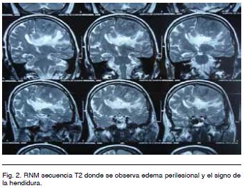
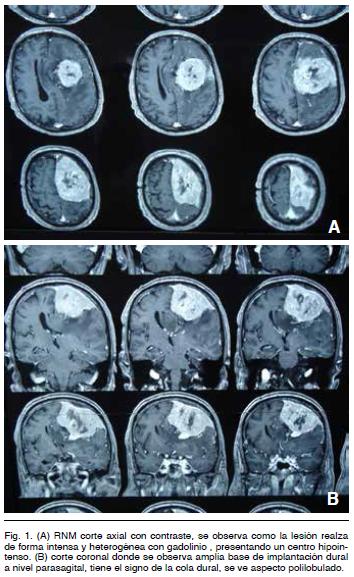
<body><![CDATA[<p style="line-height: normal;"><span style="font-size: 10pt; font-family: Verdana;" lang="ES-TRAD">Examen cl&iacute;nico al ingreso <o:p></o:p></span></p>         <p style="text-align: left; text-indent: 0cm; line-height: normal;" align="left"><span style="font-size: 10pt; font-family: Verdana;" lang="ES-TRAD">El paciente est&aacute; vigil y cumple &oacute;rdenes. Presenta una afasia mixta a predominio de la expresi&oacute;n y un d&eacute;ficit motor derecho a predominio braquial M4 en la escala del British Medical Councilie, presenta Babinski e hiperreflexia homolateral.<o:p></o:p></span></p>         <p style="text-align: left; text-indent: 0cm; line-height: normal;" align="left"><span style="font-size: 10pt; font-family: Verdana;" lang="ES-TRAD">Con la sospecha cl&iacute;nica de proceso expansivo intracraneano tumoral, se realiz&oacute; Tomograf&iacute;a de cr&aacute;neo (TC) como m&eacute;todo de screenning y luego Resonancia Nuclear Magn&eacute;tica (IRM). <o:p></o:p></span></p>         <p style="text-align: left; text-indent: 0cm; line-height: normal;" align="left"><span style="font-size: 10pt; font-family: Verdana;" lang="ES-TRAD">La misma evidenci&oacute; (<a href="#fig_1">Figura 1</a>) un tumor intracraneano extra-axial de la convexidad frontoparietal izquierda, isointenso en la secuencia T1, voluminoso, polilobulado y con extenso contacto dural que llega hasta el seno longitudinal superior y la hoz cerebral. Con la administraci&oacute;n de gadolinio hay un realce intenso y heterog&eacute;neo del tumor y su base de implantaci&oacute;n (signo de la cola dural).</span></p>       <p style="text-align: left; text-indent: 0cm; line-height: normal;" align="left">&nbsp;</p>       <p style="text-align: left; text-indent: 0cm; line-height: normal;" align="left"><span style="font-size: 10pt; font-family: Verdana;" lang="ES-TRAD"><a name="fig_1"></a><img style="width: 354px; height: 577px;" alt="" src="/img/revistas/ami/v37n1/1a07f1.jpg"></span></p>       <p style="text-align: left; text-indent: 0cm; line-height: normal;" align="left">&nbsp;</p>       <p style="text-align: left; text-indent: 0cm; line-height: normal;" align="left"><span style="font-size: 10pt; font-family: Verdana;" lang="ES-TRAD"> En T2 se observa isointenso, identific&aacute;ndose liquido cefalorraqu&iacute;deo entre el tumor y el cerebro (signo de la hendidura). Se ve tambi&eacute;n importante edema perilesional digitiforme (<a href="#fig_2">Figura 2</a>).</span></p>      <p style="text-align: left; text-indent: 0cm; line-height: normal;" align="left">&nbsp;</p>       <p style="text-align: left; text-indent: 0cm; line-height: normal;" align="left"> <font face="Verdana"><a name="fig_2"></a><img style="width: 352px; height: 271px;" alt="" src="/img/revistas/ami/v37n1/1a07f2.jpg"></font></p>       ]]></body>
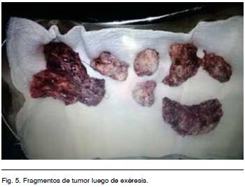
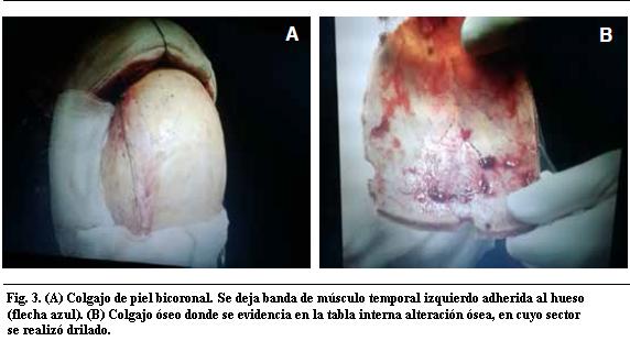
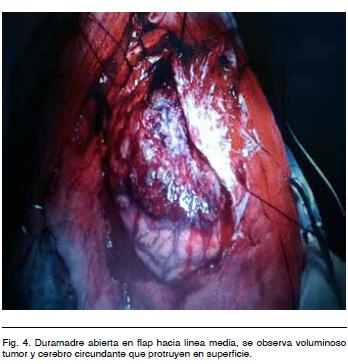
<body><![CDATA[<p style="text-align: left; text-indent: 0cm; line-height: normal;" align="left"><span style="font-size: 10pt; font-family: Verdana;" lang="ES-TRAD"><o:p></o:p></span></p>         <p style="text-align: left; text-indent: 0cm; line-height: normal;" align="left"><span style="font-size: 10pt; font-family: Verdana;" lang="ES-TRAD">Por las caracter&iacute;sticas imagenol&oacute;gicas en planteo fue de:<o:p></o:p></span></p>         <p style="margin-left: 0cm; text-align: left; text-indent: 0cm; line-height: normal;" align="left"><span style="font-size: 10pt; font-family: Verdana;" lang="ES-TRAD">&bull;<span style="">&nbsp;&nbsp;&nbsp;&nbsp;&nbsp;&nbsp;&nbsp; </span><span class="GramE">meningioma</span> <o:p></o:p></span></p>         <p style="margin-left: 0cm; text-align: left; text-indent: 0cm; line-height: normal;" align="left"><span style="font-size: 10pt; font-family: Verdana;" lang="ES-TRAD">&bull;<span style="">&nbsp;&nbsp;&nbsp;&nbsp;&nbsp;&nbsp;&nbsp; </span><span class="GramE">en</span> segundo lugar, hemangiopericitoma.<o:p></o:p></span></p>         <p style="text-align: left; text-indent: 0cm; line-height: normal;" align="left"><span style="font-size: 10pt; font-family: Verdana;" lang="ES-TRAD">Se realiz&oacute; tratamiento quir&uacute;rgico por abordaje bicoronal con exposici&oacute;n del seno longitudinal superior. Se hizo una ex&eacute;resis tumoral macrosc&oacute;picamente completa con plastia de duramadre (Figuras <a href="#fig_3">3</a>, <a href="#fig_4">4</a> y <a href="#fig_5">5</a>).</span></p>      <p style="text-align: left; text-indent: 0cm; line-height: normal;" align="left">&nbsp;</p>       <p style="text-align: left; text-indent: 0cm; line-height: normal;" align="left"> <font face="Verdana"><a name="fig_3"></a><img style="width: 572px; height: 310px;" alt="" src="/img/revistas/ami/v37n1/1a07f3.jpg"></font></p>      <p style="text-align: left; text-indent: 0cm; line-height: normal;" align="left"></p>       <p style="text-align: left; text-indent: 0cm; line-height: normal;" align="left"> <font face="Verdana"><a name="fig_4"></a><img style="width: 348px; height: 364px;" alt="" src="/img/revistas/ami/v37n1/1a07f4.jpg"></font></p>      <p style="text-align: left; text-indent: 0cm; line-height: normal;" align="left"></p>       ]]></body>
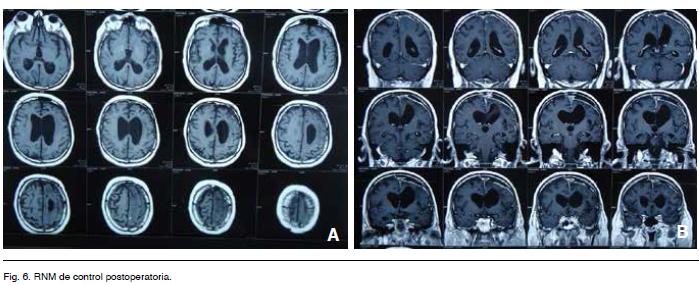
<body><![CDATA[<p style="text-align: left; text-indent: 0cm; line-height: normal;" align="left"> <font face="Verdana"><a name="fig_5"></a><img style="width: 349px; height: 267px;" alt="" src="/img/revistas/ami/v37n1/1a07f5.jpg"></font></p>         <p style="text-align: left; text-indent: 0cm; line-height: normal;" align="left"><span style="font-size: 10pt; font-family: Verdana;" lang="ES-TRAD"><o:p>&nbsp;</o:p></span></p>         <p style="line-height: normal;"><span style="font-size: 10pt; font-family: Verdana;" lang="ES-TRAD">Anatom&iacute;a patol&oacute;gica<o:p></o:p></span></p>         <p style="text-align: left; text-indent: 0cm; line-height: normal;" align="left"><span style="font-size: 10pt; font-family: Verdana;" lang="ES-TRAD">El informe histol&oacute;gico fue compatible con HPC, dada la elevada vascularizaci&oacute;n, por lo que se solicit&oacute; estudio inmunohistoqu&iacute;mico. El mismo fue negativo para CD34 pero positivo para factor VIII (de von Willebrand), presentando un &iacute;ndice de proliferaci&oacute;n del 5-10%, por lo que se concluye que corresponde a un HPC grado III (OMS 2007). <o:p></o:p></span></p>         <p style="text-align: left; text-indent: 0cm; line-height: normal;" align="left"><span style="font-size: 10pt; font-family: Verdana;" lang="ES-TRAD">El paciente presento buena evoluci&oacute;n postoperatoria con mejor&iacute;a progresiva del d&eacute;ficit motor y afasia. La RNM de control no evidencia complicaciones (<a href="/img/revistas/ami/v37n1/1a07f6.jpg">Figura 6</a>). <o:p></o:p></span></p>         <p style="text-align: left; text-indent: 0cm; line-height: normal;" align="left"><span style="font-size: 10pt; font-family: Verdana;" lang="ES-TRAD">A los 9 meses de la cirug&iacute;a est&aacute; asintom&aacute;tico (Glasgow Outcame Scale 5) y est&aacute; en control cl&iacute;nico e imagenol&oacute;gico. No se realiz&oacute; tratamiento complementario (radioterapia). <o:p></o:p></span></p>         <p style="line-height: normal;"><span style="font-size: 10pt; font-family: Verdana; text-transform: none; font-weight: normal;" lang="ES-TRAD"><o:p>&nbsp;</o:p></span></p>         <p style="line-height: normal;"><span style="font-size: 10pt; font-family: Verdana; text-transform: none;" lang="ES-TRAD">Microscop&iacute;a</span></p>      <p style="line-height: normal;">&nbsp;</p>       <p style="line-height: normal;"><font face="Verdana"><a name="fig_7"></a><img style="width: 349px; height: 614px;" alt="" src="/img/revistas/ami/v37n1/1a07f7.jpg"></font></p>       ]]></body>
<body><![CDATA[<p style="line-height: normal;"><font face="Verdana"><a name="fig_8"></a></font></p>      <p style="line-height: normal;"><font face="Verdana"><img style="width: 348px; height: 605px;" alt="" src="/img/revistas/ami/v37n1/1a07f8.jpg">&nbsp;</font></p>       <p style="text-align: left; text-indent: 0cm; line-height: normal;" align="left"><span style="font-size: 10pt; font-family: Verdana;" lang="ES-TRAD"><o:p>&nbsp;</o:p></span></p>         <p style="line-height: normal;"><span style="font-size: 10pt; font-family: Verdana;" lang="ES-TRAD">DISCUSI&Oacute;N y comentarios<o:p></o:p></span></p>         <p style="text-align: left; text-indent: 0cm; line-height: normal;" align="left"><span style="font-size: 10pt; font-family: Verdana;" lang="ES-TRAD"><o:p>&nbsp;</o:p></span></p>         <p style="text-align: left; text-indent: 0cm; line-height: normal;" align="left"><span style="font-size: 10pt; font-family: Verdana;" lang="ES-TRAD">Los hemangiopericitomas (HPC) son tumores hipervasculares poco frecuentes. <o:p></o:p></span></p>         <p style="text-align: left; text-indent: 0cm; line-height: normal;" align="left"><span style="font-size: 10pt; font-family: Verdana;" lang="ES-TRAD">Dado que se supone su m&aacute;s probable origen en c&eacute;lulas situadas en capilares y v&eacute;nulas postcapilares (pericitos de Zimmermann), pueden verse en cualquier regi&oacute;n del organismo. Sus localizaciones m&aacute;s frecuentes son el retroperitoneo, pelvis, miembros inferiores y menos frecuentemente el SNC&nbsp;<sup><a href="#2">2</a><span class="GramE">,<a href="#3">3</a>,<a href="#7">7</a>,<a name="-9"></a><a href="#9">9</a></span></sup>. <o:p></o:p></span></p>         <p style="text-align: left; text-indent: 0cm; line-height: normal;" align="left"><span style="font-size: 10pt; font-family: Verdana;" lang="ES-TRAD">Sin importar su localizaci&oacute;n los HPC presentan caracter&iacute;sticas ultraestructurales e inmunohistoqu&iacute;micas id&eacute;nticas<sup><a href="#3">3</a><span class="GramE">,<a href="#7">7</a>,<a href="#9">9</a></span></sup>.<o:p></o:p></span></p>         <p style="text-align: left; text-indent: 0cm; line-height: normal;" align="left"><span style="font-size: 10pt; font-family: Verdana;" lang="ES-TRAD">A nivel intracraneano, los HPC tienen una distribuci&oacute;n similar a los meningiomas, con una base de implantaci&oacute;n dural<a href="#4"><sup>4</sup></a>. La localizaci&oacute;n a nivel de fosa posterior es de un 15% aproximadamente<sup><a href="#1">1</a><span class="GramE">,<a href="#2">2</a>,<a name="-10"></a><a href="#10">10</a></span></sup>. <o:p></o:p></span></p>         <p style="text-align: left; text-indent: 0cm; line-height: normal;" align="left"><span style="font-size: 10pt; font-family: Verdana;" lang="ES-TRAD">Se han reportado raros casos de HPC no durales como en la regi&oacute;n pineal, as&iacute; como tambi&eacute;n puramente intraparenquimatosos, siendo a&uacute;n esto mucho m&aacute;s infrecuentes<sup><a href="#9">9</a><span class="GramE">,<a href="#10">10</a></span></sup>. Entre el 10 y 15% se ubican a nivel espinal, siendo en m&aacute;s de la mitad de los casos cervicales<a href="#10"><sup>10</sup></a>. <o:p></o:p></span></p>         ]]></body>
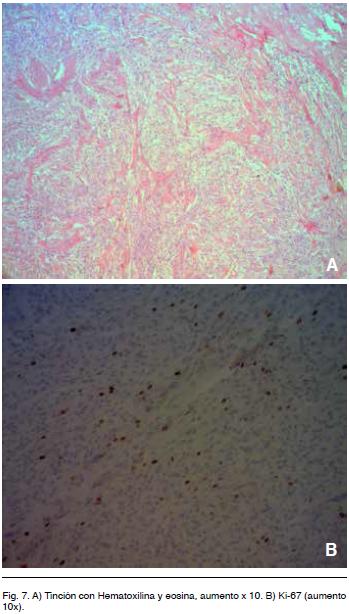
<body><![CDATA[<p style="text-align: left; text-indent: 0cm; line-height: normal;" align="left"><span style="font-size: 10pt; font-family: Verdana;" lang="ES-TRAD">Dentro de todos los tumores del SNC representan menos del 1% y casi el 3% de los vinculados a las meninges<sup><a href="#2">2</a><span class="GramE">,<a href="#3">3</a>,<a href="#7">7</a>,<a href="#9">9</a></span></sup>.<o:p></o:p></span></p>         <p style="text-align: left; text-indent: 0cm; line-height: normal;" align="left"><span style="font-size: 10pt; font-family: Verdana;" lang="ES-TRAD">Tal es su similitud con los meningiomas, que fueron considerados como una variante meningobl&aacute;stica angiomatosa o &ldquo;vasculosa&rdquo; (P&iacute;o del R&iacute;o Hortega ,1933) de los ellos. <o:p></o:p></span></p>         <p style="text-align: left; text-indent: 0cm; line-height: normal;" align="left"><span style="font-size: 10pt; font-family: Verdana;" lang="ES-TRAD">La evoluci&oacute;n cl&iacute;nica de &eacute;stos pacientes, han llevado a an&aacute;lisis gen&eacute;ticos de &eacute;stos casos, demostr&aacute;ndose no tener relaci&oacute;n histogen&eacute;tica con los tumores originados en c&eacute;lulas aracnoideas (meningiomas propiamente dichos).<o:p></o:p></span></p>         <p style="text-align: left; text-indent: 0cm; line-height: normal;" align="left"><span style="font-size: 10pt; font-family: Verdana;" lang="ES-TRAD">La clasificaci&oacute;n de la World Health Organization (WHO) desde 1993 los clasifica como un grupo aparte. Se los clasifica a su vez en 2 variantes: cl&aacute;sica, de bajo grado (grado II) y anapl&aacute;sicao (grado III)<sup><a href="#2">2</a><span class="GramE">,<a href="#3">3</a>,<a href="#6">6</a></span><a href="#6">-8</a></sup>. <o:p></o:p></span></p>         <p style="text-align: left; text-indent: 0cm; line-height: normal;" align="left"><span style="font-size: 10pt; font-family: Verdana;" lang="ES-TRAD">Por todo esto, es el estudio histopatol&oacute;gico (morfol&oacute;gico e inmunohistoquimico) el que diferencia definitivamente a los meningiomas de los HPC<sup><a href="#2">2-5</a>,<a name="-11"></a><a href="#11">11</a></sup>.<o:p></o:p></span></p>         <p style="text-align: left; text-indent: 0cm; line-height: normal;" align="left"><span style="font-size: 10pt; font-family: Verdana;" lang="ES-TRAD">Con respecto a la histolog&iacute;a, los HPC son tumores compuestos por abundantes c&eacute;lulas fusiformes, grandes, con n&uacute;cleos ovales y escaso citoplasma. Las c&eacute;lulas se disponen en torno a espacios vasculares de paredes delgadas (capilares y v&eacute;nulas postcapilares) con un patr&oacute;n caracter&iacute;sticos en &ldquo;asta de ciervo&rdquo; (<a href="#fig_7">Figura 7</a> A).<o:p></o:p></span></p>         <p style="text-align: left; text-indent: 0cm; line-height: normal;" align="left"><span style="font-size: 10pt; font-family: Verdana;" lang="ES-TRAD">La histogenesis de estos tumores es ampliamente debatible. La OMS<a href="#10"><sup>10</sup></a>, los define dentro del cap&iacute;tulo de los tumores mesenquimaticos, no meningotelial y mas presisamente &ldquo;un tumor mesenquimal altamente celular y vascularizado exhibiendo una caracter&iacute;stica <span class="GramE">mon&oacute;tona &hellip;</span> ()&hellip;.con ramificaci&oacute;n vascular &lsquo;cuerno de ciervo&rsquo;&rdquo;)<sup><a name="-12"></a><a href="#12">12</a></sup>.<sup><o:p></o:p></sup></span></p>         <p style="text-align: left; text-indent: 0cm; line-height: normal;" align="left"><span class="GramE"><span style="font-size: 10pt; font-family: Verdana;" lang="ES-TRAD">Existen</span></span><span style="font-size: 10pt; font-family: Verdana;" lang="ES-TRAD"> HPC de la OMS grado II con 2 a 3 mitosis por 10 campos de alta potencia (HPF) y tambi&eacute;n se reportan m&aacute;s de 5 por 10 HPF como grado III. En la variante anapl&aacute;sica (III OMS) tambi&eacute;n se puede encontrar necrosis, hemorragia y pleomorfismo nuclear<sup><a href="#4">4</a><span class="GramE">,<a href="#-8">8</a></span></sup>. <o:p></o:p></span></p>         <p style="text-align: left; text-indent: 0cm; line-height: normal;" align="left"><span style="font-size: 10pt; font-family: Verdana;" lang="ES-TRAD"><o:p>&nbsp;</o:p></span></p>         <p style="line-height: normal;"><span style="font-size: 10pt; font-family: Verdana;" lang="ES-TRAD">Immnunohistoqu&iacute;mica<o:p></o:p></span></p>         ]]></body>
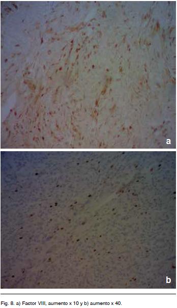
<body><![CDATA[<p style="text-align: left; text-indent: 0cm; line-height: normal;" align="left"><span style="font-size: 10pt; font-family: Verdana;" lang="ES-TRAD">Utilizando t&eacute;cnicas de inmunohistoqu&iacute;mica existen diferentes marcadores que facilitan el diagn&oacute;stico de HPC. <o:p></o:p></span></p>         <p style="text-align: left; text-indent: 0cm; line-height: normal;" align="left"><span style="font-size: 10pt; font-family: Verdana;" lang="ES-TRAD">Sundaram y col. publicaron un trabajo con 23 <span class="GramE">pacientes .</span> Los autores concluyen que:<o:p></o:p></span></p>         <p style="margin-left: 0cm; text-align: left; text-indent: 0cm; line-height: normal;" align="left"><span style="font-size: 10pt; font-family: Verdana;" lang="ES-TRAD">&bull;<span style="">&nbsp;&nbsp;&nbsp;&nbsp;&nbsp;&nbsp;&nbsp; </span>Los HPC son ant&iacute;geno epitelial de membrana (EMA) y prote&iacute;na S-100 negativos, vimentina positivos. <o:p></o:p></span></p>         <p style="margin-left: 0cm; text-align: left; text-indent: 0cm; line-height: normal;" align="left"><span style="font-size: 10pt; font-family: Verdana;" lang="ES-TRAD">&bull;<span style="">&nbsp;&nbsp;&nbsp;&nbsp;&nbsp;&nbsp;&nbsp; </span>Los meningiomas son EMA y vimentina positivos, y la respuesta es variable para S-100. <o:p></o:p></span></p>         <p style="text-align: left; text-indent: 0cm; line-height: normal;" align="left"><span style="font-size: 10pt; font-family: Verdana;" lang="ES-TRAD">El marcador CD 34 positivo esta solo en 25% de los casos de los HPC. Hay otros marcadores que se mencionan en la literatura entre los que se encuentra el factor VIIIa como ayuda para el diagnostico en los HPC<sup><a href="#11">11</a><span class="GramE">,<a name="-13"></a><a href="#13">13</a></span> </sup>(<a href="#fig_8">Figura 8</a>). <o:p></o:p></span></p>         <p style="text-align: left; text-indent: 0cm; line-height: normal;" align="left"><span style="font-size: 10pt; font-family: Verdana;" lang="ES-TRAD">Aunque no hay un solo marcador sensible y especifico, el perfil inmunohistoqu&iacute;mico es suficiente para diferenciar estos tumores de los meningiomas<a href="#13"><sup>13</sup></a>.<o:p></o:p></span></p>         <p style="text-align: left; text-indent: 0cm; line-height: normal;" align="left"><span style="font-size: 10pt; font-family: Verdana;" lang="ES-TRAD">Nuestro caso fue negativo para CD 34 pero positivo para factor VIIIa. <o:p></o:p></span></p>         <p style="text-align: left; text-indent: 0cm; line-height: normal;" align="left"><span style="font-size: 10pt; font-family: Verdana;" lang="ES-TRAD">Este hecho concuerda con la definici&oacute;n inicial del HPC y con los resultados de estudios de series de Perry, Arie et al. (1997).<o:p></o:p></span></p>         <p style="text-align: left; text-indent: 0cm; line-height: normal;" align="left"><span style="font-size: 10pt; font-family: Verdana;" lang="ES-TRAD">A pesar de la controversia en cuanto a su histog&eacute;nesis, el HPC meningeo es una entidad cl&iacute;nico patol&oacute;gica bien definida que exhiben altas tasas de recurrencia y met&aacute;stasis tard&iacute;as extracraneales. &Eacute;ste debe distinguirse de varias neoplasias benignas, especialmente:<o:p></o:p></span></p>         <p style="margin-left: 0cm; text-align: left; text-indent: 0cm; line-height: normal;" align="left"><span style="font-size: 10pt; font-family: Verdana;" lang="ES-TRAD">&bull;<span style="">&nbsp;&nbsp;&nbsp;&nbsp;&nbsp;&nbsp;&nbsp; </span>Meningioma fibroso (FM) <o:p></o:p></span></p>         ]]></body>
<body><![CDATA[<p style="margin-left: 0cm; text-align: left; text-indent: 0cm; line-height: normal;" align="left"><span style="font-size: 10pt; font-family: Verdana;" lang="ES-TRAD">&bull;<span style="">&nbsp;&nbsp;&nbsp;&nbsp;&nbsp;&nbsp;&nbsp; </span><span class="GramE">tumor</span> fibroso solitario (TFS). <o:p></o:p></span></p>         <p style="text-align: left; text-indent: 0cm; line-height: normal;" align="left"><span style="font-size: 10pt; font-family: Verdana;" lang="ES-TRAD">Para determinar el perfil immunohistoquimico se reportan numerosos trabajos donde se comparan diferentes formas de HPC, FM y TFS de las meninges como diagnosticos diferenciales. <o:p></o:p></span></p>         <p style="text-align: left; text-indent: 0cm; line-height: normal;" align="left"><span style="font-size: 10pt; font-family: Verdana;" lang="ES-TRAD">Los paneles de anticuerpos para el HPC incluyen vimentina (85% de positividad en algunas series), factor XIIIa (78% &iacute;dem), Leu-7 (70% idem), y CD34 (33% &iacute;dem). De lo que se desprende el inter&eacute;s real de efectuar factor VIIIa y m&aacute;s aun en tumores CD34 negativos. <o:p></o:p></span></p>         <p style="text-align: left; text-indent: 0cm; line-height: normal;" align="left"><span style="font-size: 10pt; font-family: Verdana;" lang="ES-TRAD">En cuanto a otros elementos de la inmunohistoqu&iacute;mica a tener en cuenta, Probst-Cousin y col. (b) encontraron que el grupo de pacientes con Ki-67 de &lt; 5% tienden a sobrevivir m&aacute;s tiempo, sin embargo no predice con exactitud el resultado cl&iacute;nico<a href="#8"><sup>8</sup></a>. Formalmete la OMS a&uacute;n no acepta este hecho.<o:p></o:p></span></p>         <p style="text-align: left; text-indent: 0cm; line-height: normal;" align="left"><span style="font-size: 10pt; font-family: Verdana;" lang="ES-TRAD">El factor de la coagulaci&oacute;n XIIIa se ha demostrado que est&aacute; presente tanto en c&eacute;lulas tumorales y en los macr&oacute;fagos asociados a tumores de diferentes neoplasias tales como la enfermedad de Hodgkin, tumor de c&eacute;lulas gigantes del hueso, histiocitoma fibroso maligno, tumores de las meninges, y el hemangiopericitoma.<o:p></o:p></span></p>         <p style="text-align: left; text-indent: 0cm; line-height: normal;" align="left"><span style="font-size: 10pt; font-family: Verdana;" lang="ES-TRAD">Los Gliomas de alto grado tales como glioblastoma, gliosarcoma, astrocitoma (grado III de la OMS), y ependimoma (III), as&iacute; como meningiomas y hemangiopericitomas men&iacute;ngeos consistentemente presentan c&eacute;lulas factor XIIIa positivas, mientras que los tumores gliales de grado bajo no lo hacen. Con la excepci&oacute;n de hemangiopericitomas, sin embargo, la principal fuente de expresi&oacute;n FXIIIa en todos estos tumores constaba de una subpoblaci&oacute;n de macr&oacute;fagos asociados al tumor, el papel exacto de los cuales sigue siendo poco claro.<o:p></o:p></span></p>         <p style="text-align: left; text-indent: 0cm; line-height: normal;" align="left"><span style="font-size: 10pt; font-family: Verdana;" lang="ES-TRAD"><o:p>&nbsp;</o:p></span></p>         <p style="line-height: normal;"><span style="font-size: 10pt; font-family: Verdana;" lang="ES-TRAD">CL&Iacute;NICA E IMAGENOLOG&Iacute;A<o:p></o:p></span></p>         <p style="text-align: left; text-indent: 0cm; line-height: normal;" align="left"><span style="font-size: 10pt; font-family: Verdana;" lang="ES-TRAD"><o:p>&nbsp;</o:p></span></p>         <p style="text-align: left; text-indent: 0cm; line-height: normal;" align="left"><span style="font-size: 10pt; font-family: Verdana;" lang="ES-TRAD">Sobre la cl&iacute;nica e imagenolog&iacute;a de los HPC hay que destacar algunos puntos.<o:p></o:p></span></p>         ]]></body>
<body><![CDATA[<p style="text-align: left; text-indent: 0cm; line-height: normal;" align="left"><span style="font-size: 10pt; font-family: Verdana;" lang="ES-TRAD">Estos tumores son ligeramente m&aacute;s frecuentes en hombres y la edad media de presentaci&oacute;n es la cuarta d&eacute;cada<sup><a href="#10">10</a><span class="GramE">,<a href="#12">12</a></span></sup>. En cuanto a la forma de presentaci&oacute;n, el s&iacute;ntoma m&aacute;s frecuente es la cefalea, luego el d&eacute;ficit focal neurol&oacute;gico, el cu&aacute;l puede ser variable seg&uacute;n la topograf&iacute;a lesional. Las crisis son relativamente poco frecuentes (16% de los tumores supratentoriales<span class="GramE">)<sup><a href="#4">4</a>,<a href="#10">10</a></sup></span>. El sangrado intratumoral es un hecho conocido pero poco frecuente<sup><a href="#3">3</a><span class="GramE">,<a href="#4">4</a></span></sup>.<o:p></o:p></span></p>         <p style="text-align: left; text-indent: 0cm; line-height: normal;" align="left"><span style="font-size: 10pt; font-family: Verdana;" lang="ES-TRAD">Con respecto a los estudios de imagen, es de elecci&oacute;n la IRM de cr&aacute;neo. Como ya se mencion&oacute;, estos tumores son imagenologicamente similares a los meningiomas, lo que hace dif&iacute;cil el diagn&oacute;stico preoperatorio. Se han descrito algunas caracter&iacute;sticas particulares que nos pueden sugerir y orientar hacia un HPC como son: 1) un contorno tumoral polilobulado con base de implantaci&oacute;n dural m&aacute;s estrecha, 2) con la administraci&oacute;n de contraste hay realce intenso pero heterog&eacute;neo, 3) en la secuencia T2 se pueden ver im&aacute;genes de vac&iacute;o de flujo intratumoral, 4) habitualmente no presentan hiperostosis, en cambio producen erosi&oacute;n &oacute;sea en mas del 50% de los casos, 5) no tienen calcificaciones intratumorales, 6) la espectroscopia podr&iacute;a diferenciar los HPC de los meningiomas, se ha sugerido que altos valores de mioinositol se pueden encontrar en los primeros<sup><a href="#4">4</a>,<a href="#10">10</a></sup>. <o:p></o:p></span></p>         <p style="text-align: left; text-indent: 0cm; line-height: normal;" align="left"><span style="font-size: 10pt; font-family: Verdana;" lang="ES-TRAD">La arteriograf&iacute;a cerebral es un estudio que debemos tener en cuenta cuando se sospecha un HPC, la presencia de alta cantidad de vasos y un drenaje venoso precoz es m&aacute;s <span class="GramE">caracter&iacute;stico</span> de los HPC a diferencia de los meningiomas. A su vez la embolizaci&oacute;n de los pediculos arteriales nutricios podr&iacute;a formar parte del tratamiento, lo que disminuir&iacute;a el sangrado intraoperatorio<a href="#10"><sup>10</sup></a>.<o:p></o:p></span></p>         <p style="text-align: left; text-indent: 0cm; line-height: normal;" align="left"><span style="font-size: 10pt; font-family: Verdana;" lang="ES-TRAD">Es de gran importancia tener en cuenta la presencia de estos tumores e intentar diferenciarlos de los meningiomas desde el preoperatorio, ya que los HPC tienen un comportamiento m&aacute;s agresivo y la ex&eacute;resis completa y amplia es un objetivo fundamental<a href="#4"><sup>4</sup></a>.<o:p></o:p></span></p>         <p style="text-align: left; text-indent: 0cm; line-height: normal;" align="left"><span style="font-size: 10pt; font-family: Verdana;" lang="ES-TRAD">Los HPC caracter&iacute;sticamente presentan un comportamiento agresivo, con marcada tendencia a recurrir localmente. A 5 y 10 a&ntilde;os hay un &iacute;ndice de recidivas de 50 y 70% respectivamente. Tambi&eacute;n pueden extenderse a lo largo de todo el SNC<sup><a href="#1">1-3</a>,<a href="#6">6</a></sup>. Los HPC grado III presentan mayor tendencia a recurrir m&aacute;s tempranamente que los grado II<a href="#13"><sup>13</sup></a>. Rutkowski y col. en un trabajo con 40 pacientes, encontraron una relaci&oacute;n directa entre el tama&ntilde;o tumoral y la probabilidad de recurrencia local: los tumores &gt; 6 cm tienen mayor recurrencia a 2 a&ntilde;os que los &lt; 6 cm, con una media de 10 a&ntilde;os (p &lt; 0,05). Esto se puede deber en parte a que cuanto m&aacute;s grande es el tumor, m&aacute;s dificultosa su ex&eacute;resis completa y a su vez tienen mayor riesgo de infiltrar sectores el par&eacute;nquima cerebral, quedando tumor residual<sup><a href="#4">4</a><span class="GramE">,<a href="#7">7</a></span></sup>. A su vez vieron que los tumores que se localizan en la base de cr&aacute;neo tienen un mayor tiempo libre de recurrencia<sup>18</sup>. Esto &uacute;ltimo podr&iacute;a deberse a la presencia de diferente comportamiento biol&oacute;gico seg&uacute;n la localizaci&oacute;n, por lo que las meninges en diferentes sectores albergan variable potencial neopl&aacute;sico, al igual que como ocurre con los meningiomas<a href="#7"><sup>7</sup></a>. <o:p></o:p></span></p>         <p style="text-align: left; text-indent: 0cm; line-height: normal;" align="left"><span style="font-size: 10pt; font-family: Verdana;" lang="ES-TRAD">Otra caracter&iacute;stica diferencial es que pueden dar met&aacute;stasis extraneurol&oacute;gicas, con m&aacute;s frecuencia en pulm&oacute;n, hueso e h&iacute;gado. El rango de aparici&oacute;n de las mismas var&iacute;a de 2 a 20 a&ntilde;os luego del diagn&oacute;stico inicial, teniendo a 5 y 15 a&ntilde;os un 30 y 70% de probabilidad respectivamente<sup><a href="#1">1-3</a>,<a href="#6">6</a></sup>. Ning&uacute;n tratamiento en la actualidad logra prevenir la aparici&oacute;n de met&aacute;stasis a distancia<a href="#7"><sup>7</sup></a>.<o:p></o:p></span></p>         <p style="text-align: left; text-indent: 0cm; line-height: normal;" align="left"><span style="font-size: 10pt; font-family: Verdana;" lang="ES-TRAD">El tratamiento de los pacientes con HPC es quir&uacute;rgico y se debe lograr la resecci&oacute;n completa del tumor, la duramadre y el hueso afectado, siempre que la topograf&iacute;a tumoral lo permita<sup><a href="#7">7</a><span class="GramE">,<a href="#10">10</a></span></sup>. Varios trabajos concluyen que la extensi&oacute;n de la resecci&oacute;n, es el factor m&aacute;s importante que determina la probabilidad de recurrencia local y el tiempo de sobrevida, por lo que es fundamental realizar una correcta planificaci&oacute;n quir&uacute;rgica<sup><a href="#2">2</a><span class="GramE">,<a href="#3">3</a>,<a href="#5">5</a>,<a href="#7">7</a></span></sup>. <o:p></o:p></span></p>         <p style="text-align: left; text-indent: 0cm; line-height: normal;" align="left"><span style="font-size: 10pt; font-family: Verdana;" lang="ES-TRAD">Una de las mayores dificultades que enfrenta el cirujano es el sangrado intraoperatorio, dado que son lesiones hipervasculares. El sangrado representa la causa m&aacute;s frecuente de muerte intraoperatoria, con un rango de mortalidad que var&iacute;a de 9 a 25%, a su vez representa uno de los mayores obst&aacute;culos para la resecci&oacute;n total del tumor<a href="#7"><sup>7</sup></a>. En los pacientes en que se sospecha un HPC, realizar una arteriograf&iacute;a con embolizaci&oacute;n preoperatoria se ha sugerido como un m&eacute;todo a tener en cuenta para limitar la p&eacute;rdida de sangre intraoperatoria<sup><a href="#4">4</a><span class="GramE">,<a href="#7">7</a>,<a href="#10">10</a></span></sup>. <o:p></o:p></span></p>         <p style="text-align: left; text-indent: 0cm; line-height: normal;" align="left"><span style="font-size: 10pt; font-family: Verdana;" lang="ES-TRAD">La radioterapia postoperatoria, con m&aacute;s de 50 Gy (siendo un tumor dosis dependiente), es considerada beneficiosa dado que prolonga el tiempo de aparici&oacute;n de recurrencia local y la sobrevida global<sup><a href="#4">4</a>,<a href="#7">7</a></sup>. Esta respuesta favorable se da tambi&eacute;n en pacientes con resecci&oacute;n total del tumor, dado que la radioterapia elimina c&eacute;lulas tumorales aisladas que puedan quedar luego de la cirug&iacute;a. La respuesta a este tratamiento oncol&oacute;gico se ve favorecida a su vez por la hipervascularizaci&oacute;n de estos tumores<sup><a href="#5">5</a><span class="GramE">,<a href="#11">11</a></span></sup>. <o:p></o:p></span></p>         <p style="text-align: left; text-indent: 0cm; line-height: normal;" align="left"><span style="font-size: 10pt; font-family: Verdana;" lang="ES-TRAD">El papel de la radiocirug&iacute;a Gama Knife tambi&eacute;n ha sido investigado en los HPC menores de 2-3 cm de di&aacute;metro, y parece tener buenos resultados en el control local del tumor<sup><a href="#4">4</a><span class="GramE">,<a href="#7">7</a></span></sup>. Puede ser considerado en pacientes que tienen tumores primarios o una recidiva menor a 3 cm y no son candidatos a cirug&iacute;a por ejemplo, por alto riesgo anest&eacute;sico comorbilidades asociadas<a href="#10"><sup>10</sup></a>.<o:p></o:p></span></p>         ]]></body>
<body><![CDATA[<p style="text-align: left; text-indent: 0cm; line-height: normal;" align="left"><span style="font-size: 10pt; font-family: Verdana;" lang="ES-TRAD">Sobre la quimioterapia, Chacko y col. mencionan que la topoisomerasa II alfa se correlaciona con el comportamiento biol&oacute;gico, siendo el riesgo de recurrencia 2,9 veces mayor en pacientes con &iacute;ndice de topoisomerasa &gt; a 5% comparados con los de menor valor. Si bien la quimioterapia en los HPC es inefectiva, podr&iacute;a tener un papel en base a agentes antitopoisomerasas en los que expresan un &iacute;ndice &gt; a 5%, si bien este hecho no est&aacute; del todo claro<sup><a name="-15"></a><a href="#15">15</a></sup>.<o:p></o:p></span></p>         <p style="text-align: left; text-indent: 0cm; line-height: normal;" align="left"><span style="font-size: 10pt; font-family: Verdana;" lang="ES-TRAD">Rutkowski y col. en un trabajo retrospectivo con 40 pacientes, evidencian que si bien una ex&eacute;resis completa del tumor con tratamiento oncol&oacute;gico adyuvante adecuado, tiene tendencia a mejorar el pron&oacute;stico global, no ha dado resultados con valores que sean estad&iacute;sticamente significativos, as&iacute; como tampoco hay evidencia que este tratamiento pueda prevenir el desarrollo de enfermedad metast&aacute;sica a distancia<sup><a name="-16"></a><a href="#16">16</a>,<a name="-17"></a><a href="#17">17</a></sup>. Por lo tanto, no hay demostraci&oacute;n clara que deba hacerse quimioterapia en estos tumores. <o:p></o:p></span></p>         <p style="text-align: left; text-indent: 0cm; line-height: normal;" align="left"><span style="font-size: 10pt; font-family: Verdana;" lang="ES-TRAD">Gran parte de la literatura publicada deja claro que la enfermedad recurrente y metast&aacute;sica es algo muy com&uacute;n a pesar de la amplia gama de estrategias terap&eacute;uticas que hay hasta el momento actual. La supervivencia promedio es de 7 a 16 a&ntilde;os aproximadamente, lo que pone en evidencia la necesidad de monitoreo con un seguimiento a largo plazo de estos pacientes<a href="#15"><sup>15</sup></a>. Se ha sugerido el empleo de la Tomograf&iacute;a por Emisi&oacute;n de Positrones (PET) para identificar met&aacute;stasis a distancia, sin embargo hace falta estudios de mayor peso estad&iacute;stico que respalden esta utilidad<a href="#4"><sup>4</sup></a>. <o:p></o:p></span></p>         <p style="text-align: left; text-indent: 0cm; line-height: normal;" align="left"><span style="font-size: 10pt; font-family: Verdana;" lang="ES-TRAD"><o:p>&nbsp;</o:p></span></p>         <p style="line-height: normal;"><span style="font-size: 10pt; font-family: Verdana;" lang="ES-TRAD">CONCLUSIONES<o:p></o:p></span></p>         <p style="text-align: left; text-indent: 0cm; line-height: normal;" align="left"><u style=""><span style="font-size: 10pt; font-family: Verdana;" lang="ES-TRAD"><o:p></o:p></span></u></p>         <p style="text-align: left; text-indent: 0cm; line-height: normal;" align="left"><span style="font-size: 10pt; font-family: Verdana;" lang="ES-TRAD">Los hemangiopericitomas del SNC son tumores poco frecuentes, con un comportamiento agresivo, tendencia a recurrir y dar met&aacute;stasis a distancia. Si bien presentan similares caracter&iacute;sticas cl&iacute;nico imagenol&oacute;gicas con los meningiomas hay ciertos elementos que nos permiten su sospecha en el preoperatorio. El estudio anatomopatol&oacute;gico nos permite realizar el diagn&oacute;stico definitivo, entendiendo en este caso el estudio immunohistoquimico (Faxtor VIIIa, CD34, Ki-67 entre otros). La cirug&iacute;a y radioterapia adyuvante es el tratamiento de elecci&oacute;n que mejora el pron&oacute;stico de estos pacientes, permiten un mayor control local de la enfermedad, si bien no inciden significativamente en la incidencia de met&aacute;stasis a distancia. Sin embargo a pesar de todas las estrategias terap&eacute;uticas sigue siendo una enfermedad recurrente y metast&aacute;sica, por lo que es de fundamental importancia el seguimiento de estos pacientes a largo plazo. <o:p></o:p></span></p>         <p style="text-align: left; text-indent: 0cm; line-height: normal;" align="left"><span style="font-size: 10pt; font-family: Verdana;" lang="ES-TRAD"><o:p>&nbsp;</o:p></span></p>     <span style="font-size: 10pt; font-family: Verdana;" lang="ES-TRAD">Colaboradores: Dr. Fernando Mart&iacute;nez, Dr. Rafael De Armas.<o:p></o:p></span><font face="Verdana">      <br>   </font>   <span style="font-size: 10pt; font-family: Verdana;" lang="ES-TRAD"><o:p>&nbsp;</o:p></span><font face="Verdana">      <br>   </font>   <span style="font-size: 10pt; font-family: Verdana;" lang="ES-TRAD"><span style="font-weight: bold;">BIBLIOGRAF&Iacute;A</span><o:p></o:p></span><font face="Verdana"> </font>      ]]></body>
<body><![CDATA[<p style="text-align: left; text-indent: 0cm; line-height: normal;" align="left"><span style="font-size: 10pt; font-family: Verdana;" lang="ES-TRAD"><o:p>&nbsp;</o:p></span></p>         <!-- ref --><p style="margin-left: 0cm; text-align: left; text-indent: 0cm; line-height: normal;" align="left"><span style="font-size: 10pt; font-family: Verdana;" lang="ES-TRAD"><a name="1"></a><a href="#-1">1</a>.<span style="">&nbsp;&nbsp;&nbsp;&nbsp;&nbsp;&nbsp; </span>Il Seo, Yun Suk Kim, Ho-Sang Kim, Jeong-Ho Kim, Myung-Ki Lee. </span><span style="font-size: 10pt; font-family: Verdana;" lang="EN-US">Hemangiopericytoma of the Posterior Fossa: A case report and review of the literature. Brain tumor Res Treat 2013; 1:95-98<o:p></o:p></span><p style="margin-left: 0cm; text-align: left; text-indent: 0cm; line-height: normal;" align="left"><span style="font-size: 10pt; font-family: Verdana;"><a name="2"></a><a href="#-2">2</a>.<span style="">&nbsp;&nbsp;&nbsp;&nbsp;&nbsp;&nbsp; </span>R. Gutierrez-Gonzalez, G. R. Boto, A. Perez-Zamarron y M. Rivero-Garvia. Hemangiopericitoma de fosa posterior: a prop&oacute;sito de un caso. Servicio de Neurocirug&iacute;a, Hospital Clinico San Caros Madrid, Espa&ntilde;a. </span><span class="GramE"><span style="font-size: 10pt; font-family: Verdana;" lang="EN-US">2008; 19:446-452.</span></span><span style="font-size: 10pt; font-family: Verdana;" lang="EN-US"><o:p></o:p></span></p>         <p style="margin-left: 0cm; text-align: left; text-indent: 0cm; line-height: normal;" align="left"><span style="font-size: 10pt; font-family: Verdana;" lang="EN-US"><a name="3"></a><a href="#-3">3</a>.<span style="">&nbsp;&nbsp;&nbsp;&nbsp;&nbsp;&nbsp; </span>Jun Maruya, Y. Seki, Morita, K. Nishimaki and T. Minkawa. <span class="GramE">Meningeal Hemangiopericytoma manifesting as massive intracranial hemorrhage &ndash; Two Caso Report.</span> <span class="GramE">Departament of Neurosurgery, <st1:placename w:st="on">Akita</st1:placename> <st1:placename w:st="on">Paed</st1:placename> <st1:placename w:st="on">Cross</st1:placename> <st1:placetype w:st="on">Hospital</st1:placetype>, <st1:city w:st="on"><st1:place w:st="on">Akita</st1:place></st1:city>.</span> Neurol. Med. Chir (<st1:city w:st="on"><st1:place w:st="on">Tokyo</st1:place></st1:city>) 46, 92-97. 2006.<o:p></o:p></span></p>         <!-- ref --><p style="margin-left: 0cm; text-align: left; text-indent: 0cm; line-height: normal;" align="left"><span style="font-size: 10pt; font-family: Verdana;"><a name="4"></a><a href="#-4">4</a>.<span style="">&nbsp;&nbsp;&nbsp;&nbsp;&nbsp;&nbsp; </span>Juan Camilo M&aacute;rquez Garc&iacute;a, Fernando Rodriguez, Armando Manuel Morales, Alfredo Miguel Fernandez de Castro. Hemangiopericytoma men&iacute;ngeo anapl&aacute;sico: presentaci&oacute;n de caso. Rev. Colomb. Radiol. 2012<span class="GramE">;23</span>(2):3510-3.    <o:p></o:p></span></p>         <!-- ref --><p style="margin-left: 0cm; text-align: left; text-indent: 0cm; line-height: normal;" align="left"><span style="font-size: 10pt; font-family: Verdana;"><a name="5"></a><a href="#-5">5</a>.<span style="">&nbsp;&nbsp;&nbsp;&nbsp;&nbsp;&nbsp; </span>Amol J. Ghia, Eric L. Chang, Pameh K. Allen, Anita Mahajan, Marta Penas-Prado, Ian E. McCutcheon, Paul D. Brown. </span><span style="font-size: 10pt; font-family: Verdana;" lang="EN-US">Intracranial Hemangiopericytoma: Patterns of Failure and the Role Radiation Therapy. Neurosurgery 73: 624-631, 2013.    <o:p></o:p></span></p>         <!-- ref --><p style="margin-left: 0cm; text-align: left; text-indent: 0cm; line-height: normal;" align="left"><span style="font-size: 10pt; font-family: Verdana;" lang="EN-US"><a name="6"></a><a href="#-6">6</a>.<span style="">&nbsp;&nbsp;&nbsp;&nbsp;&nbsp;&nbsp; </span>Jason Sheelan, D. Kondzodka, J. Flukinger, L. Dade Lunford. <span class="GramE">Radiosurgery for treatment of recurrent intracranial Hemangiopericytomas.</span> <span class="GramE">Neurosurgery Vol. 5.</span> Number 4, 905-91, October 2002.    <o:p></o:p></span></p>         ]]></body>
<body><![CDATA[<!-- ref --><p style="margin-left: 0cm; text-align: left; text-indent: 0cm; line-height: normal;" align="left"><span style="font-size: 10pt; font-family: Verdana;" lang="EN-US"><a name="7"></a><a href="#-7">7</a>.<span style="">&nbsp;&nbsp;&nbsp;&nbsp;&nbsp;&nbsp; </span>Martin J. Rutkowski, Brian J. Jian, Orin Bloch, Cheng Chen, Michael E. Sughrue, TarikTihan, Igor J. Barani, Mitchel S. Berger, Michael W. McDermott and Andrew T. Parsa. <span class="GramE">Intracranial Hemangiopericytoma.</span> <span class="GramE">Clinical experience and treatment.</span> <span class="GramE">Considerations in a modern series of 40 adult petients.</span> American Cancer Society 2012; 118:1628-36.    <o:p></o:p></span></p>         <!-- ref --><p style="margin-left: 0cm; text-align: left; text-indent: 0cm; line-height: normal;" align="left"><span style="font-size: 10pt; font-family: Verdana;" lang="EN-US"><a name="8"></a><a href="#-8">8</a>.<span style="">&nbsp;&nbsp;&nbsp;&nbsp;&nbsp;&nbsp; </span>Louis D, Ohgqki <span class="GramE">H ,</span> Otmar D et al. (1997). WHO Classification of tumours of the Central Nervous <span class="GramE">System .</span> <span class="GramE">Lyon <st1:country-region w:st="on"><st1:place w:st="on">France</st1:place></st1:country-region>.    </span><o:p></o:p></span></p>         <p style="margin-left: 0cm; text-align: left; text-indent: 0cm; line-height: normal;" align="left"><span style="font-size: 10pt; font-family: Verdana;" lang="EN-US"><a name="9"></a><a href="#-9">9</a>.<span style="">&nbsp;&nbsp;&nbsp;&nbsp;&nbsp;&nbsp; </span><st1:place w:st="on"><st1:placename w:st="on">Byoung-Joo</st1:placename> <st1:placetype w:st="on">Park</st1:placetype></st1:place>, Young-Il Kim, Yong-Kil Hong, Sin-SooJeun, Kwan-Sung Lee, Youn-Soo Lee. <span class="GramE">Clinical Analysis of intracranial Hemangiopericytoma.</span> J. Korean Neurosurg Soc. 54:309-316, 203.<o:p></o:p></span></p>         <!-- ref --><p style="margin-left: 0cm; text-align: left; text-indent: 0cm; line-height: normal;" align="left"><span class="GramE"><span style="font-size: 10pt; font-family: Verdana;" lang="EN-US"><a name="10"></a><a href="#-10">10</a>.<span style="">&nbsp;&nbsp;&nbsp;&nbsp; </span>Charles S. Cobbs and Barton L. Guthrie.</span></span><span style="font-size: 10pt; font-family: Verdana;" lang="EN-US"> <span class="GramE">Meningeal hemangiopericytomas.</span> <span class="GramE">Brain Tumors, second Edition, 2001, Cap 37, pag: 751-759.    </span><o:p></o:p></span></p>         <!-- ref --><p style="margin-left: 0cm; text-align: left; text-indent: 0cm; line-height: normal;" align="left"><span class="GramE"><span style="font-size: 10pt; font-family: Verdana;" lang="EN-US"><a name="11"></a><a href="#-11">11</a>.<span style="">&nbsp;&nbsp;&nbsp;&nbsp; </span>Nilendu C. Purandare, Sumeet G. Dua, BhratPrekhi, Sneha Shah, Anshu R-Sharma and VenkateshRangarajan.</span></span><span style="font-size: 10pt; font-family: Verdana;" lang="EN-US"> Metastatic recurrence of an intracranial Hemangiopericytoma 8 years after treatment: report of a case with emphasis on the role of PET/CT in follow-up Cancer Imaging (2010) 10, 117-120.    <o:p></o:p></span></p>         <!-- ref --><p style="margin-left: 0cm; text-align: left; text-indent: 0cm; line-height: normal;" align="left"><span style="font-size: 10pt; font-family: Verdana;" lang="EN-US"><a name="12"></a><a href="#-12">12</a>.<span style="">&nbsp;&nbsp;&nbsp;&nbsp; </span>Perry, Arie M.D.; Scheithauer, Bernd W. M.D.; Nascimento, Antonio G. M.D. The Immunophenotypic Spectrum of Meningeal Hemangiopericytoma: A Comparison <span class="GramE">With</span> Fibrous Meningioma and Solitary Fibrous Tumor of Meninges. <span class="GramE">American Journal of Surgical Pathology.</span> November 1997 - Volume 21 - Issue 11 - pp 1354-1360.    <o:p></o:p></span></p>         <!-- ref --><p style="margin-left: 0cm; text-align: left; text-indent: 0cm; line-height: normal;" align="left"><span style="font-size: 10pt; font-family: Verdana;" lang="EN-US"><a name="13"></a><a href="#-13">13</a>.<span style="">&nbsp;&nbsp;&nbsp;&nbsp; </span>Challa Sundaram, Shantveer G. Uppin, Megha S. Uppin, J. SreeRekha, Manas Kumar Panigrahi, A. K. Purohit, S. Rammurti. A clinicopathological and inmunohistochemical study of central nervous system Hemangiopericytoma. Journal of Clinical Neuroscience 17(2010) 469-472.    <o:p></o:p></span></p>         <!-- ref --><p style="margin-left: 0cm; text-align: left; text-indent: 0cm; line-height: normal;" align="left"><span style="font-size: 10pt; font-family: Verdana;" lang="EN-US"><a name="14"></a>14.<span style="">&nbsp;&nbsp;&nbsp;&nbsp; </span>Ari J. Kane, Michael E. Sughrue, Martin J. Rutkowski, GopalShangari, Shanna Frang, Michael W. McDermott, Mitchel S. Berger and Andrew T. Parsa. Anatomic location is a risk for atypical and malignant meningiomas. <span class="GramE">American Cancer.</span> Volume 117, issue 6, pages 1272-78; <span class="GramE">march</span> 2011.    <o:p></o:p></span></p>         <!-- ref --><p style="margin-left: 0cm; text-align: left; text-indent: 0cm; line-height: normal;" align="left"><span style="font-size: 10pt; font-family: Verdana;" lang="EN-US"><a name="15"></a><a href="#-15">15</a>.<span style="">&nbsp;&nbsp;&nbsp;&nbsp; </span>Chacko G, Rajshekhar V. Intracranial Hemangiopericytomas: correlation of topoisomerase II alpha expression with biologic behavior. Surg. Neurol. 2006<span class="GramE">;65:11</span>-7.    <o:p></o:p></span></p>         <!-- ref --><p style="margin-left: 0cm; text-align: left; text-indent: 0cm; line-height: normal;" align="left"><span style="font-size: 10pt; font-family: Verdana;"><a name="16"></a><a href="#-16">16</a>.<span style="">&nbsp;&nbsp;&nbsp;&nbsp; </span>Cristina Prado, Arturo Navarro-Martin, Ana Lucas, Miguel Macia, FerranGuedes. </span><span style="font-size: 10pt; font-family: Verdana;" lang="EN-US">Adyuvant fractionated radiotherapy affter resection of intracranial.    <o:p></o:p></span></p>         <!-- ref --><p style="margin-bottom: 0.0001pt;"><span style="font-size: 10pt; font-family: Verdana;" lang="EN-GB"><a name="17"></a><a href="#-17">17</a>.<span style="">&nbsp;&nbsp;&nbsp;&nbsp; </span>Probst-Cousin S1, Rickert CH, Gullotta F. Factor XIIIa-immunoreactivity in tumors of the central nervous system. </span><span style="font-size: 10pt; font-family: Verdana;" lang="ES-TRAD">Clin Neuropathol. 1998 Mar-Apr<span class="GramE">;17</span>(2):79-84.    <o:p></o:p></span></p>     </div>          ]]></body><back>
<ref-list>
<ref id="B1">
<label>1</label><nlm-citation citation-type="journal">
<person-group person-group-type="author">
<name>
<surname><![CDATA[Seo]]></surname>
<given-names><![CDATA[Il]]></given-names>
</name>
<name>
<surname><![CDATA[Kim]]></surname>
<given-names><![CDATA[Yun Suk]]></given-names>
</name>
<name>
<surname><![CDATA[Kim]]></surname>
<given-names><![CDATA[Ho-Sang]]></given-names>
</name>
<name>
<surname><![CDATA[Kim]]></surname>
<given-names><![CDATA[Jeong-Ho]]></given-names>
</name>
<name>
<surname><![CDATA[Lee]]></surname>
<given-names><![CDATA[Myung-Ki]]></given-names>
</name>
</person-group>
<article-title xml:lang="en"><![CDATA[Hemangiopericytoma of the Posterior Fossa: A case report and review of the literature]]></article-title>
<source><![CDATA[Brain tumor Res Treat]]></source>
<year>2013</year>
<volume>1</volume>
<page-range>95-98</page-range></nlm-citation>
</ref>
<ref id="B2">
<label>2</label><nlm-citation citation-type="journal">
<person-group person-group-type="author">
<name>
<surname><![CDATA[Gutierrez-Gonzalez]]></surname>
<given-names><![CDATA[R.]]></given-names>
</name>
<name>
<surname><![CDATA[Boto]]></surname>
<given-names><![CDATA[G. R.]]></given-names>
</name>
<name>
<surname><![CDATA[Perez-Zamarron]]></surname>
<given-names><![CDATA[A.]]></given-names>
</name>
<name>
<surname><![CDATA[Rivero-Garvia]]></surname>
<given-names><![CDATA[M.]]></given-names>
</name>
</person-group>
<article-title xml:lang="es"><![CDATA[Hemangiopericitoma de fosa posterior: a propósito de un caso]]></article-title>
<source><![CDATA[Neurocirugía]]></source>
<year>2008</year>
<volume>19</volume>
<page-range>446-452</page-range></nlm-citation>
</ref>
<ref id="B3">
<label>3</label><nlm-citation citation-type="journal">
<person-group person-group-type="author">
<name>
<surname><![CDATA[Maruya]]></surname>
<given-names><![CDATA[Jun]]></given-names>
</name>
<name>
<surname><![CDATA[Seki]]></surname>
<given-names><![CDATA[Y.]]></given-names>
</name>
<name>
<surname><![CDATA[Morita]]></surname>
<given-names><![CDATA[K.]]></given-names>
</name>
<name>
<surname><![CDATA[Nishimaki]]></surname>
<given-names><![CDATA[K]]></given-names>
</name>
<name>
<surname><![CDATA[Minkawa]]></surname>
<given-names><![CDATA[T.]]></given-names>
</name>
</person-group>
<article-title xml:lang="en"><![CDATA[Meningeal Hemangiopericytoma manifesting as massive intracranial hemorrhage: Two Caso Report]]></article-title>
<source><![CDATA[Neurol. Med. Chir]]></source>
<year>2006</year>
<volume>46</volume>
<page-range>92-97</page-range><publisher-loc><![CDATA[Tokyo ]]></publisher-loc>
</nlm-citation>
</ref>
<ref id="B4">
<label>4</label><nlm-citation citation-type="journal">
<person-group person-group-type="author">
<name>
<surname><![CDATA[Márquez García]]></surname>
<given-names><![CDATA[Juan Camilo]]></given-names>
</name>
<name>
<surname><![CDATA[Rodriguez]]></surname>
<given-names><![CDATA[Fernando]]></given-names>
</name>
<name>
<surname><![CDATA[Morales]]></surname>
<given-names><![CDATA[Armando Manuel]]></given-names>
</name>
<name>
<surname><![CDATA[Fernandez de Castro]]></surname>
<given-names><![CDATA[Alfredo Miguel]]></given-names>
</name>
</person-group>
<article-title xml:lang="es"><![CDATA[Hemangiopericytoma meníngeo anaplásico: presentación de caso]]></article-title>
<source><![CDATA[Rev. Colomb. Radiol]]></source>
<year>2012</year>
<volume>23</volume>
<numero>2</numero>
<issue>2</issue>
<page-range>3510-3</page-range></nlm-citation>
</ref>
<ref id="B5">
<label>5</label><nlm-citation citation-type="journal">
<person-group person-group-type="author">
<name>
<surname><![CDATA[Ghia]]></surname>
<given-names><![CDATA[Amol J.]]></given-names>
</name>
<name>
<surname><![CDATA[Chang]]></surname>
<given-names><![CDATA[Eric L.]]></given-names>
</name>
<name>
<surname><![CDATA[Allen]]></surname>
<given-names><![CDATA[Pameh K.]]></given-names>
</name>
<name>
<surname><![CDATA[Mahajan]]></surname>
<given-names><![CDATA[Anita]]></given-names>
</name>
<name>
<surname><![CDATA[Penas-Prado]]></surname>
<given-names><![CDATA[Marta]]></given-names>
</name>
<name>
<surname><![CDATA[McCutcheon]]></surname>
<given-names><![CDATA[Ian E.]]></given-names>
</name>
<name>
<surname><![CDATA[Brown]]></surname>
<given-names><![CDATA[Paul D.]]></given-names>
</name>
</person-group>
<article-title xml:lang="en"><![CDATA[Intracranial Hemangiopericytoma: Patterns of Failure and the Role Radiation Therapy]]></article-title>
<source><![CDATA[Neurosurgery]]></source>
<year>2013</year>
<volume>73</volume>
<page-range>624-631</page-range></nlm-citation>
</ref>
<ref id="B6">
<label>6</label><nlm-citation citation-type="journal">
<person-group person-group-type="author">
<name>
<surname><![CDATA[Sheelan]]></surname>
<given-names><![CDATA[Jason]]></given-names>
</name>
<name>
<surname><![CDATA[Kondzodka]]></surname>
<given-names><![CDATA[D.]]></given-names>
</name>
<name>
<surname><![CDATA[Flukinger]]></surname>
<given-names><![CDATA[J.]]></given-names>
</name>
<name>
<surname><![CDATA[Lunford]]></surname>
<given-names><![CDATA[L. Dade]]></given-names>
</name>
</person-group>
<article-title xml:lang="en"><![CDATA[Radiosurgery for treatment of recurrent intracranial Hemangiopericytomas]]></article-title>
<source><![CDATA[Neurosurgery]]></source>
<year>Octo</year>
<month>be</month>
<day>r </day>
<volume>5</volume>
<numero>4</numero>
<issue>4</issue>
<page-range>905-91</page-range></nlm-citation>
</ref>
<ref id="B7">
<label>7</label><nlm-citation citation-type="journal">
<person-group person-group-type="author">
<name>
<surname><![CDATA[Rutkowski]]></surname>
<given-names><![CDATA[Martin J]]></given-names>
</name>
<name>
<surname><![CDATA[Jian]]></surname>
<given-names><![CDATA[Brian J]]></given-names>
</name>
<name>
<surname><![CDATA[Bloch]]></surname>
<given-names><![CDATA[Orin]]></given-names>
</name>
<name>
<surname><![CDATA[Chen]]></surname>
<given-names><![CDATA[Cheng]]></given-names>
</name>
<name>
<surname><![CDATA[Sughrue]]></surname>
<given-names><![CDATA[Michael E.]]></given-names>
</name>
<name>
<surname><![CDATA[Tihan]]></surname>
<given-names><![CDATA[Tarik]]></given-names>
</name>
<name>
<surname><![CDATA[Barani]]></surname>
<given-names><![CDATA[Igor J.]]></given-names>
</name>
<name>
<surname><![CDATA[Berger]]></surname>
<given-names><![CDATA[Mitchel S.]]></given-names>
</name>
<name>
<surname><![CDATA[McDermott]]></surname>
<given-names><![CDATA[Michael W.]]></given-names>
</name>
<name>
<surname><![CDATA[Parsa]]></surname>
<given-names><![CDATA[Andrew T.]]></given-names>
</name>
</person-group>
<article-title xml:lang="en"><![CDATA[Intracranial Hemangiopericytoma: Clinical experience and treatment. Considerations in a modern series of 40 adult petients]]></article-title>
<source><![CDATA[American Cancer Society]]></source>
<year>2012</year>
<volume>118</volume>
<page-range>1628-36</page-range></nlm-citation>
</ref>
<ref id="B8">
<label>8</label><nlm-citation citation-type="">
<person-group person-group-type="author">
<name>
<surname><![CDATA[Louis]]></surname>
<given-names><![CDATA[D]]></given-names>
</name>
<name>
<surname><![CDATA[Ohgqki]]></surname>
<given-names><![CDATA[H]]></given-names>
</name>
</person-group>
<source><![CDATA[WHO Classification of tumours of the Central Nervous System]]></source>
<year>1997</year>
<publisher-loc><![CDATA[Lyon ]]></publisher-loc>
</nlm-citation>
</ref>
<ref id="B9">
<label>9</label><nlm-citation citation-type="journal">
<person-group person-group-type="author">
<name>
<surname><![CDATA[Park]]></surname>
<given-names><![CDATA[Byoung-Joo]]></given-names>
</name>
<name>
<surname><![CDATA[Kim]]></surname>
<given-names><![CDATA[Young-Il]]></given-names>
</name>
<name>
<surname><![CDATA[Hong]]></surname>
<given-names><![CDATA[Yong-Kil]]></given-names>
</name>
<name>
<surname><![CDATA[Jeun]]></surname>
<given-names><![CDATA[Sin-Soo]]></given-names>
</name>
<name>
<surname><![CDATA[Lee]]></surname>
<given-names><![CDATA[Kwan-Sung]]></given-names>
</name>
<name>
<surname><![CDATA[Lee]]></surname>
<given-names><![CDATA[Youn-Soo]]></given-names>
</name>
</person-group>
<article-title xml:lang="en"><![CDATA[Clinical Analysis of intracranial Hemangiopericytoma]]></article-title>
<source><![CDATA[J. Korean Neurosurg Soc]]></source>
<year>2013</year>
<volume>54</volume>
<page-range>309-316</page-range></nlm-citation>
</ref>
<ref id="B10">
<label>10</label><nlm-citation citation-type="">
<person-group person-group-type="author">
<name>
<surname><![CDATA[Cobbs]]></surname>
<given-names><![CDATA[Charles S.]]></given-names>
</name>
<name>
<surname><![CDATA[Guthrie]]></surname>
<given-names><![CDATA[Barton L.]]></given-names>
</name>
</person-group>
<article-title xml:lang="en"><![CDATA[Meningeal hemangiopericytomas]]></article-title>
<source><![CDATA[Brain Tumors]]></source>
<year>2001</year>
<edition>second</edition>
<page-range>751-759</page-range></nlm-citation>
</ref>
<ref id="B11">
<label>11</label><nlm-citation citation-type="journal">
<person-group person-group-type="author">
<name>
<surname><![CDATA[Purandare]]></surname>
<given-names><![CDATA[Nilendu C]]></given-names>
</name>
<name>
<surname><![CDATA[Dua]]></surname>
<given-names><![CDATA[Sumeet G]]></given-names>
</name>
<name>
<surname><![CDATA[rekhi]]></surname>
<given-names><![CDATA[Bhrat P]]></given-names>
</name>
<name>
<surname><![CDATA[Shah]]></surname>
<given-names><![CDATA[Sneha]]></given-names>
</name>
<name>
<surname><![CDATA[Sharma]]></surname>
<given-names><![CDATA[Anshu R]]></given-names>
</name>
<name>
<surname><![CDATA[Rangarajan]]></surname>
<given-names><![CDATA[Venkatesh]]></given-names>
</name>
</person-group>
<article-title xml:lang="en"><![CDATA[Metastatic recurrence of an intracranial Hemangiopericytoma 8 years after treatment: report of a case with emphasis on the role of PET/CT in follow-up]]></article-title>
<source><![CDATA[Cancer Imaging]]></source>
<year>2010</year>
<volume>10</volume>
<page-range>117-120</page-range></nlm-citation>
</ref>
<ref id="B12">
<label>12</label><nlm-citation citation-type="journal">
<person-group person-group-type="author">
<name>
<surname><![CDATA[Perry]]></surname>
<given-names><![CDATA[Arie]]></given-names>
</name>
<name>
<surname><![CDATA[Scheithauer]]></surname>
<given-names><![CDATA[Bernd W]]></given-names>
</name>
<name>
<surname><![CDATA[Nascimento]]></surname>
<given-names><![CDATA[Antonio G]]></given-names>
</name>
</person-group>
<article-title xml:lang="en"><![CDATA[The Immunophenotypic Spectrum of Meningeal Hemangiopericytoma: A Comparison With Fibrous Meningioma and Solitary Fibrous Tumor of Meninges]]></article-title>
<source><![CDATA[American Journal of Surgical Pathology]]></source>
<year>Nove</year>
<month>mb</month>
<day>er</day>
<volume>21</volume>
<numero>11</numero>
<issue>11</issue>
<page-range>1354-1360</page-range></nlm-citation>
</ref>
<ref id="B13">
<label>13</label><nlm-citation citation-type="journal">
<person-group person-group-type="author">
<name>
<surname><![CDATA[Sundaram]]></surname>
<given-names><![CDATA[Challa]]></given-names>
</name>
<name>
<surname><![CDATA[Uppin]]></surname>
<given-names><![CDATA[Shantveer G.]]></given-names>
</name>
<name>
<surname><![CDATA[Uppin]]></surname>
<given-names><![CDATA[Megha S.]]></given-names>
</name>
<name>
<surname><![CDATA[Rekha]]></surname>
<given-names><![CDATA[J. Sree]]></given-names>
</name>
<name>
<surname><![CDATA[Panigrahi]]></surname>
<given-names><![CDATA[Manas Kumar]]></given-names>
</name>
<name>
<surname><![CDATA[Purohit]]></surname>
<given-names><![CDATA[A. K.]]></given-names>
</name>
<name>
<surname><![CDATA[Rammurti]]></surname>
<given-names><![CDATA[S.]]></given-names>
</name>
</person-group>
<article-title xml:lang="en"><![CDATA[A clinicopathological and inmunohistochemical study of central nervous system Hemangiopericytoma]]></article-title>
<source><![CDATA[Journal of Clinical Neuroscience]]></source>
<year>2010</year>
<volume>17</volume>
<page-range>469-472</page-range></nlm-citation>
</ref>
<ref id="B14">
<label>14</label><nlm-citation citation-type="journal">
<person-group person-group-type="author">
<name>
<surname><![CDATA[Kane]]></surname>
<given-names><![CDATA[Ari J.]]></given-names>
</name>
<name>
<surname><![CDATA[Sughrue]]></surname>
<given-names><![CDATA[Michael E.]]></given-names>
</name>
<name>
<surname><![CDATA[Rutkowski]]></surname>
<given-names><![CDATA[Martin J]]></given-names>
</name>
<name>
<surname><![CDATA[Shangari]]></surname>
<given-names><![CDATA[Gopal]]></given-names>
</name>
<name>
<surname><![CDATA[Frang]]></surname>
<given-names><![CDATA[Shanna]]></given-names>
</name>
<name>
<surname><![CDATA[McDermott]]></surname>
<given-names><![CDATA[Michael W.]]></given-names>
</name>
<name>
<surname><![CDATA[Bergerand]]></surname>
<given-names><![CDATA[Mitchel S.]]></given-names>
</name>
<name>
<surname><![CDATA[Parsa]]></surname>
<given-names><![CDATA[Andrew T.]]></given-names>
</name>
</person-group>
<article-title xml:lang="en"><![CDATA[Anatomic location is a risk for atypical and malignant meningiomas]]></article-title>
<source><![CDATA[American Cancer]]></source>
<year>marc</year>
<month>h </month>
<day>20</day>
<volume>117</volume>
<numero>6</numero>
<issue>6</issue>
<page-range>1272-78</page-range></nlm-citation>
</ref>
<ref id="B15">
<label>15</label><nlm-citation citation-type="journal">
<person-group person-group-type="author">
<name>
<surname><![CDATA[Chacko]]></surname>
<given-names><![CDATA[G]]></given-names>
</name>
<name>
<surname><![CDATA[Rajshekhar]]></surname>
<given-names><![CDATA[V]]></given-names>
</name>
</person-group>
<article-title xml:lang="en"><![CDATA[Intracranial Hemangiopericytomas: correlation of topoisomerase II alpha expression with biologic behavior]]></article-title>
<source><![CDATA[Surg. Neurol]]></source>
<year>2006</year>
<volume>65</volume>
<page-range>11-7</page-range></nlm-citation>
</ref>
<ref id="B16">
<label>16</label><nlm-citation citation-type="">
<person-group person-group-type="author">
<name>
<surname><![CDATA[Prado]]></surname>
<given-names><![CDATA[Cristina]]></given-names>
</name>
<name>
<surname><![CDATA[Navarro-Martin]]></surname>
<given-names><![CDATA[Arturo]]></given-names>
</name>
<name>
<surname><![CDATA[Lucas]]></surname>
<given-names><![CDATA[Ana]]></given-names>
</name>
<name>
<surname><![CDATA[Macia]]></surname>
<given-names><![CDATA[Miguel]]></given-names>
</name>
<name>
<surname><![CDATA[Guedes]]></surname>
<given-names><![CDATA[Ferran]]></given-names>
</name>
</person-group>
<source><![CDATA[Adyuvant fractionated radiotherapy affter resection of intracranial]]></source>
<year></year>
</nlm-citation>
</ref>
<ref id="B17">
<label>17</label><nlm-citation citation-type="journal">
<person-group person-group-type="author">
<name>
<surname><![CDATA[Probst-Cousin]]></surname>
<given-names><![CDATA[S1]]></given-names>
</name>
<name>
<surname><![CDATA[Rickert]]></surname>
<given-names><![CDATA[CH]]></given-names>
</name>
<name>
<surname><![CDATA[Gullotta]]></surname>
<given-names><![CDATA[F]]></given-names>
</name>
</person-group>
<article-title xml:lang="en"><![CDATA[Factor XIIIa-immunoreactivity in tumors of the central nervous system]]></article-title>
<source><![CDATA[Clin Neuropathol]]></source>
<year>1998</year>
<month> M</month>
<day>ar</day>
<volume>17</volume>
<numero>2</numero>
<issue>2</issue>
<page-range>79-84</page-range></nlm-citation>
</ref>
</ref-list>
</back>
</article>