1. Introduction
Brazil plays an important role in the production and commercialization of food, especially in the cultivation of plant species that are relevant to the country's economy1. The biological control of unwanted species is one of the limiting factors for the establishment of crops, so the continuous and indiscriminate application of synthetic herbicides raises great concern in view of the negative consequences for agricultural ecosystems and possible contamination in food2.
It is necessary to develop efficient and low-cost technologies capable of minimizing negative environmental impacts. In this sense, research using natural products has stood out, mainly exploring alternative sources in the production of allelochemicals3.
Endophytic fungi are inserted in this scenario, and have received attention mainly for their competence in the production of secondary metabolites with wide applicability by the industry4. In the interaction between endophytic fungi and plant, biochemical and physiological machineries are adjusted in order to favor survival advantages for both5)(6. Endophytes can increase the competitive capacity of hosts, especially through the production of phytotoxic substances7.
For the preliminary screening of phytotoxic molecules, the germination rate and the root elongation in seeds are the established parameters8)(9. The assay using Lactuca sativa is a reliable bioindicator, particularly because it is simple, inexpensive and requires a relatively small amount of sample10.
In the search for bioactive molecules in endophytes, it is important to consider the chemical and biological history of the family, genus and species of the host plant; since fungi may have the capacity to produce secondary metabolites similar to those of their hosts11.
The vegetable genus Palicourea is known for the occurrence of several substances exhibiting toxicity for animals, and phytotoxic properties12)(13)(14. The species Palicourea corymbifera has biotechnological potential confirmed either by the occurrence of secondary metabolites and by its medicinal and toxic properties15)(16. However, studies aiming to evaluate the phytoxicity of substances produced by endophytic fungi associated to this plant have not yet been reported in the literature.
Thus, this work was carried out with the intent of exploring the biotechnological potential of two endophytic fungi isolated from Palicourea corymbifera, being: Colletotrichum dianesei and Xylaria sp., regarding the production of secondary metabolites; as well as evaluating the phytotoxic action of methanol extracts on germination and growth of Lactuca sativa.
2. Materials and methods
2.1 Isolation and cultivation of fungi
The isolated endophytic fungi were obtained from leaves of the species Palicourea corymbifera (Rubiaceae) collected in September of 2015 in the Reserva Florestal Adolpho Ducke, Manaus, AM, Brazil (2°56'58.2"S 59°57'43.1"W). The samples were transferred to the Bioprospection and Biotechnology Laboratory located at inpa.
The leaves were subjected to pre-disinfestation, being washed with neutral soap under running water. Subsequently, the disinfestation stage began, carried out in a biological safety cabinet. The disinfestation steps were: alcohol 70% (1 minute), sodium hypochlorite 2.5% (4 minutes), alcohol 70% (1 minute), and washing 3 times in sterile distilled water. 500 µL of the last water was removed from the disinfestation wash and inoculated into three plates containing Agar Sabouraud (sb) culture media, in order to verify the efficiency of the method for the elimination of the epiphytic microbiota. After disinfestation, the leaves were cut into small fragments with the aid of a scalpel and tweezers. The obtained fragments were inoculated in Petri dishes containing sb culture medium plus oxytetracycline antibiotic (125 μg/mL), and then incubated in a bod incubator at 30 °C for 48 hours.
After 48 hours of inoculation of the fragments, it was possible to begin the isolation of the emerged colonies. These were transferred to new sb culture media in order to purify the colonies. Afterwards, the isolates were preserved by the Castellani method and by cryopreservation.
Fungi were identified, by molecular methods, as Xylaria sp. and Colletotrichum dianesei. A similar genomic sequence for Xylaria at the species level in the Data Bank was not found, so it remained as sp. These were grown in Sabouraud Dextrose Broth medium with 0.2% yeast extract (sbl). For each fungus, 12 Erlenmeyer flasks of 500 mL containing 300 mL of culture medium were used. The vials were placed in a shaker incubator at 30 °C, with shaking at 120 rpm for 20 days.
No genomic sequence was found in the database at the species level within the genus Xylaria, therefore it remained as sp.
2.2 Extraction
After the growth period, extracts were obtained. First, the mycelia were separated from the growth media by filtration. Then, the mycelia were dried and mechanically milled in a pistil. To obtain the intracellular contents, the mycelia were placed in vials containing first dichloromethane (dcm), and extracted using an ultrasound bath for 20 min. The mycelia were then filtered and re-extracted with dcm by the same procedure two more times. Then mycelium was extracted with ethyl acetate (EtOAc) and finally with methanol (MeOH). All procedures repeated three times for each solvent.
The extracts of Xylaria sp. and Colletotrichum dianesei were concentrated in a rotary evaporator and subsequently dried in a fume hood. Only the methanolic extract was assayed, due to mass availability.
2.3 Germination bioassay
For the assay, methanolic extracts from Xylaria sp. and Colletotrichum dianesei were diluted in methanol at concentration of 1000 µg/mL. As substrate, filter paper discs impregnated with 2 mL of each extract were put into 9 cm Petri dishes. After all solvent evaporation, 2 mL of sterile water were added to humidify the substrate17.
Each filter paper disc received 25 seeds (pre-sterilized, selected by uniformity in size and distributed evenly) of the target species Lactuca sativa (lettuce), with an average germination rate of 97%. Four plates with 25 seeds each were used, totaling 100 seeds per concentration (1000 µg/mL) of each tested extract18.
In the control plates, an identical procedure was performed, replacing the 2 mL of extract with 2 mL of methanol, and after the solvent evaporation, adding 2 mL of sterile water.
The plates were placed into a germination room, where they stayed for an average of 10 days at a photoperiod of 16:8 hours (light/dark), at a temperature of 26 ± 2 °C. The germination percentage assessment was carried out daily and the criterion used was the presence of visible protrusion. The experiment was concluded after three consecutive days without germination.
2.4 Growth bioassay
From the total of germinated seeds in each Petri dish, 10 seedlings were randomly selected. Three days after the root protrusion, measurements of the lengthening of the hypocotyl, coleoptile and radicle of each seedling were performed, using graph paper. 40 seedlings were analyzed per concentration (1000 µg/mL).
2.5 Data analysis
The following was evaluated: pg -percentage of germination (percentage of seeds germinated in each treatment)-, gsi -germination speed index (average number of seeds germinated per day in each treatment, expressed by the following formula: gsi = (G1 / N1 ) + (G2 / N2) + ... + (Gn / Nn), where: G1 = number of seeds germinated at the first count, N1 = number of days from the first count, G2 = number of germinated seeds in the second count, N2 = number of days elapsed until the second count, n = last count)-, and growth of aerial part (hypocotyl/coleoptile) and radicle19.
The results obtained were analyzed using simple variance analysis (anova), and the Tukey test was employed to compare means at 5% probability (p < 0,05). All analyzes were performed using the GraphPad Prism program20.
2.6 Comparative thin layer chromatography and hydrogen nuclear magnetic resonance analysis
The methanolic extracts of Xylaria sp. and Colletotrichum dianesei were analyzed by tlc, using aluminum plates with silica gel with 254 uv indicator and dichloromethane/methanol 9:1 as eluent. The plates were revealed with uv lights (254 and 365 nm), iodine, anisaldehyde, ferric chloride, np-peg and Dragendorff.
All extracts were analyzed by 1H Nuclear Magnetic Resonance (300 MHz, Fourier-300, Bruker).
3. Results
It was possible to verify a reduction of 36% on germination percentage when the seeds were submitted to the extract of Colletotrichum dianesei (Figure 1). The germination speed was delayed by 63.8% for C. dianesei, and by 43.9% for Xylaria sp (Figure 2).
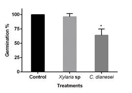
Figure 1: Percentage of Lactuca sativa seed germination under the influence of the methanolic extract of Xylaria sp. and C. dianesei
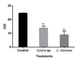
Figure 2: Germination speed index (GSI) of Lactuca sativa seeds under the influence of the methanolic extract of Xylaria sp. and C. dianesei
Root and aerial part growth (hypocotyl + coleoptile) suffered negative growth interference by the methanolic extracts of the two fungi tested (Figure 3). Growth interference can be qualitatively determined, and the presence of the beginning of necrosis in some Lactuca sativa seedlings submitted to the methanolic extract of the fungus C. dianesei is notorious (Figure 4).
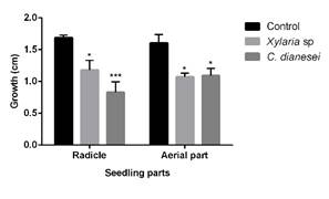
Figure 3: Root and aerial part growth of Lactuca sativa with Colletotrichum dianesei and Xylaria sp. methanolic extracts
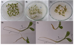
Figure 4: Interference of Colletotrichum dianesei and Xylaria sp. methanolic extracts on the growth of Lactuca sativa
tlc analysis indicates the presence of aromatic substances and alkaloids (only for C. dianesei). Terpenes were on both fungi methanolic extracts, but higher amounts on Xylaria sp. (Table 1).
Table 1: tlc comparison of Colletotrichum dianesei and Xylaria sp. methanolic extracts
| Fungus Revelator | C. dianesei | Xylaria sp. |
| UV light 254 nm | Present (Less intense) | Present (Plus intense) |
| UV light 365 nm | Present (Few spots) | Present (Few spots) |
| Draggendorff (alkaloids) | Present | Absent |
| Ferric chloride (aromatic substances) | Present | Absent |
| Anisaldehyde (general/terpenes) | Present (Less spots) | Present (Plus spots) |
| NP-PEG (flavonoids) | Absent | Absent |
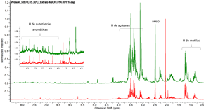
Hydrogen nuclear magnetic resonance (1H-nmr) analysis confirmed the tlc analysis, since there are signs in the region of 6 and 8.5 ppm (aromatic substances), being relatively more intense for Xylaria sp., signs between 3 and 4 ppm (sugars, showing anomeric hydrogen only for Xylaria sp.) and signs between 0.7 and 1.3 ppm, suggesting methyl groups of terpenes (Figure 5).
4. Discussion
Regarding the parameters evaluated in allelopathic activity, germination has shown to be less sensitive to allelochemicals, with greater interferences being evidenced in the speed of germination and in the growth of several species21. This corroborates with the test performed in this study, in which there was no interference in the germination of the seeds submitted to Xylaria sp., but there was a negative influence on the germination speed and growth.
Other phytotoxic evaluations have already been carried out with species of the genus Xylaria and Colletotrichum. The AcOEt extract from the culture filtrate, without cells, of Colletotrichum dematium showed phytotoxic action against Parthenium hysterophorus L.22. And the substances (3aS, 6aR) -4,5-dimethyl-3,3a,6,6a-tetrahydro-2Hcyclopenta(b) furan-2-one and myrothe-ciumone A, isolated from the ethyl acetate extract of Xylaria curta broth, presented inhibitory effects against Lactuca sativa, both of growth and germination, in concentrations below 200 μg/mL, these substances being considered phytotoxic23.
A reduction in the growth of test plants was also noted by Spiassi and collaborators24, where the fungus Fusarium graminearum, Macrophomina phaseolina and Diplodia maydis promoted a reduction on root and aerial parts growth of “amendoim bravo” (Euphorbia heterophylla).
Considering a 50% inhibition or stimulus as a satisfactory standard in the evaluation of phytotoxic activities, interesting results were evidenced for the methanolic extract of C. dianesei, in which the germination speed of L. sativa was reduced by 63.8%; and root growth inhibited by 51.1% in lettuce seedlings.
Among some of the chemical classes with great interference in plant development processes are phenolic substances, which act on enzymes that coordinate various physiological processes, and terpenes, which inhibit germination and plant growth25.


