SciELO - Scientific Electronic Library Online

 
vol.23 número2Primer registro de Palpita persimilis (Lepidoptera, Crambidae) y Oxycenus maxwelli (Acari, Eriophyidae) en el sureste de UruguayAvances en el desarrollo de un biofungicida: caracterización físico-química y actividad antifúngica de propóleos índice de autoresíndice de materiabúsqueda de artículos
Home Pagelista alfabética de revistas  

Servicios Personalizados

Revista

Articulo

Links relacionados

Compartir


Agrociencia (Uruguay)

versión impresa ISSN 1510-0839versión On-line ISSN 2301-1548

Agrociencia Uruguay vol.23 no.2 Montevideo dic. 2019  Epub 01-Dic-2019

https://doi.org/10.31285/agro.23.74 

Comunicación breve

First Report of Zonate Leaf Spot in Sorghum Caused by Microdochium sorghi in Uruguay

Primer reporte de mancha zonada de la hoja del sorgo causada por Microdochium sorghi en Uruguay

1Instituto Nacional de Investigación Agropecuaria (INIA), Programa Nacional de Cultivos de Secano. La Estanzuela, ruta 50 km 11, Colonia, Uruguay.

2Laboratorio de Bioproducción, Bioinsumos, INIA, Las Brujas, ruta 48, km 10, Canelones, Uruguay. *Email: sstewart@inia.org.uy


Abstract:

Sorghum leaves with concentric lesions, alternating purple to red and beige to cinnamon, were observed for the first time in January 2017. The same symptoms were observed later in 29 % of sorghum cultivars in official Evaluación Nacional de Cultivares inase-inia trials. The pathogen was identified as Microdochium sorghi (syn. Gloeocercospora sorghi) once pathogenicity trials, morphological and molecular descriptions were done. This is the first report of this pathogen affecting sorghum in Uruguay.

Keywords: diagnose; ITS; Koch´s postúlate; Gloeocercospora sorghi

Resumen:

En enero de 2017 se observaron por primera vez hojas de sorgo con lesiones formando manchas concéntricas, alternando color púrpura o rojo con bandas beige o canela. La misma sintomatología fue observada posteriormente en el 29 % de los materiales de los ensayos de sorgo de Evaluación Nacional de Cultivares inase-inia. Una vez realizada la prueba de patogenicidad, descripción morfológica y molecular, se identificó al patógeno como Microdochium sorghi (sin. Gloeocercospora sorghi). Este es el primer reporte de este patógeno afectando sorgo en Uruguay.

Palabras claves: diagnóstico; ITS; postulado Koch; Gloeocercospora sorghi

Introduction

Sorghum cultivation (Sorghum bicolor (L.) Moench) in Uruguay covers a total planting area of 47,000 hectares and a production of 214,000 tons1. This figure only considers dry grain sorghum harvest, wet grain or forage sorghum are not taken into account, so it is estimated that the crop would cover thousands of hectares more in the country.

In January 2017, a sample of sorghum, cultivar F1497, with concentric necrotic lesions on the leaves enters the laboratory. Symptomatology is associated with the sorghum disease called zonate leaf spot caused by the fungus Microdochium sorghi (d.c. Bain & Edgerton ex Deighton) U. Braum (sin. Gloeocercospora sorghi d.c. Bain & Edgerton ex Deighton, Ramulispora andropogonis Miura, Titaeospora andropogonis (Miura) f.l. Tai). In the same growing season, the disease was observed in 29%of the evaluated materials in the sorghum official trials of Evaluación Nacional de Cultivares inase-inia at La Estanzuela and Young2.

Zonated leaf spot is common in wet areas of southern USA3 and may affect other crops, such as corn, sugar cane and numerous pastures3)(4. The pathogen is present in South America: Argentina, Brazil, and Venezuela5, but it has not been reported in Uruguay. The objective of this study was to identify and report the causative agent of this leaf spot in our country.

Material and Methods

Isolation

The pathogen was isolated from conidia produced in the sporodochia on the upper side of the leaf with a loop, and transferred to Petri dishes containing PDA (extract of 200 g potato, 10 g dextrose, 20 g agar in 1000 ml of dH2O).

Identification

The identification was carried out by observing the characteristics of the lesion and the sporodochia, and measuring conidia under a microscope. In addition, growth and morphology of the structures in PDA culture medium were observed.

Genomic dna was extracted for molecular identification with DNeasy® Plant Mini Kit (Qiagen Inc.). The amplification was performed by conventional pcr using the primers ITS 5 (52 -ggaagtaaaagtcgtaacaagg) and ITS 4 (52 -tcctccgcttattgatatgc)6. The mixing and cycling conditions were carried out according to Abreo and others7. pcr products were sequenced using the standard service of Macrogen Inc., South Korea (http://www.macrogen.com). The obtained sequence was manually corrected using the program MEGA version 68 and aligned with similar sequences obtained from GenBank9. A phylogenetic analysis was performed by the maximum parsimony method with the MEGA software10. The new isolate was deposited into INIA Las Brujas’ collection with the code ilb 356.

Pathogenicity Tests

The fungus was inoculated in Petri dishes with PDA. Ten days later, the medium containing microsclerotia and mycelium was macerated, passing it several times through a syringe. The paste that resulted from the maceration of three plates was diluted in 100 ml of sterile water. Four pots with 5 plants, each of forage sorghum cultivar Estanzuela Comiray, were inoculated by spraying at third leaf sheath visible stage, covered with nylon bags for 24 hours and kept in a growth chamber at 24 °C. Two sorghum pots were used as control, with the same treatment, except that the spraying was performed with macerated PDA without the fungus. Once the lesions developed, the fungus was reisolated from the inoculated plants.

Results

Symptomatology and identification

Leaf lesions were circular or semicircular, alternating purple to red bands with brown to cinnamon bands, resulting in a concentric or zonate lesion (Figure 1). Several coalescing lesions affected most of the leaf surface.

Figure 1: Symptom of the zonate leaf spot in sorghum in field samples. 

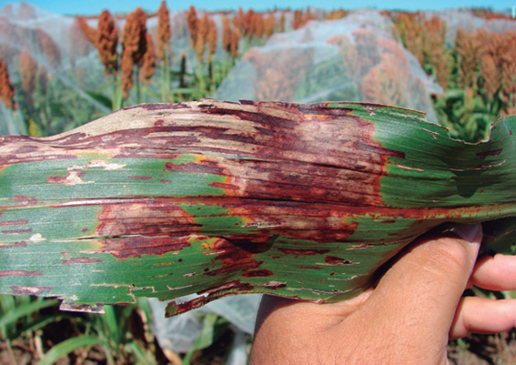
The fungus produces a lot of conidia over the lesions, which emerge from stomata as a pink gelatinous mass, product of sporodochia sporulation. Under a microscope, long filiform, curved conidia of variable size between 1.5-3.5 x 20-190 µm were observed, presenting up to 10 septa. On senescent tissue and PDA medium, black microsclerotia with 0.1 to 0.2 mm in diameter were observed.

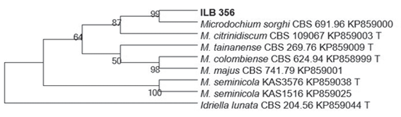
Phylogenetic analysis, based on the alignment of GenBank related sequences, allowed to group ilb 356 strain with an isolate of Microdochium sorghi (accession KP85900)11 with a Bootstrap value of 99 %, sharing 521 of the 522 bp (Figure 2). The ITS sequence was deposited in GenBank with the number MH38491012.

Figure 2: Phylogenetic tree obtained by maximum parsimony based on the analysis of ILB 356 sequence and related sequences obtained from GenBank, including sequences of strains type (t). A sequence of Idriella lunata was included as root. The Bootstrap value 1000 is shown in the nodes. 

Pathogenicity Tests

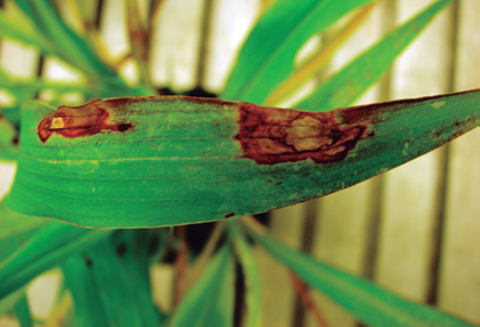
Three days after inoculation small aqueous lesions were observed on the leaves. Five days later, typical lesions of the disease were observed on the leaves (Figure 3), while the control did not develop symptoms.

The pathogen reisolated from the inoculated leaves presented the same morphological characteristics as the inoculated isolate.

Figure 3: Symptom in plants inoculated in growth chamber 5 days after inoculation. 

Conclusion

The observed symptomatology coincides with the description of the sorghum disease called zonate leaf spot4)(13. The symptoms were reproduced in plants inoculated in growth chamber and the fungus was reisolated from the lesions, fulfilling Koch postulate. The morphological characters and the molecular analysis allowed to confirm the identity of the pathogen causing the zonate spot as Microdochium sorghi. It should be noted that, although the taxonomy and phylogeny of this fungal group were revised11, this sorghum pathogen is still named in literature as Gloeocercospora sorghi13. Although symptoms had been previously observed in the field2, this study represents the first laboratory-confirmed report of the causal pathogen of zonate leaf spot in Uruguay.

References:

1. Ministerio de Ganadería, Agricultura y Pesca, DIEA (UY). Encuesta agrícola invierno 2017 (Internet). Montevideo: MGAP; 2017 (cited 2019 May 28). 26 p. Available from: Available from: http://www.mgap.gub.uy/sites/default/files/enc_agricola_inv2017.pdf . [ Links ]

2. Cuitiño MJ, Malaniski S, Stewart S, Vera M, Morales X, Cardozo V. Resultados. In: Resultados experimentales de la evaluación nacional de cultivares de sorgo granífero: Período 2016. Montevideo: INASE; INIA; 2017. p.17-41. [ Links ]

3. Edmunds LK, Zummo N. Sorghum diseases: In the United States and their control. Washington (DC): USDA; 1975. p. 22-4. (Agricultural Handbook; no. 468). [ Links ]

4. Frederiksen R, Odvody GN. Compendium of sorghum diseases. 2nd ed. St. Paul (MN): The American Phytopathological Society; 2000. 78 p. [ Links ]

5. CABI. Gloecercospora sorghi (zonate leaf spot). In: Invasive Species Compendium (Internet). Wallingford (UK): CAB International; 2018 (cited 2018 may 25). (about 3 screens). Available from: Available from: https://www.cabi.org/isc/datasheet/25332 . [ Links ]

6. White TJ, Bruns T, Lee S, Taylor J. Amplification and direct sequencing of fungal ribosomal RNA genes for phylogenetics. In: Innis MA, Gelfand DH Sninsky JJ, White TJ, editors. PCR protocols: A guide to methods and applications. New York: Academic Pres; 1990. p. 315-22. [ Links ]

7. Abreo E, Vaz-Jaur P, Nuñez L, Stewart S, Mattos N, Dini B, Altier N. Pathogenicity of Pythium spp. obtained from agricultural soils and symptomatic legume seedlings in Uruguay. Australas. Plant Dis Notes. 2017;12:35. [ Links ]

8. Tamura K, Stecher G, Peterson D, Filipski A, Kumar S. MEGA6: Molecular Evolutionary Genetics Analysis version 6.0. Mol Biol Evol. 2013;30:2725-9. [ Links ]

9. Benson DA, Cavanaugh M, Clark K, Karsch-Mizrachi I, Lipman DJ, Ostell J, Sayers EW. GenBank. Nucleic Acids Res. 2013;41(DI):D36-D42. [ Links ]

10. Kumar S, Tamura K, Nei M. MEGA: Molecular Evolutionary Genetics Analysis Software for Microcomputers. Comput App Biosci. 1993;10:189-91. [ Links ]

11. Hernández-Restrepo M, Groenewald JZ, Crous PW. Taxonomic and phylogenetic re-evaluation of microdochium, monographella and idriella. Persoonia. 2016;36:57-82. [ Links ]

12. Stewart S. First report of zonate leaf spot in sorghum caused by Gloeocercospora sorghi in Uruguay. (2018?) (cited 2019 Aug 12). In: GenBank (Internet). Bethesda (MD): National Center for Biotechnology Information (U.S.). (1982) - (about 1 screen). Available from: Available from: https://www.ncbi.nlm.nih.gov/nuccore/MH384910 . Accesion: MH384910.1. [ Links ]

13. Jiang Y, Xu J, Hu L, Liu KJ, Xu XD, Liu Z, Meng WL. First report of sorghum zonate leaf spot caused by Gloeocercospora sorghi in China. Plant Dis. 2018;102:1033. [ Links ]

Authors’ contribution: SS: pathogen isolation and greenhouse tests, interpretation of results, writing, RM: pathogen isolation and greenhouse tests, writing, MN: DNA collection and amplification and phylogenetic analysis, AE: interpretation of results, DNA collection and amplification and phylogenetic analysis, participation in writing.

Received: June 11, 2018; Accepted: March 13, 2019

Creative Commons License This is an open-access article distributed under the terms of the Creative Commons Attribution License