


Servicios Personalizados
Revista

Articulo
 texto en
  Inglés (pdf)
 Articulo en XML
 Referencias del artículo
Links relacionados
Compartir

 Permalink
Odontoestomatología
versión impresa ISSN 0797-0374versión On-line ISSN 1688-9339
Odontoestomatología vol.24 no.40 Montevideo dic. 2022 Epub 01-Dic-2022
https://doi.org/10.22592/ode2022n40e411
Case report
Oral condylomatosis in a 9-year-old girl. Clinical case report

1Servicio de Odontología. Hospital del día “Nova Clínica El Recreo”. Quito, Ecuador. richardfdavilat4@gmail.com
2Servicio de Medicina. Hospital del día “Nova Clínica El Recreo” Quito, Ecuador
A clinical case of a 9-year-old patient who seeks care for elevated lesions of the oral mucosa is presented. A disease caused by HPV was diagnosed. This case might potentially involve child abuse due to modes of disease transmission. On clinical examination, an ulcerated papillary lesion of approximately 0.5 cm was observed in the right lip corner. Multiple raised warty lesions were seen on both cheeks and the lower labial mucosa. We conclude about the importance of a good clinical history and a complete oral examination to implement the right treatment and follow-up of pathologies.
Keywords: papilloma; condyloma acuminata; child abuse
Se presenta un caso clínico de una paciente de 9 años que acude a la consulta por lesiones sobre elevadas de la mucosa oral. Se diagnosticó una enfermedad causada por HPV. Debido a sus formas de transmisión, podría indicar un posible caso de abuso infantil. Al examen clínico se observó en la comisura labial derecha una lesión papilar ulcerada de aproximadamente 0,5 cm. Múltiples lesiones elevadas con características verrugosas se observaron en ambas mejillas y la mucosa labial inferior. Se concluye en la importancia de una buena historia clínica y un examen oral completo para realizar un correcto tratamiento y el seguimiento de las patologías.
Palabras clave: papiloma; condiloma acuminado; abuso infantil
Apresenta-se o caso clínico de uma paciente de 9 anos de idade que chega ao ambulatório devido a lesões elevadas da mucosa oral. Foi diagnosticada uma doença causada pelo HPV. Devido às suas formas de transmissão, pode indicar um possível caso de abuso infantil. Ao exame clínico, observou-se lesão papilar ulcerada de aproximadamente 0,5 cm em canto labial direito. Múltiplas lesões elevadas com características verrucosas foram observadas em ambas as bochechas e na mucosa labial inferior. Conclui-se sobre a importância de uma boa história clínica e um exame oral completo para a realização de um tratamento correto e o acompanhamento das patologias.
Palavras-chave: papiloma; condiloma acuminado; abuso infantil
Introduction
Defining the etiology of a condition can make a difference when making a diagnosis. Therefore, it is interesting to contrast the literature on pathologies with similar clinical characteristics, but different modes of transmission, origin, prognosis, and treatment since each pathology affects patients differently.
In pathologies related to ill-treatment or sexual abuse, the timely identification of signs and symptoms compatible with child abuse can determine the physician-patient approach. This is because it allows professionals to identify findings ranging from poor hygiene, lack of resources, and other oral health care aspects, as a right of the child or adolescent. Health professionals are in a privileged position to detect and act in cases of child abuse. Reporting this situation timely to the relevant authorities contributes positively to society.1
The differential diagnosis of intraoral papillary lesions should be guided by clinical features, clinical history, and associated risk factors.
The Human Papilloma Virus (HPV) has been linked to several pathologies. HPV lesions can be benign or malignant, depending on their serotype.2 Other lesions have similar characteristics, such as oral condyloma (OC) and multifocal epithelial hyperplasia (MEH).3 These pathologies share similar characteristics depending on their stage of presentation. To this end, we need an accurate dental history for diagnostic purposes, a histopathology exam, and the most suitable treatment.
Background and case description
A 9-year-old girl sought care accompanied by her father. She was reluctant to be examined, inhibited, and distant towards the professionals. The patient reported that she had had some painless lesions in the oral cavity for approximately two years without apparent cause, and the lesions caused her discomfort when chewing.
She reported no personal pathological history or allergies. She was not under medical treatment.
Hereditary and family history: The patient’s mother, who had died in a traffic accident five years before, had the same lesions in the mouth and genitals.
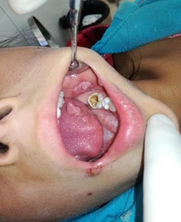
On inspection, there were raised lesions similar in color to the patient’s gum and located on the right lip commissure, tongue, and cheeks. They had different morphology at the buccal mucosa. The lesion on the right labial commissure was papillary, ulcerated, and measured approximately 0.5 cm.
We found multiple raised warty lesions in the oral cavity on both cheeks and the lower-lip mucosa (Figure 1).
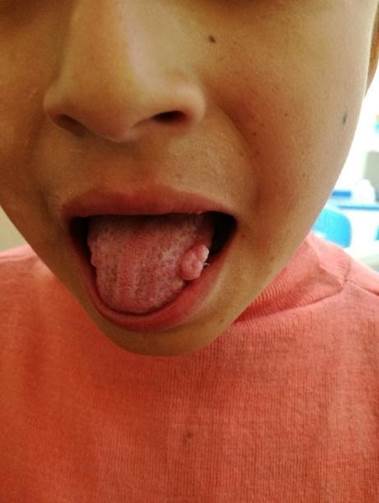
The papillary lesions on the tongue edges were soft and multiple. There was a confluent cauliflower-shaped lesion approximately 1 cm in diameter. It was not painful to the touch and had irregular borders, which had appeared about eight months before. (Figure 2.)
Complementary tests were run. Their results were not significant. Blood biometry Hb:13 mg/dl; hematocrit: 39%; leukocytes: 6500; platelets: 233000; glucose: 82 mg/dl; urea: 12 mg/dl; creatinine: 0.5 mg/dl. Rapid HIV and VDRL tests were negative. We decided to follow the condyloma acuminata protocol, which included the surgical excision and histopathology test of the lesions. However, this protocol could not be implemented due to the father’s refusal because of his financial situation. Subsequent appointments with a psychologist were planned to determine if this was a child abuse case.
The probable diagnosis was based on the clinical features of the lesions. Child abuse was suspected due to the context of the physical examination: the patient was reluctant to be examined and had poor hygiene. These are indicators of a lack of parental care. Furthermore, these lesions are potentially compatible with a viral disease that is primarily sexually transmitted. The case is now in court because the hospital’s social work department decided to intervene and investigate the situation. The team found signs of child abuse, as the girl and her siblings were left alone for long periods without the care of a responsible adult. The neighbors were interviewed, and they described the children as abandoned. The patient is now undergoing psychological treatment, and the family has been followed up as part of the family medicine and social welfare protocol.
Discussion
Human papillomavirus is thought to infect about 6.2 million people each year in the United States. HPV is one of the most common sexual infections. Dunne et al. conducted a study between 2003 and 2004 in a group of women aged between 14 and 59. They determined that the highest HPV prevalence was between ages 20 and 24.2) These data correspond to sexually active individuals. These lesions are rarely found in children. In some cases, they may be associated with child abuse. Therefore, the family, school, and social environments should be investigated, in all cases, as risk factors that may support an alleged abuse. (3
There are over 200 HPVs characterized by genotype. Most are low risk and associated with benign papillomatous lesions with low malignancy potential progression. Genotypes 13 and 32 are exclusive to the oral cavity and cause multifocal epithelial hyperplasia. Genotypes 6 and 11 are linked to squamous papilloma; 2 and 4 to verruca vulgaris; and 6, 11, 16, and 18 to condyloma acuminata.4,5
Condyloma acuminata appears as anogenital warts and is commonly considered a sexually transmitted disease. This lesion is uncommon in the oral cavity.5 Associated genital and oral lesions suggest the presence of a sexually transmitted disease.
Multifocal epithelial hyperplasia is characterized by multiple well-defined mucosal lesions measuring 5 mm in diameter. Its growth is slow and mainly occurs in children. These lesions manifest as papules or nodules involving the lip, gingival, and lingual mucosa. It rarely affects the gingiva or the hard palate. 7,8
These two diseases have similar characteristics in the early stages. Multifocal epithelial hyperplasia has two presentations: papulonodular and papillomatous. The papillomatous variant is less common and is usually located on the tongue and gums.7 Oral condylomatosis is papillary, soft, and sessile. The lesions vary in size and develop into a cauliflower shape in more advanced stages.6
Condyloma acuminata presents several modes of transmission. The primary mode is sexual transmission in people of reproductive age. Vertical transmission from mother to child during birth and transmission due to parental cutaneous HPV has also been reported in children.9 Multifocal epithelial hyperplasia is said to be related to predisposing genetic factors.10,11
Focal epithelial hyperplasia is highly prevalent in some native communities in South America and Africa.12 In Brazil, a study of 587 people from the Waimiri-Atroari community found that multifocal epithelial hyperplasia is the second most common oral mucosa disease.13 In Colombia, it was reported that 13% of children in the Embera-Chami community presented multifocal epithelial hyperplasia.11 In contrast, a study conducted in Mexico reports a very low incidence: 0.026% of the population.14) Multifocal epithelial hyperplasia is more common in women, which would be associated with the poor living conditions they are exposed to in certain ethnic groups. (15
A presumptive diagnosis can be made based on the clinical observation of the lesions. Location, shape, size, color, evolution, and clinical history are considered. In this case, the lesion located on the tongue was compatible with condyloma acuminata. However, the clinical features and their histopathology study will allow us to make an accurate diagnosis to guide the treatment.16
Finally, we must remember that HPV-related oral cavity lesions include different pathologies that share similar clinical and histological features with each other and with other pathologies. While HPV is considered the most common sexually transmitted infection, the virus can be transmitted non-sexually through skin-to-skin, skin-to-mucosa, and mucosa-to-mucosa routes.5
Conclusions
It is essential to take a thorough dental history in the dental appointment because there might be a history of intrafamilial child abuse when children or adolescents have these types of lesions.
Primary care needs protocols to identify potential cases of abuse that should be reported to the competent authorities. It is essential to make differential diagnoses to identify clinically similar diseases and to follow up on the pathologies correctly, especially if they are potentially compatible with child abuse. An accurate diagnosis will positively influence the patient’s physical health and psychosocial development.
REFERENCES
1. Dunne EF, Nielson CM, Stone KM, Markowitz LE, Giuliano AR. Prevalence of HPV infection among men: A systematic review of the literature. J. Infect. Dis. 2006;194 (8): 1044-57. [ Links ]
2. Dunne EF, Unger ER, Sternberg M, McQuillan G, Swan DC, Patel SS, Markowitz LE. Prevalence of HPV infection among females in the United States. J Am Med Assoc 2007; 297(8): 813-9. [ Links ]
3. López A, Basurto C, Salazar R. VPH en cavidad oral: condiloma. Rev Tamé 2019; 7(21): 838-41. [ Links ]
4. Estrada G, Márquez M, González E, Nápoles M, Ramón R. Infección por virus del papiloma humano en la cavidad bucal. Medisan 2015; 19(3): 55-78. [ Links ]
5. Betz SJ. HPV-related papillary lesions of the oral mucosa: A review. Head Neck Pathol. 2019;13(1): 80-90. doi: 10.1007/s12105-019-01003-7. [ Links ]
6. Cháirez Atienzo P, Vega Memíje ME, Zambrano Galván G, García Calderón AG, Maya García IA, Cuevas González JC. Presencia del virus papiloma Humano en la cavidad oral: Revisión y actualización de la Literatura. Int. J. Odontostomatol. 2015; 9(2) :233-8. [ Links ]
7. González M, Suarez R, Canul J, Conde L, Eljure N. Multifocal epithelial hyperplasia in a community in the Mayan area of Mexico. Int. J. Dermatol. 2011; 50(3): 304-9. doi:10.1111/j.1365-4632.2010.04718. [ Links ]
8. Yarmuch P, Chaparro X, Fischer C, Benveniste S. Enfermedad de Heck: A propósito de un caso. Rev. Chilena Dermatol.2012; 28(4):431-4. [ Links ]
9. Obalek S, Jablonska S, Favre M, Walczak L, Orth G. Condylomata acuminata in children: frequent association with human papillomaviruses responsible for cutaneous warts. J. Am. Acad. Dermatol. 1990; 23 (2 Pt 1):.205-13. [ Links ]
10. Bennett LK, Hinshaw M. Heck's disease: diagnosis and susceptibility. Pediatr. Dermatol. 2009; 26(1): 87-9. [ Links ]
11. González LV, Gaviria AM, Sanclemente G, Rady P, Tyring SK, Carlos R, Correa LA, Sanchez GI. Clinical, histopathological and virological findings in patients with focal epithelial hyperplasia from Colombia. Int. J. Dermatol. 2005; 44(4): 274-9. [ Links ]
12. Harris AM, Van Wyk CW. Heck's disease (focal epithelial hyperplasia): a longitudinal study. Community Dent. Oral Epidemiol. 1993; 21(2): 82-5. [ Links ]
13. Dos Santos PJ, Bessa CF, De Aguiar MC, Do Carmo MA. Cross-sectional study of oral mucosal conditions among a central Amazonian Indian community, Brazil. J. Oral Pathol. Med. 2004;3(1):.7-12. [ Links ]
14. Ledesma-Montes C, Vega-Memije E, Garcés-Ortiz M, Cardiel-Nieves M, Juárez-Luna C. Hiperplasia multifocal del epitelio: Reporte de nueve casos. Med. Oral Patol. Oral Cir. Bucal. 2005 ;10(5):.394-401. [ Links ]
15. Nartey NO, Newman MA, Nyako EA. Focal epithelial hyperplasia: report of six cases from Ghana, West Africa. J. Clin. Pediatr. Dent. 2002; 27(1): 63-6. [ Links ]
16. Luciano R, Oviedo J. Virus del papiloma humano y cáncer bucal. Acta Odontol. Venez., 2013; 51(1): 1-3. [ Links ]
Conflict of interest declaration: The authors have no conflict of interest regarding the publication of this paper
Authorship contribution 1. Conception and design of study 2. Acquisition of data 3. Data analysis 4. Discussion of results 5. Drafting of the manuscript 6. Approval of the final version of the manuscript RFDT has contributed in: 1, 2, 4, and 6 JEPL has contributed in: 3, 4, 5, and 6
Received: March 12, 2022; Accepted: June 21, 2022
 Este es un artículo publicado en acceso abierto bajo una licencia Creative Commons
