


Servicios Personalizados
Revista

Articulo
 texto en
  Inglés (pdf)
 Articulo en XML
 Referencias del artículo
Links relacionados
Compartir

 Permalink
Odontoestomatología
versión impresa ISSN 0797-0374versión On-line ISSN 1688-9339
Odontoestomatología vol.23 no.38 Montevideo 2021 Epub 30-Sep-2021
https://doi.org/10.22592/ode2021n37e208
Research
A comparative in vitro study of apical leakage with three obturation techniques

1Catedra de Endodoncia, Facultad de Odontología, Universidad de Guayaquil, Ecuador. endaramarylou1@gmail.com
Objective:
To compare the in vitro apical microleakage of teeth sealed with three techniques: lateral condensation, vertical condensation, and Obtura II.
Methods:
A total of 75 single-root teeth were divided into five groups: Group 1 - lateral condensation, Group 2 - vertical condensation, Group 3 - Obtura II system, Group 4 - positive control, Group 5 - negative control. The roots were subjected to apical leakage through indirect diffusion with Chinese ink, diaphonized, and observed under a Vasconcellos microscope. Descriptive statistical tests were conducted with GraphPad Prism 8.
Results:
Group 1 showed greater microleakage (0.6603 ± 0.5063) with a statistically significant difference compared to group 3 (p < 0.05). However, there was no gutta-percha extrusion, unlike in the other groups.
Conclusion:
The Obtura II technique showed better sealing, which led to decreased apical microleakage, thus proving to be more effective than the other techniques studied.
Keywords: In Vitro Techniques; Root Canal Obturation; Dental Leakage
Objetivo:
Comparar la microfiltración apical de dientes obturados con tres diferentes técnicas: condensación lateral, condensación vertical y sistema obtura II in vitro.
Métodos:
Se utilizaron 75 dientes uniradiculares, divididos en 5 grupos, Grupo 1: condensación lateral, Grupo 2: condensación vertical, Grupo 3: sistema obtura II, Grupo 4: control positivo, Grupo 5: control negativo. Las raíces fueron sometidas a filtración apical por difusión indirecta con tinta china, diafanizadas y observadas con un microscopio Vasconcellos. Se realizaron pruebas estadístico descriptivo con el programa GraphPad prism 8.
Resultados:
El grupo 1 presentó mayor filtración (0,6603±0,5063) con una diferencia estadísticamente significativa en comparación al grupo 3 (p<0.05), sin embargo, no hubo extrusión de la gutapercha a diferencia de los otros dos grupos.
Conclusiones:
La técnica de sistema Obtura II presentó un mejor sellado apical, lo que condujo a una disminución en la filtración apical, demostrando ser más efectiva en comparación a las otras técnicas utilizada en el estudio.
Palabras clave: Técnicas in vitro; Obturación del Conducto Radicular; Filtración Dental
Objetivo:
Comparar a microfiltração apical de dentes obturados com três diferentes técnicas: condensação lateral, condensação vertical e sistema obtura II.
Métodos:
foram utilizados 75 dentes unirradiculares, divididos em 5 grupos, Grupo 1: condensação lateral, Grupo 2: condensação vertical, Grupo 3: sistema obtura II, Grupo 4: controle positivo, Grupo 5: controle negativo. As raízes foram submetidas à filtração apical por difusão indireta com tinta da Índia, diafanizadas e observadas ao microscópio Vasconcellos. Os testes estatísticos descritivos foram realizados com o programa GraphPad prism 8.
Resultados:
O Grupo 1 apresentou maior filtração (0,6603 ± 0,5063) com diferença estatisticamente significante em relação ao Grupo 3 (p <0,05), porém não houve extrusão de guta-percha ao contrário dos outros grupos onde havia.
Conclusões:
A técnica do sistema Obtura II apresentou melhor selamento apical, o que levou à diminuição da filtração apical, mostrando-se mais eficaz em relação às outras técnicas utilizadas no estudo.
Palavras-chave: Técnicas in vitro; Obturação do Canal Radicular; Infiltração Dentária
Introduction and background
Root canal anatomy is a complex set of many variations; therefore, the challenge of endodontic obturation is to ensure the clean and tight sealing of the root canal to maintain or restore the health of periradicular tissues 1,2.
Microleakage of crown and apical tissue fluids is the most common cause of endodontic treatment failure. These fluids are colonized by microorganisms and their byproducts, causing the inflammation of periradicular tissues 3,4).
Canal sealing is often evaluated according to the filtration of biological fluids in various obturation techniques to determine which method is most straightforward and effective in relation to the relevant procedure. This procedure should be fast, less complicated, and affordable for both dentist and patient 5.
Many techniques have been developed over the years to prevent or reduce this type of root canal treatment failure. This has resulted in various obturation methods, but none of them has successfully covered all clinical cases 6.
Currently, there is no universally accepted procedure to evaluate apical microleakage, but the most widely used method is dye penetration with solutions such as methylene blue and Chinese ink, given its sensitivity and ease of use 7.
The last decade has witnessed a debate about obturation quality and sealing capacity using different techniques such as single cone, lateral condensation, and the more recent injectable obturation techniques 2. This study aimed to compare the in vitro apical microleakage of teeth filled with three different techniques: lateral condensation, vertical condensation, and Obtura II.
Methods
This study was conducted at the School of Dentistry of Universidad de Guayaquil. Samples were obtained from 75 fresh human single-root teeth with roots without fractures and a curvature lower than 20 degrees. The study was performed by an operator following this protocol; the teeth were rinsed with plenty of water and immersed in 5.25% sodium hypochlorite for two days after extraction. Initial X-rays were taken to determine the PWL (provisional working length). However, further root clearing showed the gutta-percha under the microscope, so final periapical X-rays were no longer necessary, and the initial ones were used only for reference. The X-rays were taken, and the samples were kept in distilled water (changed every 6 hours) at 37 ºC until instrumentation (Fig. 1). This was done to keep the teeth in conditions similar to the oral cavity.
Tooth preparation
Each tooth had the crown removed with a Diatech (Coltene) diamond bur with a low-speed handpiece. All the roots maintained a total length of 19 mm, measured with a Zipperer 25 mm 06 C-Pilot file. The file was placed inside the canal until its apical exit was detected. The working length of the canal was established at 18 mm by subtracting 1 mm to that measurement. The teeth were placed in plastic trays, which were deposited in a water bath container to obtain 100% humidity and 37 degrees.
Canal preparation
The opening was started with a small head Kavo turbine and a (CB33) Cepamed medium fissure taper carbide bur. For canal preparation, the crown-apical instrumentation technique was applied with Profile .04 Maillefer files with Tecnika motor by Dentsply, using a rotation of 250 rpm and a speed reducer of 16:1. A Nº 10 Maillefer K-File was used to seal the canals, and when the tip of the file reached the anatomical apex, 1 mm was subtracted to work with an 18 mm length.
A 19 mm Maillefer orifice shaper was used to open and shape the canal. This file was placed on the Tecnika motor, in phase F2, at 350 degrees of torque with a 15 mm cap (a measurement used only with this file). Then we used profile .04 lilac ProFile (No. 10) and a white ProFile (No. 15) of the first group using the Tecnika motor, phase F1, at 350 degrees of torque. The files in the second group were then used: the yellow ProFile (No.20). It was placed in the Tecnika, the F2 program was set, and we continued working with the same torque. Then the files of the third group, red ProFile (No. 25) and blue ProFile (No. 30), were used successively, the F3 program was set, and the same torque applied, always reaching 18mm. Glyde was used with each file. The canal was irrigated with 5% sodium hypochlorite after each instrumentation process. After the canals were prepared, they were dried with 70% alcohol.
Study groups
Once the canals were prepared, the teeth were divided and distributed as follows:
Group 1: 20 teeth filled using lateral condensation.
Group 2: 19 teeth filled using vertical condensation.
Group 3: 20 teeth filled using Obtura II.
Group 4: 8 teeth as positive controls.
Group 5: 8 teeth as negative controls.
Lateral obturation technique (Group 1)
A fine-medium gutta-percha point (Hygenic) was used as the main cone and two Fine-Fine cones (Hygenic) as accessories. They were adapted to the canal by cutting 2 mm from the tip to avoid possible extrusion. Top Seal/Maillefer was prepared for sealing in a 1:1 ratio, with a “thread” consistency. The necessary amount was used-as a drop-to cover approximately 1.5 mm of the tip of the gutta-percha cone, which was then placed in the apical third.
Vertical condensation technique (Group 2)
A Fine-Medium (Hygenic) cone was used as the main cone, which was adapted to the canal, cutting 2 mm off the tip to avoid possible extrusion. It was then retested in the canal. The sealer was prepared as used in the lateral obturation technique with one difference: the master cone was cut in the apical third with Touch & Heat (SybronEndo. Touch & Heat Mod 5004). The master cone was condensed with Hu-Friedy N. 9.5 obturator, the gutta-percha was heated in the apical third and condensed again; then Obtura II was placed in two stages to complete obturation.
Obtura II technique (Group 3)
The technique used was Obtura II - Heated Gutta Percha System. mod. 823600 at 180º: 23-gauge needles were used for the Obtura II System, and gutta-percha was used to seal the canals. It contained Zinc Oxide, gutta-percha, barium sulfate, and dyes manufactured by Obtura Spartan. Top Seal (Maillefer) was used as a sealer. It was placed in the apical third of the canal with a Nº 30 gutta-percha cone. The cone was removed, and once the gun reached 180°C, the gutta-percha was injected in two stages, the first one from the apical third to mid working length. After filling, the tip was removed and a Nº 9.5 Hu-Friedy condenser was used to compact the gutta-percha cone vertically. This process was repeated once more, from half the middle third to the cervical third, condensing with Nº 10 Hu-Friedy obturator, leaving 2 mm free in the cervical area for provisional cementing (occlusal closing).
Control group (Groups 4 and 5)
Each experimental group had its positive control (group 4). The canals were instrumented and obturated according to each experimental group. However, Maillefer Top Seal was not used. Additionally, the canals in the negative control group (group 5) were instrumented, red sticky wax was placed on the apical and occlusal surfaces, and then covered with two layers of nail varnish on the remaining root surface. A mark was made at 3 mm from the anatomical apex in occlusal direction. Another mark was made towards the occlusal surface at 3 mm between the first and second marks. A double layer of nail varnish was placed (Fig. 2). These teeth were used to confirm that ink in the canal shows the penetration of the unvarnished apical foramen and not a different route or access point.
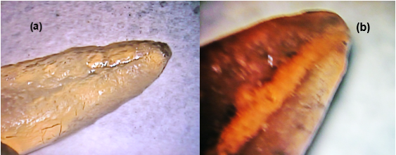
Fig. 2: Negative control where no ink penetration is observed (a) 24x magnification (b) 25x magnification.
Finally, all the roots were wrapped in moistened gauze and placed in the humidifier at 37º for 5 days for the sealant to set completely. After completing obturation, 4 mm of gutta-percha was removed from the cervical-radicular third and filled with Zinc oxide and Eugenol (3 mm) and sticky wax (1 mm).
Dye penetration (indirect or passive ink leakage)
The ink microleakage stage was implemented indirectly: the samples were left in a container with Chinese ink so that it would enter the samples passively.
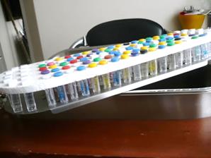
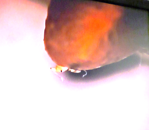
Once all the root groups were obturated, the entire root surface was painted with two coats of nail varnish, except for the three most apical millimeters. The roots were placed on a horizontal platform with holes. Each root was fixed with yellow wax to keep them upright. All the apices were exposed to black Pelikan Chinese ink filtration from the anatomical apex until only 3 mm was covered (Fig. 3). After five days of exposure to the ink, the samples were rinsed with plenty of water, and the nail varnish and sticky wax were removed with a Hu-Friedy Nº 12 curette.
Diaphonization
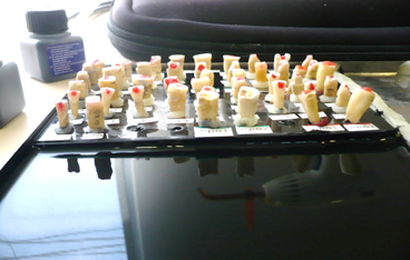
This phase was standardized at the beginning of the study using different concentrations of nitric acid, ethyl alcohol, and methyl salicylate to detect the best results in terms of color and transparency (Fig. 4).
Once the staining phase was completed, the teeth were de-calcified and cleared (modified Tagger method), using 5% nitric acid for 5 days, shaking 3 times a day (8 p.m., 9 a.m., and 1 p.m.), and the acid was changed once a day. Finally, the nitric acid was discarded, and the teeth were washed with plenty of water for four hours to then begin dehydration with 80% ethyl alcohol for one day. It was shaken three times a day (8 p.m., 9 a.m., and 1 p.m.). Then the alcohol was changed to 90% for 1 hour and then to 100% (98%) for 3 hours (shaking every hour). Then, it was cleared with methyl salicylate for one day. The samples remained in salicylate before and after taking microscopy measurements to avoid changes in transparency. The cleared roots made it possible to inspect the root surface and visually determine ink penetration.
Filtration evaluations were performed with a DF Vasconcellos MUM 119 microscope at 24x and 25x magnifications. Measurements were taken with a flexible metal ruler marked every 5 mm, and the filtration results for each sample (expressed in mm) were recorded in a Microsoft Excel spreadsheet. GraphPad Prism 8 statistical software was used for descriptive statistical analysis and graphics.
Results
In this study, the relative amount of ink penetration was measured through the apex of teeth filled with three different techniques: lateral condensation (group 1), vertical condensation (group 2), and Obtura II (group 3) to compare apical microleakage.
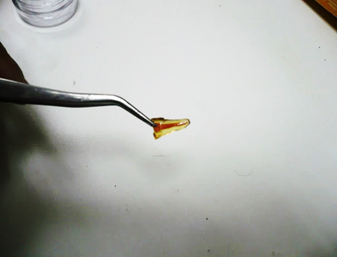
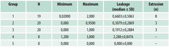
Table 1 shows descriptive data such as the minimum and maximum leakage in each study group. It shows that some teeth did not present leakage in the vertical condensation and Obtura II groups. However, both groups presented gutta-percha extrusion (three teeth in each group) Fig. 5, while all the samples in the lateral condensation group presented leakage of up to 2 mm. However, unlike in the other experimental groups, there was no gutta-percha extrusion.
Additionally, when comparing the study groups, a statistically significant difference was found between the lateral technique and Obtura II (p=0.015), with greater filtration in the lateral technique group, which was significantly different from the negative control group (p = 0.005). Similarly, there was a statistically significant difference when comparing all the study groups (lateral, vertical, Obtura II, and negative control) to the positive control group (p < 0.0001) (Fig. 6).
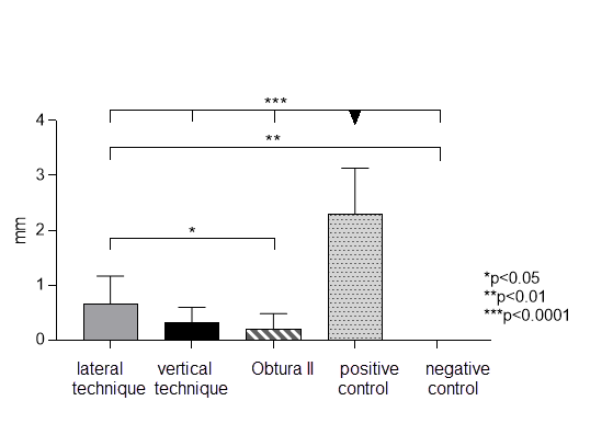
The bars represent the average microleakage observed in each technique and the control group. The asterisks represent statistical differences between techniques when performing a one-way ANOVA test for independent samples with a significance value of p < 0.05.
Discussion
Avoiding apical microleakage is one of the main endodontic challenges. This is achieved by obturating the root canals correctly; this aims to guarantee the surgical preparation conditions to preserve the health of periradicular tissues 1.
Several techniques are used to study in vitro microleakage. However, dye penetration with methylene blue or Chinese ink is one of the most widely used methods. Chinese ink was used in this study because it remains stable during diaphonization process and yields good results 7.
Monterde et al. 8 compared five obturation techniques, lateral condensation, SoftCore®, Obtura II Guttaflow®, and Resilon. They used ink and transparency in the samples as we did, and concluded that the Soft-Core® technique yielded an average leakage of 1.16 mm, while the other techniques had lower values: Guttaflow® (0.49 mm), lateral condensation (0.33 mm), Obtura II® (0.21 mm) and Resilon® (0.15 mm).
Noblecilla 9 found in vitro apical microleakage using the lateral condensation and vertical condensation techniques but no significant differences when comparing the experimental groups. However, greater leakage was observed in the lateral condensation group, which is consistent with the results of this study.
Samson et al. 6. compared the in vitro apical sealing capacity of three different obturation techniques: lateral condensation, Obtura II, and Thermafil. They concluded that the Thermafil obturation technique showed minimal mean apical dye penetration compared to the Obtura II and lateral condensation groups. Furthermore, lateral condensation showed the maximum mean apical dye penetration compared to the other groups. However, the study showed no significant differences between the apical dye penetration of lateral condensation and Obtura II. Our results are largely consistent with the findings of the study described above, as lateral condensation had the highest apical leakage values compared to Obtura II.
Condensation is considered the gold standard in endodontics regarding canal obturation techniques as it is very popular and clinically effective. However, its ability to adapt to internal root canal surfaces has been questioned due to gaps, space between the root walls and the obturation material, incomplete fusion of gutta-percha cones, and lack of surface adaptation 10.
It must be said that the Obtura II System better meets the requirements of a root canal filling since it provides homogeneous sealing (6). The Obtura II System can be used in complex cases such as internal resorptions, curved canals, open apex, perforations, and canal calcifications. However, length control is a disadvantage as this system increases the chances of obturations being longer than the root 11.
This study found that 4% of the teeth obturated presented extrusion with the Obtura II technique and 1.33% with the vertical condensation technique. In the other groups, there was no extrusion. This finding could be consistent with the disadvantage described above regarding the Obtura II and the vertical condensation techniques. Not being able to control the length of the obturation material and the extrusion into the periapical tissues is a major disadvantage 12.
Similarly, our results coincide with those described by Chu Ch et al. 13 concerning lateral condensation. They consider this technique has the advantage of enabling the clinician to control the filler’s length. Controlling the filler prevents gutta-percha extrusion, which we demonstrated in this study.
While gutta-percha extrusion is a problem to be considered when selecting a filling technique, the decision will also depend largely on the operator’s skill when applying any filling technique. The results of this study suggest that the use of warm gutta-percha techniques such as Obtura II is more favorable since they can better adapt to the root canal walls, preventing canal microbial filtration or contamination during endodontic treatment.
Despite detecting less microleakage with Obtura II, it might be a good option to use the vertical technique combined with Obtura II, as in this study, because we observed low leakage levels and less gutta-percha extrusion compared to the Obtura II group.
Conclusion
Of the three techniques used, the Obtura II system technique presented a better apical seal, which led to lower apical leakage, proving to be more effective compared to the other techniques used in the study.
Future studies should be conducted. However, as in vitro research has its limitations, in vivo clinical studies should also be conducted to support results and make them conclusive.
REFERENCES
1. Corro Salazar E, Florean Pérez HJ, Cueto Sánchez Y, Cantarini C, Goldberg F. Estudio comparativo del sellado apical de dos técnicas de obturación endodóntica en conductos curvos simulados. Rev Asoc Odontol Argent. 2018;106:19-24. [ Links ]
2. Shetty KP, Satish SV, Luke AM, Badade AR, Kilaru KR. In vitro Interrelationship between Apical Fill and Apical Leakage Using Three Different Obturation Techniques. J Int Soc Prev Community Dent. 2018 Nov-Dec;8(6):503-507. doi: 10.4103/jispcd.JISPCD_436_17. Epub 2018 Nov 29 [ Links ]
3. Mejía Arredondo Gilmar. Filtración apical y coronal. Odont Moder. 2011;8(88):4. [ Links ]
4. Lahor-Soler E, Miranda-Rius J, Brunet-Llobet L, Farré M, Pumarola J. In vitro study of the apical microleakage with resilon root canal filling using different final endodontic irrigants. J Clin Exp Dent. 2015 Apr 1;7(2):e212-7. doi: 10.4317/jced.51755 [ Links ]
5. Paucar-Gutiérrez H, Maldonado-Huamaní L, Palomares-Bustamante P, Cáceres-Monzón S, Salcedo-Moncada D, Mallqui-Herrada L. Microfiltración apical en dientes obturados empleando la técnica de condensación lateral, cono único y nueva técnica propuesta. Odontol Sanmarquina. 2016;19(1):12-5. [ Links ]
6. Samson E, Kulkarni S, Kumar C S, Likhitkar M. An In-Vitro Evaluation and Comparison of Apical Sealing Ability of Three Different Obturation Technique - Lateral Condensation, Obtura II, and Thermafil. J Int oral Heal 2013;5(2):35-43. [ Links ]
7. Labarta A, Serpone R, Gualtieri A, Sierra L. Evaluación de la filtración apical de la obturación mediante técnica de diafanización. Rev Fac Odon UBA. 2017;32(73):25-33. [ Links ]
8. Monterde M, Pallarés A, Cabanillas C, Zarzosa I, Victoria A. A Comparative in Vitro Study of Apical Microleakage of Five Obturation Techniques. Acta stomatol Croat. 2014;48(2):123-131. [ Links ]
9. Noblecilla MT. Valoración de la filtración apical in vitro en unirradiculares utilizando las técnicas de obturación lateral y vertical. Rev. Cient. Univ. Odontol. Dominic. 2016; 3 (2): 63-72. [ Links ]
10. Guigand M, Glez D, Sibayan E, Cathelineau G, Vulcain JM. Comparative study of two canal obturation techniques by image analysis and EDS microanalysis. Br Dent J. 2005;198:707-11. [ Links ]
11. Bilal Bakht Ansari, Fahad Umer, Farhan Raza Khan. A clinical trial of cold lateral compaction with Obtura II technique in root canal obturation. J Conserv Dent. 2012 Apr-Jun; 15(2): 156-160. [ Links ]
12. Lea CS, Apicella MJ, Mines P, Yancich PP, Parker MH. Comparison of the obturation density of cold lateral compaction versus warm vertical compaction using the continuous wave of condensation technique.J Endod. 2005Jan;31(1):37-9. [ Links ]
13. Chu CH, Lo EC, Cheung GS. Outcome of root canal treatment using Thermafil and cold lateral condensation filling techniques. IntEndod J. 2005;38:179-85. [ Links ]
Authorship contribution 1. Conception and design of study 2. Acquisition of data 3. Data analysis 4. Discussion of results 5. Drafting of the manuscript 6. Approval of the final version of the manuscript. M.E. has contributed in 1, 2, 3, 4, 5, 6.
Received: November 07, 2020; Accepted: February 03, 2021
 Este es un artículo publicado en acceso abierto bajo una licencia Creative Commons
