


Serviços Personalizados
Journal

Artigo
 texto em
  Inglês (pdf)
 Artigo em XML
 Referências do artigo
Links relacionados
Compartilhar

 Permalink
Odontoestomatología
versão impressa ISSN 0797-0374versão On-line ISSN 1688-9339
Odontoestomatología vol.22 no.35 Montevideo 2020 Epub 01-Jun-2020
https://doi.org/10.22592/ode2020n35a4
Research
Human dentin bond strength of a chlorhexidine containing universal adhesive system used in total-etch and self-etch mode
1
 http://orcid.org/0000-0001-6259-042X
2
http://orcid.org/0000-0003-4631-9116
3
http://orcid.org/0000-0002-3326-2547
4
http://orcid.org/0000-0002-1561-2283
5
http://orcid.org/0000-0001-6733-4484
1Cátedra de Materiales Dentales, Facultad de Odontología, Universidad de la República, Uruguay
2Cátedra de Materiales Dentales, Facultad de Odontología, Universidad de la República, Uruguay
3Cátedra de Materiales Dentales, Facultad de Odontología, Universidad de la República, Uruguay
4Cátedra de Materiales Dentales, Facultad de Odontología, Universidad de la República, Uruguay
5Cátedra de Materiales Dentales, Facultad de Odontología, Universidad de la República, Uruguay
6Laboratorio de Materiales Dentales, Área académica de Odontología, Universidad Autonoma del Estado de Hidalgo, México
7Cátedra de Materiales Dentales, Facultad de Odontología, Universidad de la República, Uruguay. ggrazioli@gmail.com
Objectives:
To evaluate the microtensile bond strength to human dentin of a chlorhexidine containing universal adhesive system applied in the total-etch and self-etch modes.
Methods:
20 third molars were randomly divided into 4 groups according to the universal adhesive system (Single Bond Universal ®, 3M ESPE and Peak Universal Bond®, Ultradent) and application mode used (total-etch and self-etch). Specimens were performed and subjected to a microtensile bond strength using a MTS SANS universal testing machine.
Results:
No statistically significant differences were found in the microtensile bond strength between the four groups studied.
Conclusions:
The bond strength of a composite resin to human dentin was not affected by the use of a universal adhesive system containing chlorhexidine in its composition applied in the total-etch and self-etch mode.
Palavras-chave: Clorexidina; Microtração; Metaloproteinases; Camada híbrida.
Key words: Chlorhexidine; Microtensile; Metalloproteinases; Hybrid layer
Objetivos:
Evaluar la resistencia de unión a la microtracción en dentina humana de un sistema adhesivo universal con clorhexidina en su composición, en modo de grabado y lavado en 2 pasos, y en modo de autograbado.
Metodología:
20 terceros molares divididos aleatoriamente en 4 grupos según el tipo de sistema adhesivo utilizado (Single Bond Universal®, 3MESPE y Peak Universal Bond®, Ultradent Products) y modo de uso (grabado total y autograbado). Se confeccionaron cuerpos de prueba sometidos al ensayo de microtracción utilizando una máquina de ensayos universales.
Resultados:
No se encontraron diferencias estadísticamente significativas entre los cuatro grupos estudiados.
Conclusiones:
La resistencia de unión de una resina compuesta a dentina humana no fue afectada por el uso de un sistema adhesivo universal que contiene clorhexidina en su composición aplicado en los modos de grabado total y autograbado.
Palabras clave: Clorhexidina; Microtracción; Metaloproteinasas; Capa híbrida.
Objetivos:
Avaliar a resistência de união á microtração na dentina humana de um sistema adesivo universal com clorexidina na sua composição, no modo de condicionamento total em dois passos e no modo autocondicionante.
Metodologia:
20 terceiros molares foram divididos aleatoriamente em 4 grupos, de acordo com o tipo de sistema adesivo utilizado (Single Bond Universal®, 3MESPE e Peak Universal Bond®, Ultradent Products) e modo de uso (condicionamento total e autocondicionante). Os corpos de prova criados foram submetidos ao teste de microtração utilizando uma máquina de ensaios universal.
Introduction
The demand for aesthetic adhesive restorations has increased since Buonocore introduced the acid etching technique in 19551. This has led to the development of different adhesive strategies2) based on the preservation/integration of the smear layer3-4: smear layer removal or etch-and-rinse technique5-8, and self-etch, which integrates the smear layer. These adhesive techniques behave differently over dentin. Although the etch-and-rinse technique is the most widely used system, its effectiveness is questioned because the adhesive monomers do not always manage to cover all the etched surfaces, leaving a hybrid layer with empty spaces and unsupported collagen fibers9. Additionally, with the self-etch technique, it is possible to remove minerals from the smear layer surface and replace them simultaneously with resin monomers10-11. The adhesion process includes a phenomenon called nanofiltration, which occurs due to the presence of nanometric empty spaces that take fluids from the dental pulp to the external surface of dentin14. These spaces appear because of an uncomplete infiltration of the resinous monomers into the spaces created between the collagen fibers. This creates a demineralized dentin zone under the hybrid layer, that eventually causes degradation of the resin-dentin bond by hydrolysis11,15. In addition, unprotected collagen fibers (not surrounded by resin or dentin) can be degraded by endogenous proteolytic enzymes found in dentin, called matrix metalloproteinases (MMPs). These enzymes lead to degradation of the extracellular matrix in different physiological processes11,15. At the hybrid layer, these enzymes are responsible for a decrease in the long-term bond strength of resinous materials to dentin16.
The use of different agents to counteract the effect of MMPs has been suggested, such as riboflavin17, natural extracts17 and chlorhexidine11,15,17. Chlorhexidine is a synthetic chemical compound with remarkable antibacterial properties; it is widely used in various medical, surgical and dental procedures11,15. In restorative dentistry, chlorhexidine was initially introduced as a cavity disinfectant used before applying restorative materials in order to eliminate bacteria that could irritate the dental pulp11,15. In addition, MMPs -2, -8 and -9 have shown an inhibitory effect against proteolytic activity, thus contributing to increased durability of the resin-dentin bond11,15.
In order to simplify decision making and the clinical technique implemented, nowadays, the market offers universal adhesives, which are highly versatile (they can be applied as part of the etch-and-rinse or self-etch techniques) and are compatible with multiple substrates (teeth, metals and/or ceramics)3,5,14.
In addition, adhesive systems with chlorhexidine are available on the market. This could contribute to adhesive stability by decreasing adhesive bond degradation, increasing the durability and success of the adhesive plastic restorations (11,15. The recent introduction of this universal adhesive with chlorhexidine into the market raises questions about its effectiveness, advantages and properties for consumers (dentists) as there is not enough evidence proving that adding chlorhexidine does not affect its performance.
In this context, this work aims to compare the dentin-bond strength of a new universal commercial adhesive system with chlorhexidine and to compare the values obtained with a universal commercial adhesive system without chlorhexidine in the etch-and-rinse and self-etch techniques.
The hypotheses proposed are that the adhesive system with chlorhexidine will have similar bond strength values compared to the adhesive system without chlorhexidine. In addition, there will be no statistically significant differences between the etch-and-rinse and the self-etch techniques.
Methodology
Obtaining samples
The sample included 20 healthy third molars regardless of root development status. The teeth were obtained from the surgical block service of the School of Dentistry. Patients signed a written informed consent of donation for research (Protocol approved by the Ethics Committee of the School of Dentistry - UdelaR - File number 278/16), where it was stated that tooth removal was indicated for reasons unrelated to this investigation (e.g., orthodontics). The procedure for the collection, cleaning and storage of teeth is protocolized by the Human Tooth Bank of the School of Dentistry (File 091100-000933-11, Council of the School of Dentistry - UdelaR).
Once removed, the teeth were stored in 0.5% Chloramine-T for 7 days and subsequently stored at 3° to 5°C until the study was conducted, for a maximum of 3 months.
The sample was randomly divided into four groups, with five molars each:
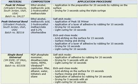
Group 1: Universal adhesive, Single Bond Universal (3M ESPE; ST PAUL, MN, USA), with etch-and-rinse technique (SBU TE).
Group 2: Universal adhesive, Single Bond Universal (3M ESPE; ST PAUL, MN, USA), with self-etch technique (SBU SE).
Group 3: Universal adhesive with 0.2% chlorhexidine, Peak Universal Bond (Ultradent Products; South Jordan, Utah, USA), with etch-and-rinse technique (PBU TE).
Group 4: Universal adhesive with 0.2% chlorhexidine, Peak Universal Bond (Ultradent Products; South Jordan, Utah, USA), with self-etch technique (PBU SE).
Sample preparation for the microtensile test
Each tooth was worn down transversely, using a wet trimmer, removing enamel from the occlusal face, exposing a flat area of coronal dentin without pulp exposure. The coronal dentin was subsequently worn down with silicon carbide sandpaper with 600 grit size by making gentle eight-shaped movements and supplying copious irrigation to standardize the smear layer. The corresponding adhesive treatment was applied on the exposed dentinal surface according to the group that the sample was part of and following the manufacturer’s instructions (Table 1). The selected adhesives were used according to the manufacturer’s instructions. After the adhesive procedures, a 4 mm block of 3M Filtek™ Z250 XT composite resin (3M THICK; ST PAUL, MN, USA) was created, handling the material as directed by the manufacturer. Polymerization was performed using an Optilight Max light-curing unit (Gnatus; Ribeirao Preto, Brazil). The irradiance of the light-curing unit was tested with a Bluephase Meter (Ivoclar Vivadent; Schaan, Linchenstein, Germany), which ensured it was working properly.
Once prepared, the samples were stored for 24 hours in distilled water at 37°C. After that period, the teeth were sectioned using a Gellings-Hamco micro trimmer (Hamco Machines INC.; Rochester, NY, USA), to obtain 1 mm2 sectioned rods (Fig. 1), (approximately eight rods per tooth) which were half dentin and half composite resin. Only five rods obtained from the central region of each tooth (n=25) were selected for the microtensile test.
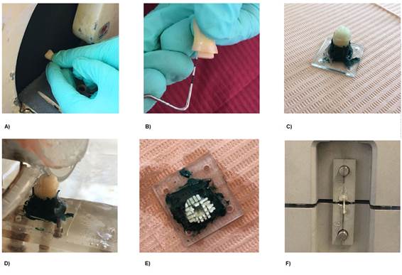
A device with two sliding stainless steel plates was used for this test. Each rod was glued from both ends with cyanoacrylate to said plates, leaving the tooth-restoration interface free between both plates. In this way, the tensile strength was transmitted to the rod18.
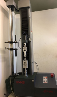
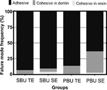
The microtensile test was performed using an MTS SANS CMT 2000 5KN universal testing machine (MTS Systems Corporation; Shanghai China). The specimens were subjected to microtensile tests, according to ISO 11405 (Fig. 2)19. Once fractured, the specimens were analyzed under the microscope at a 40x magnification to classify the type of fracture into adhesive, dentin cohesive and resin cohesive.
Statistical analysis
The results obtained were analyzed by performing a two-way ANOVA test using the material factors (Peak Universal Bond or Single Bond Universal) and mode (etch-and-rinse or self-etch) to compare bond strength across various adhesive systems. Variability in the frequency of distribution of fracture types was analyzed by performing the Chi-square test. A significance level of α=0.05 was used for all the analyses.
Results
The values (mean and standard deviation) of microtensile bond strength in dentin are shown in Figure 3. According to the chart, Peak Universal Bond (Ultradent) system applied with the self-etch technique yielded the highest bond strength values, while the Single Bond Universal (3M ESPE) self-etch system yielded the lowest bond strength values on average. However, no statistically significant differences were observed among any of the four groups studied after performing a two-way ANOVA test (material factor p=0.537, mode factor p=0.864, factor interaction p=0.27).
Regarding the types of failure, all groups had mainly adhesive failures, and to a lesser extent, cohesive failures in dentin. However, the group where Peak Universal Bond adhesive system was used and the self-etch technique was applied exhibited more cohesive failures in dentin (Fig. 4). No mixed failures were observed. The variability in the frequencies of failure types when comparing different groups was not statistically significant (p=0.121).
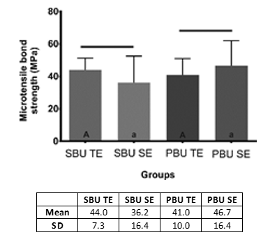
Fig. 3: Chart: Microtensile bond strength according to adhesive system (Single Bond Universal - SBU or Peak Bond Universal - PBU) and mode of use (etch-and-rinse - TE or self-etch - SE). The same capital letters indicate no difference between the adhesive systems used in the etch-and-rinse mode. Table: Arithmetic mean andstandard deviation of microtensile bond strength
Discussion
This study found no statistically significant differences in the bond strength values between the two adhesive systems used, as well as no statistically significant differences between the etch-and-rinse and self-etch techniques. Therefore, our working hypothesis can be accepted.
These results are consistent with various studies that show that nowadays, thanks to technological and scientific advances, statistically similar values are achieved with both etch-and-rinse and self-etch adhesive systems. Traditionally, some papers defended the superiority of etch-and-rinse systems over self-etch in dentin4-5,13. A few current studies show that the dentin self-etch technique allows forhigher adhesion values than the traditional etch-and-rinse technique12.
It has been shown that dentin bond strength values obtained through microtensile testing using two-step self-etch and total-etch adhesive systems presented statistically similar values, with the exception of one-step self-etch, which deviates from the values8. It is reported in the literature that one-step self-etch systems do not have good results in bond strength tests, possibly due to the combination of hydrophilic and hydrophobic acids and monomers in a single solution22.
One study concluded that, regardless of the substrate on which the systems are applied (superficial or deep dentin), bond strength was higher in the two-step self-etch system than in the one-step or conventional total-etch system22. In addition, self-etch adhesive systems are less reliable regarding bond strength and nanofiltration compared to three-step etch etch-and-rinse systems, which remain the gold standard for comparative studies1. Among self-etch systems, two-step systems have better clinical and laboratory performance than one-step systems. Additionally, self-etch adhesive systems perform better in dentin adhesion than in enamel23.
The use of universal adhesives is indicated for the total-etch and self-etch techniques, and according to the manufacturer, they are equally effective in any of the modes used. Recent studies have shown that during the acid-etch technique, matrix metalloproteinase enzymes (MMPs) are released from the dentin and can degrade the hybrid layer. In order to control the effect of MMPs, the use of different agents, including chlorhexidine, has been studied. This compound has antibacterial properties that inhibit the proteolytic activity of MMPs at very low concentrations, thus inhibiting hybrid layer degradation and achieving greater resin-dentin bond durability11,15.
This study compared the adhesive efficacy of two universal adhesive systems, one of which contains chlorhexidine in its composition. The results showed that the use of chlorhexidine does not influencebond strength24. These results suggest that adhesive systems containing 0.2% chlorhexidine can be used. Despite this result, it must be noted that the group that used the Peak Universal Bond as an adhesive system in self-etch mode showed a greater number of cohesive failures in dentin, which indicates a possible greater interaction of this adhesive system with the tooth substrate compared to that of other groups studied.
Although the use of chlorhexidine does not directly affect immediate bond strength, long-term outcomes may differ given its role as an MMP inhibitor. Another review studies including chlorhexidine in the first of some two-step self-etch adhesives. The authors found that during the adhesion protocol, it inhibits the action of MMPs to a certain extent and has no adverse effects on the immediate bond strength to the dental substrate 23.
In summary, both the aforementioned reviews and this study agree that chlorhexidine plays a major role in inhibiting MMPs without having adverse effects on the bond strength regardless of the adhesive system used, as the technique selected, etch-and-rinse or self-etch, shows no difference in bond strength. In order to expand the knowledge already created with this in vitro study, the authors recommend further in vitro studies in the medium and long term to monitor the presence of chlorhexidine in the adhesive interface and its effect on bond strength. Furthermore, once they are completed, we recommend conducting randomized clinical trials that can confirm the clinical performance of these adhesive systems to verify restoration durability and the bond interface obtained with the various systems.
It should be noted that this study has limitations in terms of the number of samples studied, since a greater n could decrease the standard deviation, which might allow us to detect a statistically significant difference. In addition, we must consider that bond strength, as the only factor taken into account does not provide comprehensive knowledge about the performance of an adhesive system; there are other in vitro methodologies such as microfiltration, bond strength tests after aging, and electron microscopy analysis of adhesive interfaces, among others, that would make it possible to have a complete notion of the laboratory performance of an adhesive system. Additionally, the strengths of this study are its methodological compliance with current ISO regulations, the standardization of samples and protocols, and the use of specific equipment, as well as data verification, as they are consistent with the existing literature, supporting the reliability of the published data.
Conclusions
Despite the limitations of this study, the performance of universal adhesive systems does not depend on how they are used in dentin. In addition, including chlorhexidine in a universal adhesive system did not significantly affect its performance.
Considering that chlorhexidine plays a major role in inhibiting MMPs, the use of such adhesives may prevent degradation of the hybrid layer and, therefore, increase the durability of the resin-dentin bond. Further studies should be conducted to confirm this.
Acknowledgments:
This study was funded by the Student Research Support Program (PAIE) of the Sectoral Commission for Scientific Research (CSIC).
REFERENCES
1. Correa MB, Peres MA, Peres KG, Horta BL, Barros AD, Demarco FF. Amalgam or composite resin? Factors influencing the choice of restorative material. J Dent. 2012; 40 (9): 703-10. [ Links ]
2. Bedran-Russo A, Leme-Kraus AA, Vidal CMP, Teixeira EC. An Overview of Dental Adhesive Systems and the Dynamic Tooth-Adhesive Interface. Dent Clin North Am. 2017; 61 (4): 713-731. [ Links ]
3. Sofan E, Sofan A, Palaia G, Tenore G, Romeo U, Migliau G. Classification review of dental adhesive systems: from the IV generation to the universal type. Ann Stomatol (Roma). 2017; 8 (1):1-17. [ Links ]
4. Tsujimoto A, Iwasa M, Shimamura Y, Murayama R, Takamizawa T, Miyazaki M. Enamel bonding of single-step self-etch adhesives: Influence of surface energy characteristics. J Dent. 2010; 38 (2):123-30. [ Links ]
5. Mandri MN, Aguirre Grabre de Prieto A, Zamudio ME. Sistemas adhesivos en Odontología Restauradora. Odontoestomatologia. 2015; 17 (26): 50-6. [ Links ]
6. Van Meerbeek B, Peumans M, Poitevin A, Mine A, Van Ende A, Neves A, De Munck J. Relationship between bond-strength tests and clinical outcomes. Dent Mater. 2010; 26 (2): 100-21. [ Links ]
7. Atash Biz Yeganeh L, Seyed Tabai E, MohammadiBasir M. Bonding Durability of Four Adhesive Systems. J Dent (Tehran). 2015; 12 (8): 563-70. [ Links ]
8. Dourado Loguercio A, Reis A. Sistemas Adhesivos. RevOperDent y BiomaterDent y Biomater. 2006; 1 (2): 13-28 [ Links ]
9. Hernández JM. Aspectos prácticos de la adhesión a dentina Practicalaspects in dentinaladhesion. AvOdontoestomatol. 2004; 20 (1): 19-32. [ Links ]
10. Ceballos L, Camejo DG, Victoria Fuentes M, Osorio R, Toledano M, Carvalho RM, et al. Microtensile bond strength of total-etch and self-etching adhesives to caries-affected dentine. J Dent. 2003; 31 (7): 469-77. [ Links ]
11. Carrilho MRO, Geraldeli S, Tay F, de Goes MF, Carvalho RM, Tjäderhane L, et al. In vivo preservation of the hybrid layer by chlorhexidine. J Dent Res. 2007; 86 (6): 529-33. [ Links ]
12. Oliveira SSA, Pugach MK, Hilton JF, Watanabe LG, Marshall SJ, Marshall GW. The influence of the dentin smear layer on adhesion: A self-etching primer vs. a total-etch system. Dent Mater. 2003; 19 (8): 758-67. [ Links ]
13. Scherrer SS, Cesar PF, Swain M V. Direct comparison of the bond strength results of the different test methods: a critical literature review. Dent Mater. 2010; 26 (2): e78-93. [ Links ]
14. Bader Mattar M, Ibáñez Musalem M. Evaluación de la interfase adhesiva obtenida en restauraciones de resina compuesta realizadas con un sistema adhesivo universal utilizado con y sin grabado ácido previo. Rev Clínica Periodoncia, Implantol y Rehabil Oral. 2014; 7 (3): 115-22. [ Links ]
15. Pomacóndor-Hernández C. Papel de la clorhexidina en la odontología restauradora. OdontolSanmarquina. 2010; 13 (2): 46-9. [ Links ]
16. Tekçe N, Tuncer S, Demirci M, Balci S. Do matrix metalloproteinase inhibitors improve the bond durability of universal dental adhesives? Scanning. 2016; 38 (6): 535-44. [ Links ]
17. Vola J. Influencia de los inhibidores de las metaloproteinasas, agentes reticuladores y remineralizaciónbiomimética en la longevidad de la unión adhesiva. Actas Odontológicas. 2014; XI (2): 10-21. [ Links ]
18. Pashley DH, Carvalho RM, Sano H, Nakajima M, Yoshiyama M, Shono Y, et al. The microtensilebond test: a review. J Adhes Dent. 1999; 1 (4): 299-309. [ Links ]
19. International Organization for Standarization. ISO 4049:2009 Dentistry Polymer based restorative materials. 2009. [ Links ]
22. Sanchez F, Osorio R, Toledano M. Control del colapso del colágeno: sistemas autograbadores. AvOdontoestomatol. 2004; 20 (4): 175-83. [ Links ]
23. Parra Lozada M, Rayo HG. Sistemas adhesivos autograbadores, resistencia de unión y nanofiltración: una revisión self-etchingadhesivesystems, bond strength and nanofiltration: a review. Rev Fac Odontol Univ Antioquia. 2012; 24 (24): 133-150. [ Links ]
24. Roopa D ARS, D R. Comparative Evaluation of the Bonding Efficacy of Seventh Generation Bonding Agent and Peak Universal Bond: An In-Vitro Study. JBR J Interdiscip Med Dent Sci. 2015; 03 (02): 1-4. [ Links ]
Authorship contribution
1.Conception and design of study
2.Acquisition of data
3.Data analysis
4.Discussion of results
5.Drafting of the manuscript
6.Approval of the final version of the manuscript
RT has contributed in: 1, 2, 4, 5
CS has contributed in: 1, 2, 4, 5
PV has contributed in: 1, 2, 4, 5
MM has contributed in: 4, 5
AG has contributed in: 4, 5
CECS has contributed in: 3, 4
GG has contributed in: 1, 3, 4, 6
Received: September 20, 2019; Accepted: January 30, 2020
 Este es un artículo publicado en acceso abierto bajo una licencia Creative Commons
