Servicios Personalizados
Revista
Articulo
Links relacionados
Compartir
Odontoestomatología
versión impresa ISSN 0797-0374versión On-line ISSN 1688-9339
Odontoestomatología vol.21 no.33 Montevideo jun. 2019 Epub 01-Jun-2019
https://doi.org/10.22592/ode2019n33a10
Case Report
Cephalometric comparison between an acromegalic patient and his twin brother
1Práctica Privada, Santiago, Chile, tomasfreundlich@gmail.com
2 Práctica Privada, Santiago, Chile
3 Unidad de Física, Instituto de Investigación en Ciencias Odontológicas y Centro de Análisis Cuantitativo en Antropología Dental, Facultad de Odontología, Universidad de Chile. Departamento de Antropología, Facultad de Ciencias Sociales, Universidad de Chile, Santiago, Chile
4 Servicio de Cirugía Máxilo Facial, Hospital San Borja Arriarán. Departamento del Niño y Ortopedia Dento Maxilar, Facultad de Odontología, Universidad de Chile. Centro de Análisis Cuantitativo en Antropología Dental, Facultad de Odontología, Universidad de Chile, Santiago, Chile
Acromegaly is characterized by a slowly progressive somatic disfigurement caused by the overproduction of growth hormone (GH) and insulin-like growth factor 1 (IGF 1), mainly associated with a pituitary adenoma. The most evident facial manifestation is mandibular prognathism due to excessive growth of the jaw. This work aimed to perform a craniofacial morphological comparison through cephalometric analysis and cephalometric superimposition of a patient diagnosed with acromegaly and his twin brother without the disease. Our results showed that the acromegalic patient has a significant increase in the size of the sella turcica, an anterior displacement of the maxilla and mandible, the mandibular displacement being more marked. The morphological change of the mandible in acromegaly is mainly attributed to the growth of the mandibular ramus due to an increase in the condylar unit.
Keywords: acromegaly; twins; cephalometry; mandibular condyle
La acromegalia es una enfermedad caracterizada por una desfiguración somática de progresión lenta causada por la sobreproducción de hormona de crecimiento (GH) y factor de crecimiento insulinoide tipo 1 (IGF-1), predominantemente asociada con un adenoma hipofisario. La manifestación más evidente a nivel facial es un prognatismo mandibular por exceso de crecimiento de la mandíbula. El propósito del presente trabajo fue realizar una comparación morfológica craneofacial mediante análisis cefalométrico y superposición cefalométrica entre un paciente con diagnóstico de acromegalia y su hermano gemelo que no presenta la enfermedad. Nuestros resultados mostraron que en el hermano con acromegalia existe un significativo aumento del tamaño de la silla turca, un desplazamiento hacia anterior del maxilar y mandíbula, siendo más marcado el desplazamiento mandibular. El cambio morfológico que experimenta la mandíbula en la acromegalia es atribuido principalmente al crecimiento de la rama mandibular por aumento de la unidad condilar.
Palabras clave: acromegalia; gemelos; cefalometría; cóndilo mandibular
Introduction
Acromegaly is a low-prevalence disease that affects between 40 and 70 out of 1,000,000 people. It begins in adulthood and is characterized by a slowly progressive somatic disfigurement caused by the overproduction of growth hormone (GH) and insulin-like growth factor 1 (IGF 1), mainly associated with a pituitary adenoma1-2. Growth hormone is produced in the anterior portion of the pituitary gland and stimulates IGF-1 production, mainly in the liver. Both hormones stimulate the growth of tissues3. The disease may occur in an isolated form or be part of a syndromic presentation such as McCune Albright syndrome4. It is associated with premature mortality if not properly treated5.
The clinical manifestations of acromegaly are both local and general. In the first case, because of the increase in size of the pituitary gland, neighboring tissues are compressed, which may trigger headaches and visual alterations6. At a general level, the increase in GH and IGF-1 secretion can lead to cardiovascular, respiratory and metabolic systemic complications7-9. Morphologically, there is an increase in the size of the extremities, widening of the fingers, thickening of the skin, protuberance of the forehead and thickening and increase of the size of the lips, nose and ears8-9.
The most evident facial manifestation is mandibular prognathism due to excessive growth of the jaw. It is not clear which area of the jaw might be most affected. It has been pointed out that the main growth might occur in the ramus10-11. However, in addition to the ramus, the body and chin might also show greater growth12. Cephalometrically, a decrease in the S-Ar-Go angle, an increase in the SNB angle and an increase in the goniac angle are described in acromegalic patients10-11,13. Apparently, the maxilla would not show significant changes14. It has been proposed, through experimentation in rats, that excessive jaw growth could be the result of an IGF-1 stimulation on the cartilage of the mandibular condyle15.
A clinical finding normally seen in this disease is macroglossia16-18. There appears to be a greater sensitivity of the tongue muscles to IGF-1 compared to other muscles and tissues15. Postmortem studies show that there would be an enlargement of muscle fibers in acromegalic patients compared to their control counterparts19. Diastemas and increased tooth mobility have also been reported as characteristic signs20-21. However, these clinical signs appear to be secondary to the enlargement of the tongue20,22.
This work aims to expand the knowledge on acromegaly and its related facial morphological changes by comparing a patient diagnosed with acromegaly and his twin brother without the disease. We conducted a morphological radiographic comparison between the twins.
Method
A 27-year-old male patient was referred by the Endocrinology Department with a diagnosis of acromegaly by pituitary adenoma to the Maxillofacial Surgery Service of the San Borja Arriarán Hospital in Santiago de Chile for corrective treatment of facial deformity. In the medical history, the patient said he had a twin brother. For this reason, both brothers were asked for consent to conduct this study. The twin brother voluntarily agreed to a lateral craniofacial radiograph. Both standardized tests were performed under the same conditions and with the same radiological equipment (Planmeca Promax).
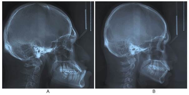
Both radiographs were obtained (Fig. 1), and a cephalometric analysis was performed using some angular measures commonly used in orthodontics13. Subsequently, two cephalometric superimpositions were conducted: one general and one mandibular, both based on the method proposed by Björk23-25. This allowed us to see the areas with the greatest craniofacial morphological change in the acromegalic patient with respect to his twin brother.
Results
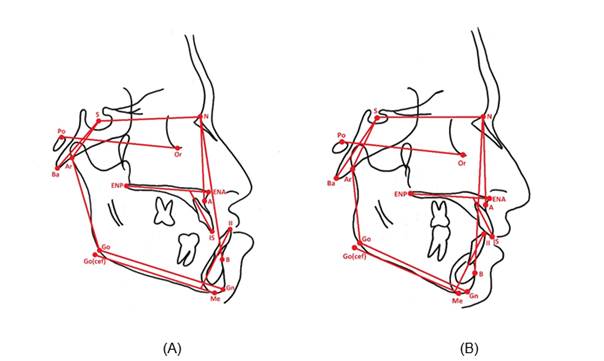
Table 1 summarizes the cephalometric values of both twins and the differences between them. The Ba-S-N angle was larger in the acromegalic patient (136°) compared to his twin brother (126°). Something similar occurred with the SNB values (96° vs. 85°). There were no significant changes in the SNA angle (89° vs. 90°). The S-Ar-Go angle was reduced in the acromegalic patient (120°) compared to his brother (146°). The ANB angle was greatly altered in the acromegalic patient (-7º) with a difference of 12° compared to his twin brother (5º). The S-N/Go-Gn angle was reduced by 5° compared to his brother (20° vs. 25°). Finally, the goniac angle was increased by 5° (125° vs. 120°). Dentally, there was a greater proinclination of the upper incisors in the acromegalic patient (130° vs. 126°). Fig. 2 shows the cephalometric tracings of both the acromegalic patient and his twin brother.
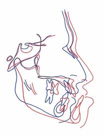
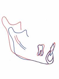
Fig. 3 shows the general superimposition based on Björk’s method, using the anterior skull base as a reference, where the best fit was sought between the anterior wall of the Turkish chair, the anterior clinoid process and the cribous lamina of the ethmoid. This superimposition shows a significant increase in the size of the sella turcica in the acromegalic patient with a deepening of the floor and excavation of the anterior wall. Both the maxilla and the mandible present an anterior displacement in the acromegalic patient, the mandibular displacement being more pronounced, causing the characteristic mandibular prognathism. As for the maxilla, the superimposition shows an increase in the distance between the anterior and posterior nasal spine. There is also an increase in the size of the glabellar area and a moderate anterior and inferior displacement of the nasal fronts, nasal bones and upper maxilla. The soft tissues are prominent in the acromegalic patient in relation to his brother. Fig. 4 shows a mandibular superimposition, also based on Björk’s method, but this time looking for the best fit between the anterior contour of the mentonian symphysis (foremost point), the inferior contour of the internal cortex of the mentonian symphysis (lowest point) and the canal of the inferior dental nerve. In this superimposition, we can see an increase in the size of the ramus, the coronoid process and the mandibular condyle.
Table 1: Cephalometric values of both twins and the differences between them
| Measures | With acromegaly | Without the disease | Difference |
|---|---|---|---|
| Ba-S-N | 126º | 136º | +10º |
| SNA | 90º | 89º | -1º |
| S-N/ENA-ENP | 8º | 9º | +1º |
| SNB | 85º | 96º | +11º |
| S-Ar-Go | 146º | 120º | -26º |
| ANB | 5º | -7º | -12º |
| S-N/Go-Gn | 25º | 20º | -5º |
| Po-Or/Me-Go(cef) | 20º | 12º | -8º |
| Ar-Go-Gn | 120º | 125º | +5º |
| Po-Or/Me-Go(cef) | 18º | 12º | -6º |
| IS - ENA-ENP | 126º | 130º | +4º |
| Po-Or/Me-Go(cef) | 95º | 95º | 0º |

Fig. 3: General cephalometric superimposition between the brothers based on the method proposed by Björk. The red lines represent the acromegalic brother; the blue ones, the brother without the disease
Discussion
This work aimed to make a craniofacial morphological comparison through cephalometric analysis of a patient diagnosed with acromegaly and his twin brother without the disease. According to the review of the relevant literature, this work is the first study with this objective.
It assumes that there is a high phenotypic concordance between twins. It is generally known that the phenotype is determined by a gene-environment interaction26. Therefore, in our case, although they are twins, they could still present certain morphological differences attributed to the environment27. Nevertheless, there are reports such as Sidlauskas et al 28 where 90 monozygotic twins were studied and a high correlation was found between them in the total mandibular size and the body and ramus size. Likewise, Manjusha et al. 29 found a craniofacial morphological correlation between monozygotic twins, concluding that the genetic component plays a dominant role in craniofacial morphology. Based on these reports of phenotypic concordance, we performed a cephalometric comparison between these two twins. For this reason, we believe that the results obtained in this study are attributable to the effect that acromegaly has on facial morphology. Although we could not categorically rule out morphological differences between the two brothers before the diagnosis of the disease, our work contributes to the description of acromegalic facial deformities
This increase in the size of the sella turcica shown in the general superimposition is caused by a deepening of the floor and excavation of the anterior wall, and is undoubtedly due to the increase in the size of the pituitary gland given the existing adenoma 30. This would explain the increase of the Ba-S-N angle observed in the acromegalic patient, since this deepening of the sella turcica would be conditioning a lower location of the sella point (S) and consequently this angle would increase.
The excessive growth of the jaw and consequent prognathism observed in the acromegalic patient compared to his twin brother was reflected in an increase in the SNB angle, a decrease in the S-Ar-Go angle and an increase in the goniac angle. This is broadly in line with the literature10-11,13. The mandibular superimposition showed a growth of the ramus, the condylar unit and the coronoid unit. Interestingly, the mandibular notch moved upward along with the condyle and the coronoid process. Fariña et al.31 reported a similar finding for condylar hyperplasia, in which the mandibular notch accompanies the growth of the condyle on the affected side. Although there are autopsy studies of acromegalic patients, these focus on other organs or tissues and do not account for what happens with the mandibular condyle19,32. Likubo et al. 15 suggest, through experimentation in rats, that this increase in the mandibular size is the result of an IGF-1 simulation on the cartilage of the condyle. The role of the tongue as stimulating mandibular growth factor in the acromegalic patient has also been discussed by some authors33-35, who considered certain syndromes such as Beckwith-Wiedemann Syndrome, where the excessive growth of the jaw would be a consequence of macroglossia36. When macroglossia occurs in growing children, the tongue would act as a mechanical stimulus on the cartilage of the mandibular condyle causing prognathism37. However, unlike what happens in some syndromes during growth, in the acromegalic patient, macroglossia should not mean a mechanical stimulus leading to an increased size of the mandible, since in adulthood, the cartilage of the condyle would be inactive2. Excessive growth of the jaw in acromegaly could be explained by the presence of cartilaginous cells in the adult condyle that can potentially be reactivated by an IGF-1 increase. However, we cannot discard that once these cells have been stimulated, they cannot receive an additional stimulus from macroglossia.
The general superimposition showed a characteristic change of acromegaly over the middle third of the facial skeleton that consists in the increase of the glabellar frontal area, which coincides with what Balos Tuncer et al.38) reported. Together with this, there was an anterior and inferior displacement of the nasal bones and the upper jaw, which could be explained by the response of the cartilage of the nasal septum to the IGF-139, causing a growth stimulus of the middle third of the face. This anterior displacement of the maxilla was accompanied by an increase in the distance between the anterior and posterior nasal spine in the acromegalic patient, which would entail the real growth of the maxillary bone. This agrees with what was reported by Balos Tuncer et al.38. However, these authors also found that the SNA angle did not undergo significant changes. Other authors, using this same angle as an indicator, conclude that the maxilla does not present morphological changes in the acromegalic patient12,14. The SNA angle is normally considered indicative of an anterior displacement of the maxilla during the growth. In our work, the brothers’ SNA angles were similar. If we had used only the SNA angle, we would have concluded that acromegaly does not cause changes in the maxilla. However, the general superimposition clearly shows an anterior displacement of this structure. The almost null variation of the SNA angle between both twins is explained by the fact that both the N point (frontonasal suture) and the A point experienced an anterior displacement, which tends to maintain the SNA angle at similar values.
Conclusion
The morphological change of the mandible in acromegaly is mainly attributed to the growth of the mandibular ramus due to an increase in the condylar unit. It is believed that the excessive growth of the jaw could be attributed to IGF-1 stimulation on the condylar cartilage. There is no full consensus on what happens with the maxilla
Referencias
1. Holdaway IM, Rajasoorya C. Epidemiology of acromegaly. Pituitary. 1999; 2 (1): 29-41. [ Links ]
2. Chanson P, Salenave S. Acromegaly. Orphanet J Rare Dis. 2008; 3: 17. [ Links ]
3. Rozario KS, Lloyd C, Ryan F. GH and IGF-1 Physiology In Childhood. En: De Groot LJ, Chrousos G, Dungan K. Endotext. Internet. South Dartmouth (MA): MDText.com, Inc. 2000. Actualizado: 2015 Nov 20; Acceso: 2018 Ago 14. Disponible en: https://www.ncbi.nlm.nih.gov/books/NBK343487/. [ Links ]
4. Salenave S, Boyce AM, Collins MT, Chanson P. Acromegaly and McCune-Albright síndrome. J Clin Endocrinol Metab. 2014; 99 (6): 1955-69. [ Links ]
5. Galoiu S, Poiana C. Current therapies and mortality in acromegaly. J Med Life. 2015; 8 (4): 411-5. [ Links ]
6. López-Macía A, Picó-Alfonso A. Clínica de la acromegalia: presentación, cuadro clínico y comorbilidades. Endocrinol Nutr. 2005; 52 Supl 3: 18-22. [ Links ]
7. Melmed S. Acromegaly. N Engl J Med. 1990; 322 (14): 966-77. [ Links ]
8. Chanson P, Salenave S, Kamenicky P, Cazabat L, Young J. Pituitary tumours: acromegaly. Best Pract Res Clin Endocrinol Metab. 2009; 23 (5): 555-74. [ Links ]
9. Vilar L, Vilar CF, Lyra R, Lyra R, Naves LA. Acromegaly: clinical features at diagnosis. Pituitary. 2017; 20 (1): 22-32. [ Links ]
10. Bruwier A, Albert A, Beckers A, Limme M, Poirrier R. Acromegaly and sleep apnea: cephalometric evaluations. Ann Endocrinol (Paris). 2011; 72 (3): 211-7. [ Links ]
11. Karakis D, Aktas-Yilmaz B, Dogan A, Yetkin I, Bek B. The bite force and craniofacial morphology in patients with acromegaly: a pilot study. Med Oral Patol Oral Cir Bucal. 2014; 19 (1): e1-7. [ Links ]
12. Künzler A, Farmand M. Typical changes in the viscerocranium in acromegaly. J Craniomaxillofac Surg. 1991; 19 (8): 332-40. [ Links ]
13. Dostálová S, Sonka K, Smahel Z, Weiss V, Marek J. Cephalometric assessment of cranial abnormalities in patients with acromegaly. J Craniomaxillofac Surg. 2003; 31 (2): 80-7. [ Links ]
14. Pelttari L, Polo O, Rauhala E, Vuoriluoto J, Aitasalo K, Hyyppä MT, Kronholm E, Irjala K, Viikari J. Nocturnal breathing abnormalities in acromegaly after adenomectomy. Clin Endocrinol (Oxf). 1995; 43 (2): 175-82 [ Links ]
15. Likubo M, Kojima I, Sakamoto M, Kobayashi A, Ikeda H, Sasano T. Morphological and histopathological changes in orofacial structures of experimentally developed acromegaly-like rats: an overview. Int J Endocrinol. 2012; 2012: 254-367 [ Links ]
16. Agrawal M, Maitin N, Rastogi K, Bhushan R. Seeing the unseen: diagnosing acromegaly in a dental setup. BMJ Case Rep. 2013. [ Links ]
17. Smith CB, Waite PD. Surgical management of obstructive sleep apnea in acromegaly with mandibular prognathism and macroglossia: a treatment dilemma. J Oral Maxillofac Surg. 2012; 70 (1): 207-10. [ Links ]
18. Kashyap RR, Babu GS, Shetty SR. Dental patient with acromegaly: a case report. J Oral Sci. 2011; 53 (1): 133-6. [ Links ]
19. Wittmann AL. Macroglossia in acromegaly and hypothyroidism. Virchows Arch A Pathol Anat Histol. 1977; 373 (4): 353-60. [ Links ]
20. Lima DL, Montenegro RM Jr, Vieira AP, Albano MF, Rego DM. Absence of periodontitis in acromegalic patients. Clin Oral Investig. 2009; 13 (2): 165-9. [ Links ]
21. Herrmann BL, Mortsch F, Berg C, Weischer T, Mohr C, Mann K. Acromegaly: a cross-sectional analysis of the oral and maxillofacial pathologies. Exp Clin Endocrinol Diabetes. 2011; 119 (1): 9-14. [ Links ]
22. Cortet-Rudelli C. The mouth of patients with acromegaly. Presse Med. 2017; 46 (9): 831-837. [ Links ]
23. Björk A. Variations in the growth pattern of the human mandible: longitudinal radiographic study by the implant method. J Dent Res. 1963; 42 (1) Pt 2: 400-11. [ Links ]
24. Skieller V, Björk A, Linde-Hansen T. Prediction of mandibular growth rotation evaluated from a longitudinal implant sample. Am J Orthod. 1984; 86 (5): 359-70. [ Links ]
25. Roldan SI, Carvajal CM, Rey D, Buschang PH. Método de superposición estructural de Björk para evaluar crecimiento y desarrollo craneofacial. Rev. CES Odont. 2013; 26 (2): 127-133. [ Links ]
26. Griffiths AJF, Miller JH, Suzuki DT, Lewontin RC, Gelbart WM. An Introduction to Genetic Analysis. 7 ed. New York, 2000. [ Links ]
27. Fraga MF, Ballestar E, Paz MF, Ropero S, Setien F, Ballestar ML, Heine-Suñer D, Cigudosa JC, Urioste M, Benitez J, Boix-Chornet M, Sanchez-Aguilera A, Ling C, Carlsson E, Poulsen P, Vaag A, Stephan Z, Spector TD, Wu YZ, Plass C, Esteller M. Epigenetic differences arise during the lifetime of monozygotic twins. Proc Natl Acad Sci U S A. 2005; 102 (30): 10604-9. [ Links ]
28. Šidlauskas M, Šalomskienė L, Andriuškevičiūtė I, Šidlauskienė M, Labanauskas Ž, Šidlauskas A. Mandibular morphology in monozygotic twins: a cephalometric study. Stomatologija. 2014; 16 (4): 137-43. [ Links ]
29. Manjusha KK, Jyothindrakumar K, Nishad A, Manoj KM. Growth and Development of Dentofacial Complex influenced by Genetic and Environmental Factors using Monozygotic Twins. J Contemp Dent Pract. 2017; 18 (9): 754-758. [ Links ]
30. Chang HP, Tseng YC, Chou TM. An enlarged sella turcica on cephalometric radiograph. Dentomaxillofac Radiol. 2005; 34 (5): 308-12. [ Links ]
31. Fariña R, Bravo R, Villanueva R, Valladares S, Hinojosa A, Martinez B. Measuring the condylar unit in condylar hyperplasia: from the sigmoid notch or from the mandibular lingula?. Int J Oral Maxillofac Surg. 2017; 46 (7): 857-860. [ Links ]
32. Gershberg H, Heinemann HO, Stumpf HH. Renal function studies and autopsy report in a patient with gigantism and acromegaly. J Clin Endocrinol Metab. 1957; 17 (3): 377-85. [ Links ]
33. Ardran GM, Kemp FH. The tongue and mouth in acromegaly. Clin Radiol. 1972; 23 (4): 434-44. [ Links ]
34. Benda C. Akromegalie. Deutsche Klinik am Eingange Des Zwanzigsten Jahrhunderts. 1902; 3: 261. [ Links ]
35. Chalk WO. Partial dislocation of the lower jaw from an enlarged tongue. Transactions of the Pathological Society. 1856; 8: 305-308. [ Links ]
36. Kawafuji A, Suda N, Ichikawa N, Kakara S, Suzuki T, Baba Y, Ogawa T, Tsuji M, Moriyama K. Systemic and maxillofacial characteristics of patients with Beckwith-Wiedemann syndrome not treated with glossectomy. Am J Orthod Dentofacial Orthop. 2011; 139 (4): 517-25. [ Links ]
37. Argandoña J, Pantoja R, Cortés J. Rol de la lengua en la génesis de Dismorfosis Maxilares (I parte). Revista Dental de Chile. 1998; 89 (1): 37-42. [ Links ]
38. Balos Tuncer B, Canigur Bavbek N, Ozkan C, Tuncer C, Eroglu Altinova A, Gungor K, Akturk M, Balos Toruner F. Craniofacial and pharyngeal airway morphology in patients with acromegaly. Acta Odontol Scand. 2015; 73 (6): 433-40. [ Links ]
39. Vetter U, Zapf J, Henrichs I, Gammert C, Heinze E, Pirsig W. Human nasal septal cartilage: analysis of intracellular enzyme activities, glycogen content, cell density and clonal proliferation of septal chondrocytes of healthy adults and acromegalic patients. Connect Tissue Res. 1989; 18 (4): 243-54 [ Links ]
Received: October 08, 2018; Accepted: December 12, 2018


 texto en


