


Serviços Personalizados
Journal

Artigo
 texto em
  Inglês (pdf)
 Artigo em XML
 Referências do artigo
Links relacionados
Compartilhar

 Permalink
Odontoestomatología
versão impressa ISSN 0797-0374versão On-line ISSN 1688-9339
Odontoestomatología vol.20 no.31 Montevideo jun. 2018
https://doi.org/10.22592/ode2018n31a10
Case report
Skeletonized body identified by analysis of frontal sinus morphology and characteristics of osteosynthesis material: a forensic case report
1
 http://orcid.org/0000-0002-3680-7020
2
http://orcid.org/0000-0002-1290-3755
3
http://orcid.org/0000-0001-5256-9811
4
http://orcid.org/0000-0002-6218-4015
5
http://orcid.org/0000-0001-9250-9282
1 Professor of Forensic Dentistry, School of Dentistry, Universidad Federal de Goiás. Criminal Expert at the Scientific Police of Goiás (Goiânia, Goiás, Brazil).
2 Undergraduate Student, School of Dentistry, Universidad Federal de Goiás (Goiânia, Goiás, Brazil)
3 Undergraduate Student, School of Dentistry, Universidad Federal de Goiás (Goiânia, Goiás, Brazil)
4 Postgraduate Student, School of Dentistry, Universidad Federal de Goiás (Goiânia, Goiás, Brazil)
5 Specialist in Dental Radiology, Brazilian Dentistry Association, Goiás Section (Goiânia, Goiás, Brazil)
6 Associate Professor at the Patient Admission and Registry Department, School of Dentistry, Universidad de la República (Montevideo, Uruguay)
7 PhD Student of Anatomy, School of Dentistry of Piracicaba, Universidad Estadual de Campinas (Sao Paulo, Brazil)
Forensic dentistry is essential for the identification of highly decomposed and charred bodies, as well as skeletal remains. This study reports a case of human identification by analyzing the morphology of the frontal sinuses and osteosynthesis material. In the anthropological assessment of skeletal remains a surgical plate used for osteosynthesis was detected in the periorbital regions. Relatives of the potential victim provided ante-mortem (AM) radiographs which revealed the presence of an osteosynthesis plate. Post-mortem (PM) imaging exams were performed to reproduce the AM data. Similarities were observed between the AM and PM radiographs, especially regarding the morphology of the frontal sinuses and the position and outline of the surgical plate used for osteosynthesis. The comparison of AM and PM images made it possible to identify the victim and to aid the criminal investigation. It also highlighted the role of radiographs and anatomical characteristics in the process of human identification.
Keywords forensic dentistry; forensic anthropology; frontal sinus; radiography
La odontología legal resulta fundamental para la identificación de víctimas en descomposición avanzada, carbonizadas o esqueletizadas. Este trabajo relata un caso de identificación humana, por medio de análisis morfológico del seno frontal y material de osteosíntesis. Un cuerpo esqueletizado fue sometido a examen antropológico, evidenciándose la existencia de tres placas de osteosíntesis en ambas regiones periorbitarias. Parientes de la supuesta víctima aportaron radiografías ante-mortem, en las que se aprecia una sola placa de osteosíntesis. Empero, tras la realización de exámenes imagenológicos del esqueleto, con la misma incidencia de los efectuados ante-mortem, se constató una clara semejanza morfológica del seno frontal y de la única placa de osteosíntesis presente en las imágenes ante-mortem y post-mortem. Así, fue posible identificar positivamente a la víctima, allanando el camino a la investigación criminal y realzando el rol de las imágenes radiográficas y caracteres anatómicos en el proceso de identificación humana.
Palabras-clave Odontología Legal; Antropología Forense; Seno Frontal; Radiografía
Introduction
With the rise of urban violence, traffic accidents and crime, the number of charred, mutilated or highly decomposed bodies and skeletal remains that reach forensic medicine departments around the world has increased significantly1,2.
The cause of death and the identity of the victim could be difficult to determine in such conditions, as when soft tissues are destroyed it is not possible to conduct a fingerprint analysis1,3. This is why forensic anthropology and forensic dentistry play a major role in criminal investigations, where the relatives of the missing person will be interviewed and asked to provide medical and dental records, such as clinical records, radiographs, CT scans, cast models and pictures to aid in the human identification process1,3-6.
Among the many methods available it is worth mentioning those comparing AM and PM radiographic imaging of the skull4, as the morphological variability of its many anatomical structures, like the frontal sinus, is very useful in forensics to determine a person’s identity7.
The specialized literature presents cases, although few, where human remains were identified by analyzing the frontal sinus7,8, comparing images of the anatomical characteristics of the sinus and those of the osteosynthesis material9. The aim of this study is to present a forensic case report of a skeletonized body identified by analysis of frontal sinus morphology and the type and location of the material used to reduce trauma-induced fractures.
Case description
A skeletonized body was found in a forested region in the State of Goiás, Central-West region of Brazil. The forensic anthropological analysis (biological profiling) and cause of death determination were conducted after the examination at the scene had taken place and the remains were transported to the Institute of Forensic Medicine (IML) in the state capital.
During the external examination of the body we observed the pelvis and skull had male characteristics10, closed medial clavicular epiphysis11, no osteophytes in the vertebral bodies12, sagittal suture with few points of synostosis and third molars with full rhizogenesis, so we determined the age to be between 30 and 40. We estimated the height (between 1.67 m and 1.74 m) by measuring the length of the long bones of the upper and lower limbs and by using Trotter and Gleser’s13 1952 table. There were three osteosynthesis metal plates on the skull, fixed with screws, which suggests the occurrence of AM trauma (characteristics and location shown in Table 1 and Fig. 1).
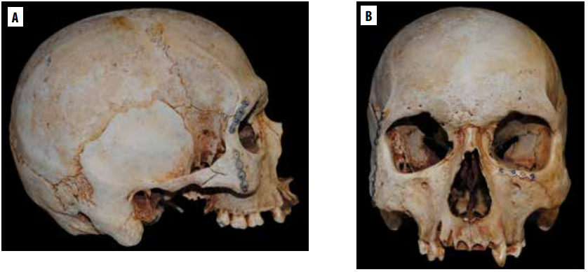
Fig. 1: right-hand side (A) and front (B) of the skull, with male anthropological features and plates, fixed with screws, in the periorbital region, bilaterally.
Table 1: Characteristics of the osteosynthesis material found in the skull
| Plate: | Anatomical Region | Shape of the plate | Number of fixing screws | Presence of bone on the plate |
|---|---|---|---|---|
| 1 | Lateral wall of the right orbital ridge | Linear | 4 | No |
| 2 | Body of the right malar bone | Linear | 4 | Yes |
| 3 | Left infraorbital rim | Linear | 3 | Yes |
The cause of death was determined to be traumatic brain and face injury by means of a perforating, blunt instrument. There were transfixing wounds caused by firearms, with entry holes in the occipital and right jaw regions (Fig. 2).
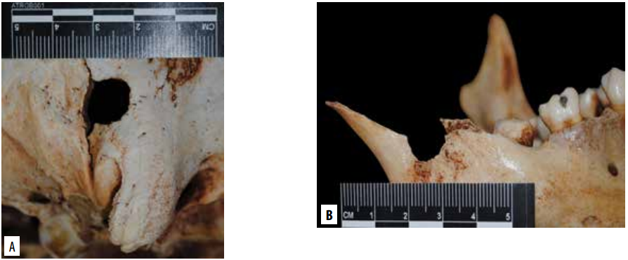
Fig. 2: Entry gunshot holes on the right-hand side of the occipital region (A) and right ramus (B), fractured.
In addition to expert examinations, police investigations revealed that the victim was probably a 29-year old man who had been kidnapped seven months earlier. The AM documentation submitted entailed a medical form and two posteroanterior (PA) skull radiographs (one presurgical and one immediately after surgery), taken seven years before his disappearance, which showed a fracture and an osteosynthesis plate in the right periorbital region. These also showed the morphology of the frontal sinus: expanded to both sides of the midline (Fig. 3).
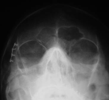
Fig. 3: AM postsurgical PA radiograph of the skull, taken immediately after the first osteosynthesis plate was fitted.
We decided to conduct PM exams, a PA radiograph and a cranial cone-beam CT (Fig. 4). This was done at a radiological laboratory that has signed an agreement with the IML. InVesalius® software was used to review the PM CT. The cranial image was reconstructed, rotated and processed in the same position as the AM immediate postsurgical PA radiograph.
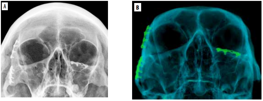
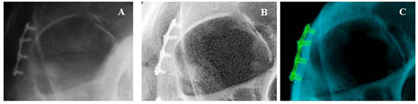
After that, we compared the AM immediate postsurgical PA radiograph to the PA radiograph and PM CT. We found morphological similarity of the frontal sinus in terms of its area of expansion, number and location of the septa and lobes (Table 2) and the osteosynthesis plate present in the lateral wall of the right orbit, anatomic location, type and fixing screws (number, design, length, arrangement and fixing angle), as shown in Fig. 5. This made it possible to positively identify the missing person.
Table 2: Comparison of frontal sinus characteristics in AM and PM radiographs.
| Feature | AM radiograph | PM radiograph | Similarity |
|---|---|---|---|
| Bilateral Expansion | Yes | Yes | Yes |
| Greater expansion area | Left-hand side | Left-hand side | Yes |
| Number of lobes on the right-hand side | 2 | 2 | Yes |
| Number of lobes on the left-hand side | 3 | 3 | Yes |
| Presence of median septum | Yes | Yes | Yes |
| Presence of intermediate septa | No | No | Yes |
Discussion
Radiograph images are widely used for identification purposes, especially when a lophoscopy analysis cannot be performed, and are very useful in cases of charred, decaying and skeletonized bodies4,5,9,14.
This is further enhanced by the number of skull x-rays performed on a daily basis, as the population has increased access to dental care, to diagnose, plan and/or check different clinical treatments5,15).
In addition, the skull is highly resistant to environmental conditions and thus has demonstrated great forensic potential. In fact, PA radiographs make it possible to study the anatomical features (dental or bone), pathological and/or therapeutic characteristics, as well as the type of a possible osteosynthesis material9.
However, we should remember that PA radiographs are not the best alternative for a detailed dental analysis, if compared to panoramic5 or intraoral1) radiographs. These should be used with caution to avoid misleading or inconsistent conclusions. In return, they become valuable in a morphologic, qualitative and quantitative assessment of the frontal sinus, bearing in mind that this expansive cavity is unique and does not change in adults7-9,16,17.
In this case, the morphological similarity between AM and PM x-ray images of the frontal sinus, comparative procedure widely supported and described in the literature(7-9,16.17), made the positive identification of the victim possible, paving the way for the criminal investigation to arrest and prosecute those involved in his death. The presence of the osteosynthesis plate on the lateral wall of the right orbit was an additional element for comparison, with results consistent with those of the frontal sinus9.
Finally, it should be mentioned that this method is faster and less expensive than a DNA test, showing how important it is to have a radiology service in the IMLs or, if this were not possible, to enter into agreements with public or private radiological laboratories. It is also necessary to have forensic dentists with the right expertise and training in imaging techniques, especially extraoral x-rays and CTs, usually used for criminal investigation purposes.
Referencias
1. Silva RF, Almeida SM, Daruge Júnior E, Pereira SDR, Oliveira RN. Identificação de cadáver carbonizado utilizando documentação odontológica. Rev. Odonto Ciên. 2008; 23 (1): 90-93. [ Links ]
2. Tavares R, Catalan VDB, Romano PMM, Melo EM. Homicídios e vulnerabilidade social. Ciên. Saúde Coletiva. 2016; 21 (3): 923-934. [ Links ]
3. Silva RF, Portilho CDM, Reges RV, Leles CR, Freitas GC, Daruge Júnior E. Importância pericial dos registros odontológicos decorrentes de tratamento restaurador. Rev. Dental Press Estét. 2007; 4 (4): 32-38. [ Links ]
4. Silva RF, Botelho TL, Prado FB, Kawagushi JT, Daruge Júnior E, Bérzin F. Human identification based on cranial computed tomography scan: a case report. Dentomaxillofac. Radiol. 2011; 40 (4): 257-61. [ Links ]
5. Scoralick RA, Barbieri AA, Moraes ZM, Francesquini Júnior L, Daruge Júnior E, Naressi SCM. Identificação humana por meio do estudo de imagens radiográficas odontológicas: relato de caso. Rev. Odontol. UNESP. 2013; 42 (1): 67-71. [ Links ]
6. Silva RF, Mundim MBV, Picoli FF, Franco A. Dental identification of a mummified body using dental cast and prosthesis. J Forensic Investigation. 2015; 3 (2): 3. [ Links ]
7. Silva RF, Prado FB, Caputo IG, Devito Kl, Botelho Tl, Daruge Júnior E. The forensic importance of frontal sinus radiographs. J. Forensic Leg. Med. 2009; 16 (1): 18-23. [ Links ]
8. Silva RF, Franco A, Dias PME, Gonçalves AS, Paranhos LR. Interrelationship between forensic radiology and forensic odontology - A case report of identified skeletal remains. J. Forensic Radiol. Imaging. 2013; 1 (4): 201-206. [ Links ]
9. Prado FB, Freire AR, Rondon BCS, Costa ST, Rossi AC, Daruge Júnior E. Analysis of the frontal sinus morphology and the titanium plates shape in skull fracture for human identification. Int. J. Odontostomat. 2016; 10 (2): 303-308. [ Links ]
10. Buikstra JE, Ubelaker DH. Standards for data collection from human skeletal remains: proceedings of a seminar at the field museum of natural history. 44ª Edição. Fayetteville: Arkansas Archeological Survey; 1994. 218p. [ Links ]
11. Mckern TW, Stewart TD. Skeletal Age Changes in Young American Males. Natick, USA: Quartermaster Research and Development Command Technical Report EP-45, Headquarters, Quartermaster Research and Development Command; 1957 [ Links ]
12. Watanabe S, Terazawa K. Age estimation from the degree of osteophyte formation of vertebral columns in Japanese. Leg. Med (Tokyo). 2006; 8 (3): 156-60. [ Links ]
13. Trotter M, Gleser GC. Estimation of stature from long bones of American whites and negroes. Am. J. Phys. Anthropol. 1952; 10 (4): 463-514. [ Links ]
14. Silva RF, Franco A, Mendes SDSC, Picoli FF, Marinho DEA. Human identification through the patella - Report of two cases. Forensic Sci. Int. 2014; 238: e11-4. [ Links ]
15. Davila S, Barbosa KGN, Bernardino IM, Nóbrega LM, Bento PM, Ferreira EF. Facial trauma among victims of terrestrial transport accidents. Braz. J. Otorhinolaryngol. 2016; 82 (3): 314-320. [ Links ]
16. Silva RF, Paranhos LR, Martins EC, Fernandes MM, Daruge Júnior E. Associação de duas técnicas de análise radiográfica do seio frontal para identificação humana. RSBO. 2009; 6 (3): 310-315. [ Links ]
17. Gonçalves AS, Marcelino JC, Prado MM, Silva RF. Identificação humana utilizando radiografia PA de seios maxilares: Relato de caso. Rev. Bras. Odont. Leg. RBOL. 2014; 1(1): 30-39. [ Links ]
Received: July 08, 2017; Accepted: February 28, 2018
 Este es un artículo publicado en acceso abierto bajo una licencia Creative Commons
