Servicios Personalizados
Revista
Articulo
Links relacionados
Compartir
Odontoestomatología
versión impresa ISSN 0797-0374versión On-line ISSN 1688-9339
Odontoestomatología vol.19 no.spe Montevideo set. 2017
https://doi.org/10.22592/ode2017n.esp.p77
Articles
Use of osseointegrated oral implants for orbital prosthesis: a clinical case
1Servicio de Prótesis Buco Maxilo Facial. Integrante Departamento de Implantología Oral y Maxilofacial. Facultad Odontología. Universidad de la República. Uruguay. consultorioborgia@gmail.com
22Cátedra de Cirugía Buco-Maxilo-Facial I. Facultad de Odontología. Universidad de la República
33Servicio de Prótesis Buco-Maxilo-Facial. Facultad de Odontología. Universidad de la República
4Cátedra de Rehabilitación, Prostodoncia Fija y Trastornos Témporo Mandibulares. Director del Departamento de Implantología Oral y Maxilofacial. Director de las Carreras de Especialización en Prostodoncia y en Implantología Oral y Maxilofacial. Facultad de Odontología. Universidad de la República, Uruguay.
Abstract: The constant evolution of implantology in recent years has made osseointegrated implants an effective and safe anchorage tool for oral-maxillofacial prostheses. High success rates in clinical studies confirm that osseointegrated implants are the treatment of choice for certain patients. The aim of this paper is to present a clinical case in which oral implants were placed to anchor an orbital prosthesis. The patient was treated at the School of Dentistry of Universidad de la República, at the Oral-Maxillofacial Prostheses Service, jointly with the Department of Oral and Maxillofacial Implantology
Keywords: oral-maxillofacial prosthesis; dental implants; orbital prosthesis; extraoral implants
Resumen: La constante evolución de la Implantología en los últimos años, ha logrado que el implante oseointegrado, sea un medio de anclaje eficaz y seguro, para las prótesis buco-maxilo-faciales. Los altos índices de éxito, que surgen de los estudios clínicos, confirman que los implantes oseointegrados son el tratamiento de elección para determinados pacientes1. El objetivo de este trabajo es la presentación de un caso clínico, en el que se instalaron implantes orales, para anclaje de una prótesis orbitaria. El paciente fue atendido en la Facultad de Odontología de la Universidad de la República, en el Servicio de Prótesis Buco Maxilo Facial, conjuntamente con el Departamento de Implantología Oral y MaxiloFacial
Palabras clave: prótesis buco-maxillo-facial; implantes dentales; prótesis orbitaria; implantes extraorales
Introduction
Research on the use of osseointegration to anchor oral-maxillofacial prostheses points to a significant improvement in the patients’ quality of life. Osseointegrated implants are an extraoral alternative for the rehabilitation of patients suffering from facial mutilation due to trauma, cancer or congenital diseases. Treatment planning must be handled by a multi and interdisciplinary team1. Although reconstructive surgery treatments have evolved, prosthetic rehabilitation is still the treatment of choice for many patients. The literature shows extraoral implants have a high success rate, although lower than intraoral implants. Such difference could be due to the local conditions of the peri-implant soft tissue1.
Patient selection
General conditions: According to J. Wolfaardt2, the main benefits of implant-supported oral-maxillofacial prosthesis treatments over reconstructive surgery are:
-surgery is shorter
-surgical risk is lower
-the procedure can be performed under local anesthesia
-the results are more predictable than with autogenous grafts
-there is no donor site
-there is lower morbidity
-it is possible to monitor the tumor resection site to diagnose and provide early treatment in case of potential relapse.
Parel and Tjellström3 raise a number of considerations for such procedures in irradiated patients. They state patients should be selected carefully as they show a lower success rate than non-irradiated patients. Radiotherapy causes primary and secondary changes in soft and hard tissues. The secondary changes will depend on the type and intensity of the radiation. Seignemartin et al.4 recommend that all surgical procedures should be performed at least six months after radiotherapy.
Based on the studies conducted by Granström et al.5 in Gothenburg, Sweden, some authors recommend hyperbaric oxygen therapy. They state that the failure rate for the osseointegration of implants in the maxilla and orbit placed after irradiation and with no hyperbaric oxygen therapy (between 1983 and 1990) was 58%; while in patients who had undergone irradiation and hyperbaric oxygen therapy (between 1988 and 1990), the failure rate was 2.6%. However, there is significant disagreement in the scientific literature about the real benefits of hyperbaric oxygen therapy4. There is agreement on the fact that these treatments are particularly suitable for patients who need monitoring, due to the tumor relapse risk, and for patients where autogenous reconstruction has failed. According to Wolfaardt2, these would be contraindicated in the case of: psychiatric illnesses and untreated addictive behaviors, failure to keep implants in proper hygienic conditions (which jeopardizes the prognosis) and when the patient is not easily accessible in order to sustain a proper maintenance therapy.
Specific considerations for patients requiring prosthesis oculo-palpebral
The treatment options for patients who need oculo-palpebral prosthesis are:
Eye patch: This treatment is not aesthetically pleasing, nor does it offer proper protection against cold temperatures.
Surgical reconstruction: it has high morbidity, it is not aesthetically pleasing and it blocks tumor relapse monitoring in cancer patients. In addition, there is no surgical procedure to rehabilitate the eyeball6.
Prostheses retained by adhesive, mechanical or anatomical means:
Mechanical: Eyeglass frames and acrylic pieces are the most commonly used retention devices for facial prostheses. Eyeglass frames are excellent for nasal and oculo-palpebral prostheses retention. However, many patients become distressed when they have to remove the prosthesis together with the frame.
Anatomical: These may be used as retainers in anatomical cavities through soft material extensions or acrylic structures, when the support tissues around the cavity are healthy.
Adhesives : Skin adhesives may cause contact allergies, lose adhesion with perspiration and have poor effectiveness, depending on the prosthesis size and weight. In addition, some patients report difficulties when refitting the prostheses.
Prosthesis on implants. It is the treatment of choice for defects of this nature. The use of implants has reduced the number of problems associated to the integrity of the prosthesis edges, the difficulties patients experience when repositioning them and they also make it possible to camouflage their borders. Therefore, the advantages of prostheses over implants are: excellent retention, simple connection for the patient, no skin damage, longer lifespan and better aesthetic results7.
Pre-surgical prosthetic planning
1.Pre-surgical information. It is important to collect as much information as possible about the patient’s physical features before the surgical procedure. This can be done by observing pictures, using models, profiling instruments, etc.6.
2.Pre-surgical and pre-prosthetic psychological assessment. It is essential to evaluate the patient’s expectations and to determine the suitability of the treatment6.
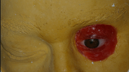
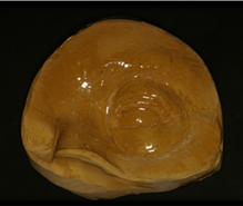
3.Model analysis. Before placing the implants it is essential to develop models for diagnostic study (Fig. 1) and to make the surgical guides (Fig. 2). These can be made out of resin or vinyl-acetate based on the wax sculptures8. Some guides can be obtained by 3D modeling and attached to bone tissue. The less invasive ones can be attached to the teeth in the upper jaw9.
4. Sculpture Before making the sculpture it is necessary to define the extension, which should be as limited as possible, so the prosthesis is unnoticeable. Then, a wax film is tailored to the model and the operator may use wax for the entire sculpture or modeling clay, as recommended by the Sao Paulo Brazilian school10. This is a replication stage, where we must consider facial proportions and apply our knowledge of artistic anatomy, physical anthropology and facial mapping. It is important to consider every detail on the healthy side, or whatever source of information selected, whether it be pictures of the patient, relatives, etc.6).
5. Imaging studies11. To place extraoral implants it is essential to have a CT scan. The scan makes it possible to produce a prototype, through a modeling process (stereolithography) that integrates different technologies. This enables us to obtain a three-dimensional materialization of the structures identified in the CT scan in a life-size 1:1 scale, which is very useful to map the surgery.
Surgical technique
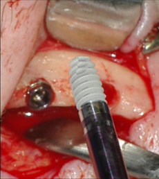
General surgical considerations: According to Badie12, the three main sites to place extraoral implants are: the temporal region, the oculo-palpebral region and the naso-maxillary region. The supraorbital margin is typically used in the oculo-palpebral region, as it is possible to place 3-4 mm-long implants, or even longer ones sometimes, depending on the assessment previously made based on the X-rays or CT scans. The author says that the success rate for the oculo-palpebral region is lower than for the temporal region, as the margin is thin and there is low bone density12. Wolfaardt recommends using implants with no expanded platform in this area, since the risk of exposure is high as the bone is thin. He advises surgeons to be particularly careful not to affect the appearance of the eyebrow when repositioning the flap2. Based on this, dental implants were the option selected in this clinical case as their design is more suitable for the orbital region. Nowadays, there are tapered implants available with a smaller diameter and more robust threads to compensate low bone density. This makes it possible to achieve good primary stability (Fig. 3).
It is essential to apply the right surgical technique to achieve good prosthetic results and to preserve the health of the peri-implant tissue. Soft tissue needs to be handled effectively. In preparation, hair follicles need to be removed from the area surrounding the implants. Subcutaneous tissue must be removed in approximately 10 mm around the implant area13. Therefore, the ideal peri-implant tissue will be thin, motionless, with no hair follicles and no ridges14. Potential implant sites in this region are: the upper, lateral and lower orbital ridge, where it is possible to place 3-4 mm-long implants, as previously stated. About three to four implants are necessary to retain these prostheses. The longitudinal axis of the implant should be positioned towards the center of the orbit. The surgical guide will be essential to identify the position of the implants, so these do not interfere with that of the ocular prosthesis. Transepithelial pillars must be kept within the limits of the orbital cavity so as not to affect the prosthesis. The use of surgical guides is fundamental (Fig. 2), and so is the presence of the rehabilitation professionals during the procedure2.
The regular protocol for tapered implants of reduced diameter (Biomet 3i) was followed in this clinical case:
-Four mg of dexamethasone was prescribed one hour before surgery.
-The patient was administered 2 g amoxicillin as a single dose one hour before the surgical procedure.
-Perioral antisepsis in the area with 2% chlorhexidine.
-Isolation of the operative field with sterile surgical field.
-Terminal infiltration anesthesia (3.6 cc) with 2% mepivacaine with epinephrine 1:100.000
-Incision and separation performed with surgical guide as reference
-Placement of surgical guide
-Cortical drilled with a round bur (3i® Biomet, USA.), under cooling, at a maximum speed of 1.200 rpm
-Complete the drilling sequence based on the implant diameter chosen during the planning stage, which will warrant an optimal three-dimensional position.
-Tapered implant placement (Biomet-3i Osseotite NT mini, 3.25 x 8.5 long)
-Primary stability measured with surgical torque wrench and Osstell
-Fitting of healing caps
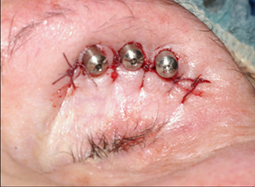
-Repositioning the flap and 5.0 silk suture; application of medicinal dressing folded eight times (Fig. 4).
Medication and post-operative care. The patient was administered 875 mg of oral amoxicillin every 12 hours for 7 days. As analgesia, 600 mg of oral ibuprofen every 8 hours for 3 days was prescribed. Postoperative assessment took place seven days after the surgical procedure, when sutures and dressing were removed, and then monthly for 4-6 months. After this period, the osseointegration of the implants was assessed and the prosthesis was made (Fig. 5). The criteria used to assess osseointegration were: absence of pain, dysesthesia and immobility15).
Imaging studies. An initial digital tomography was performed before the surgery (T0) for diagnosis and planning. A second control tomography will be performed two years after the procedure.
Discussion
The high success rates achieved in the clinical studies confirm that osseointegrated implants are the treatment of choice for patients who need oral-maxillofacial prostheses1. However, the success rate for the orbital region is lower than for the temporal region, as the margin is thin and there is low bone density12. Therefore, in this study the authors recommend using dental implants following a design seemingly more suitable for these anatomical features. Studies show that the most common complication in this area is the inflammation of the peri-implant tissue16,17. Implants surfacing through the skin is the main problem of extraoral implants. However, after over forty years of experience, the results reported in the scientific literature are very good, with very few cases with signs and symptoms of inflammation14. The surfacing of the implant disrupts the body’s first biological protective barrier. The same happens in the mouth, but the mucosa allows for better epithelium-titanium adhesion. The formation of non-keratinized tissue with a higher concentration of inflammatory cells has been observed in the skin. This could compensate for the absence of epithelial adhesion14. According to Telljeström, the predisposing factors for skin inflammation are: extraoral implant designs with platform, the patient’s local and systemic health, skin movement and thickness around the implant and poor hygiene. Abu-Serriah17) considers the problem to be multifactorial, and says that some predisposing causes are: disruption to skin integrity, poor prosthesis ventilation and difficulties to achieve good hygiene. According to Toljanic18, in order to control infection it is essential to: carefully select the patient, achieve proper prosthesis ventilation and follow an adequate hygiene protocol. Despite efforts to achieve good hygiene, some studies have observed the presence of opportunistic pathogen microorganisms. These did not show a direct correlation between hygiene and peri-implant tissue inflammation18. Other studies found staphylococcus aureus in patients with tissue inflammation. Researchers believe that this shows how essential patient hygiene is19. According to Abu-Serriah20, since there is no predominant microorganism in the infected sites, microorganism culture studies and sensitivity studies should guide the treatment of infections. Watson21 argues that hygiene problems arise when the soft tissue is too thick and when there is not enough space between pillars. This will depend on adequate prosthetic planning and a proper surgical technique. Nishimura22 reported few complications in patients with thin and motionless peri-implant tissue and good hygiene. Holgers et al.4 classified skin reaction around extraoral implants as:
-0: healthy tissue;
-1: slight tissue redness (mild inflammation);
-2: formation of granulation tissue with local secretion
-3: exuberant granulation tissue and discharge;
-4: infection.
Patients should be instructed on hygiene practices using different means from the beginning of the treatment, depending on each case. The patient may use gauze, swabs, dental floss or tape, brushes, etc. with water and liquid soap three times a day. It is also advisable to use hydrogen peroxide once a day4. Allen’s4 hygiene protocol involves: cleaning pillars with swabs soaked in 50:50 water and hydrogen peroxide, using interdental brushes and expanding dental floss, cleaning the prosthesis with soap and water with a soft brush and removing the prosthesis while sleeping to ventilate the skin and reduce the risk of tissue reaction. Patients must also comply with a number of check-up appointments which will depend on the risk assessment conducted. In these check-ups, each patient’s risk factors will be reassessed and the necessary treatments will be performed depending on their needs. Following Holgers’ classification, treatment in each case would entail:
-Holgers type 1: Encourage the patient to improve hygiene practices and increase the number of check-ups;
-Holgers type 2: Same procedure as Holgers 1 plus the administration of terracortril and polymyxin B, both antibiotics, antifungals and steroidal anti-inflammatory drugs. These must be applied three times a day for one week;
-Holgers type 3: Same procedure as Holgers 2 plus a medicinal B-soaked tampon. It is also possible to remove the bar and refit the healing collars. Surgery would be indicated if there is no improvement, in order to remove the peri-implant granulation tissue;
-Holgers type 4: Remove pillars and allow for second intention healing.
The clinical case described herein presented mild inflammation (Holgers 1) in the second biannual check-up appointment. Therefore, the patient was encouraged to improve hygiene practices. The patient was very cooperative, thus there was good peri-implant tissue response, with no complications, two years after the implants had been placed. The successful outcome of this clinical case by placing an implant-supported orbital prosthesis is consistent with what the abovementioned authors have reported in terms of a tapered dental implant design, with a reduced platform. These implants are suitable for a low density and limited width region such as this.
Conclusions
Using dental implants for rehabilitation with oral-maxillofacial prostheses is a suitable treatment option, as their design has advantages for some extraoral sites like the orbital region. The design of tapered dental implants, with a reduced platform, is suitable for this low density and limited width region. The treatment must be carefully selected by a multi and interdisciplinary team, and monitoring and maintenance should be conducted based on individual needs. Patients must know the risks and the medium and long term prognosis of both the implants and the prosthesis. In addition, they need to know what their responsibilities are regarding hygiene practices and attending scheduled check-up appointments
Referencias
1. Tolman E, Desjardins RP, Jackson T, Bränemark PI. Complex Cranio-facial Reconstruction Using an Implant-Supported Prosthesis: Case Report With Long-Term Follow-up.Int J Oral Maxillofac Implants 1997; 12:243-251. [ Links ]
2. Wolfaardt J,Sugar A, Wilkes G. Advanced Technology and the Future of Facial Prosthetics in Head and Neck Reconstruction. Int J Oral Maxillofac Surg. 2003;32:121-123 [ Links ]
3. Parel SM, Tjellström A. The United States and Swedish experience with osseointegration and facial prostheses. Int. J. Oral Maxillofac. Implants. 1991 Spring;6(1):75-9. [ Links ]
4. Seignemartin CP, Dib LL, Oliveira, JM Piras de. A reabilitaçao facial com próteses convencionais e sobre implantes osseointegrados: Congresso Internacional de Osseointegraçao da APCD, Cap. 13, 2004, p:251-268 [ Links ]
5. Granström G, Jacobsson M, Tjellström A. Titanium implants in irradiated tissue: benefits from hiperbaric oxigen Int. J. Oral Maxillofac. Implants 1992, 7: 15-25. [ Links ]
6. Jankielewicz I. et al. Rehabilitación Buco-Maxilofacial con Prótesis en Implantes Oseointegrados. Barcelona: Quintessence 2003. p407-414. [ Links ]
7. Seals RR Jr, Cortes AL, Parel SM. Fabrication of facial prostheses by applying the osseointegration concept for retention. J Prosthet Dent 1989 Jun;61(6):712-6. [ Links ]
8. Arcuri MR, Rubenstein JT. Facial implants. Dent Clin North Am 1998 Jan; 42(1): 161-75. [ Links ]
9. Eggers G, Mühling J, Marmulla R. Templated-Based Registration for Image-Guided Maxillofacial Surgery. J Oral Maxillofac. Surg 2005, 63:1330-1336. [ Links ]
10. De Rezende JR, Pirras de Oliveira J, Brito e Dias R,-Prótese facial-Prótese buco-maxilo-facial conceitos básicos e práticas de laboratorio. Sao Paulo: Savier, 1986. p51-69 [ Links ]
11. Todescan FF, Bechelli A, Romanelli H. Implantología Contemporánea- Cirugía y Prótesis. Sao Paulo: Artes Médicas, 2005. [ Links ]
12. Badie-Modiri B, Kaplanski P. Extra-oral implants: principal areas of implantation. Rev Stomatol Chir Maxillofac. 2001 Aug;102 (5):229-33. [ Links ]
13. Gary JJ, Donovan M. Retention designs for bone-anchored facial prostheses. J Prosthet Dent. 1993 Oct; 70(4):329-32. [ Links ]
14. Bränemark PI, Tolman DE. Osseointegration in Craniofacial Reconstruction. Chicago: Quintessence Publishing, 1998. p387-417. [ Links ]
15. Albrektsson T, Zarb G, Worthington P, Eriksson AR. The long-term efficacy of currently used dental implants: a review and proposed criteria of success. Int J Oral Maxillofac Implants 1986; 1(1): 11-25. [ Links ]
16. Allen PF, Watson G, Stassen l, Mcmillan AS. Peri-implant soft tissue maintenance in patients with craniofacial implant retained prostheses. Int J Oral Maxillofac Surg 2000; 29(2): 99-103. [ Links ]
17. Abu-Serriah MM, McGowan DA, Moos KF, Bagg J. Extra-oral endosseous craniofacial implants: current status and future developments. Int J Oral Maxillofac Surg. 2003; Oct; 32(5):452-8. [ Links ]
18. Toljanic A, Morello J.A, Moran W.J, Panje WR, May EF. Microflora Associated With Percutaneous Craniofacial Implants Used for Retention of Facial Prostheses: A pilot Study. Int J Oral Maxilofac Implants 1995; 10:578-582. [ Links ]
19. Gitto CA, Plata WG, Schaaf NG. Evaluation of the Peri-implant Epithelial Tissue of Percutaneous Implant Abutments Supporting Maxillofacial Prostheses. Int. J Oral Maxillofac. Implants, 1994; 9:197-206. [ Links ]
20. Abu-Serriah MM, Bagg J,McGowan DA, Mackenzie D. The microflora associated with extra-oral endosseous craniofacial implants: a cross-sectional study.Int. J. Oral Maxillofac. Surg. 2000;29: 344-350. [ Links ]
21. Watson RM; Coward TJ; Forman GH. Results of treatment of 20 patients with implant-retained auricular prostheses. Int J Oral Maxillofac Implants, 1995 Jul-Aug; 10(4):445-9. [ Links ]
22. Nishimura RD, Roumanas E, Moy PK, Sugai T. Nasal defects and osseointegrated implants: UCLA experience. J. Prosthet. Dent. 1996 Dec;76(6):597-602 [ Links ]
Received: June 20, 2017; Accepted: August 03, 2017


 texto en

