


Services on Demand
Journal

Article
 text in
  English (pdf)
 Article in xml format
 Article references
Related links
Share

 Permalink
Odontoestomatología
On-line version ISSN 1688-9339
Odontoestomatología vol.16 no.24 Montevideo Nov. 2014
Applicability of the Moyers prediction tables at 75% on Mapuche-Huilliche patients, Chile
Carrasco Marcelo**,
Rioseco Juan*** ,
Bizama Gabriel****,
Fierro Claudia*****
*MA in Pediatric
Dentistry, Associate Professor of Pediatric Dentistry, Facultad de Odontología,
Universidad de Concepción
mperezf@udec.cl
** Dental Surgeon,
Hospital Quilacahuín, San Pablo, Osorno
*** Dental Surgeon,
Cecof 8 de Mayo, Talcahuano.
**** Dental Surgeon,
CESFAM Los Alamos.
***** Specialist in Pediatric Dentistry, Associate Professor of
Pediatric Dentistry, Facultad de Odontología, Universidad de Concepción.
ABSTRACT
Objective. To determine the applicability of the
Moyers prediction tables at 75% on patients from the Mapuche-Huilliche
population, Chile. Materials and
Methods. A descriptive, observational study which evaluated the Moyers
prediction tables at 75% in a Mapuche‑Huilliche population aged between 11 and
17 (25 men and 25 women). The sum of the lower incisors, and the sum of the
canines and premolars of each quadrant was compared with each of the predictive
values. Results. Increased number of
cases in the range from 23.5 mm to 25.2 mm in the sum of lower incisors, which
represent 60% of the sample. The sum of canines and premolars showed in maxilla
a mean of 23.7 mm in men and 23.1 mm in women, and in mandible a mean
of 22.9 mm in men and 22.1 mm in women. A negative discrepancy of 60%
was found in men and of 40% in women. For women, the positive discrepancy
significantly exceeded the negative discrepancies with percentages of 88% and
8%. Conclusions. The Moyers method
at 75% was applicable in maxilla and mandible on Huilliche men, and partially
applicable on women of the same ethnic group.
Keywords: Mapuches, mixed dentition analysis, incisor, Moyers prediction tables.
Received on: 31 Mar 2014 - Accepted on: 24 Aug 2014
Introduction
The analysis of mixed dentition is a key element of
orthodontic evaluation: it helps to determine the space available for permanent
teeth, and it is also necessary to take decisions in relation to eruption,
serial extractions, space maintenance or recovery, among other things (1–4).
It is argued that the Moyers
prediction tables are the most widely used method to
predict the size of permanent canines and premolars according to their
correlation with the mesio-distal width of lower permanent incisors (5–7). This is the case because the systematic error
is usually minimal, it can be used by beginners and experts with the same level
of reliability, it does not require a complex clinical opinion and it saves
time. It requires no specific equipment or radiographic projections; it can be
used in both arches and, although it is better to apply it on dental models, it
can be used in mouth with reasonable precision (3).
Although the Moyers prediction tables have advantages, they were developed in a
Caucasian population. The application of this method to other ethnic
populations has been studied and also questioned on several occasions (6–9).
The population of Latin America is basically a mixture of
European and indigenous people. Thus, diverse groups live all around the
continent with the anatomic characteristics of each ethnic group (10).
According to the 2002 Census of Chile, 604,349 people in
Chile stated that they belonged to the Mapuche population, approximately 4% of
the total population, and 87.3% of the total indigenous population. They live
mainly in the Araucania Region (33.6%) and in the Metropolitan Region (30.3%),
and in smaller numbers in the regions of Biobío (8.8%), Los Lagos and Los Ríos
(16.7% for the two together). The Region of Los Lagos is the third region of
the country regarding the number of members of the ethnic group. It has a total
of 100,327 individuals, which accounts for 16.6% of the total Mapuche
population. Out of these, 2,121 individuals live in the commune of San Pablo,
which amounts to 21% of the total population there (11,
12).
The aim of this study is to determine the applicability of
the Moyers prediction tables at the 75 percentile confidence level on
the Mapuche-Huilliche population of the rural/coastal area of the Commune of
San Pablo, Province of Osorno, 10th Region, Chile.
Materials
and methods
This is a descriptive observational study whose universe is
the indigenous Mapuche-Huilliche population of the coastal area of the Commune
of San Pablo, Osorno, Chile.
We had a convenience sample of 50 patients (25 men and 25
women) aged between 11 and 17, who complied with the following criteria for
inclusion:
-
At least one of their surnames was of indigenous origin (13,
14)
-
A complete set of permanent teeth from the first molar to
its contralateral tooth in both maxilla and mandible
-
No proximal restorations and/or interdental caries
-
Residence and place of studies in the coastal area of the
commune
The criteria for exclusion were the following:
-
Foreign surnames
-
Dental anomalies of size, number and shape
-
Previous orthodontic treatment
-
Craniomandibular dysfunctions, fractures and/or attritions
-
Syndromes
Parents and/or guardians were asked to sign a consent form
so that their children could be included in the study. Additionally, the
consent of the participants was requested before records were taken.
Casts of the maxilla and the mandible were taken using
Jeltrate Orthodontic Chromatic (Dentsply) fast-set alginate and Jeltrate pre-shaped
stainless steel metallic trays. To reduce possible distortions of the
impression material when making the study models, the trays were emptied using
stone cast immediately after the impressions had been taken. The greatest
mesio-distal width of the 4 mandibular incisors and of canines and premolars of
both arches were measured independently using a Dentaurum dental vernier analog
caliper with fine tips (0.1 mm). Three alternate measurements were taken
by the same professional in relation to the other teeth of the arch under
study. The modal value was set as the final value, and the dental caliper was
kept perpendicular to the long axis of the tooth crown and parallel to the
buccal and occlusal surfaces. The procedure was conducted with natural light from
a source located opposite the professional and within the same physical space.
The predictive analysis was conducted using the Moyers prediction tables at the
75 percentile confidence level. The differences between the real dimensions and
the predictive value for the sum of the mesio-distal diameters of canines and
premolars in each quadrant were determined. The following values were
calculated: mean, range, discrepancy between real values and predictive values,
and standard deviation. The real values of the sum of mesio-distal diameters of canines and premolars were compared with the predictive
values of the Moyers tables at 75%.
When
Student's t-distribution was applied to verify the differences between the
predictive measurements of Moyers 75% and the real values observed, the
following values were obtained: in men, a value p=0.667 in maxilla and p=0.474
in mandible (without statistical significance); and in women, p=0.191 in
maxilla (without statistical significance), and p=0.000 in mandible, where the
difference was statistically significant.
Results
Most
values of the total sum of lower incisors fall within the 23.5 mm to
25.2 mm range, which represent 60% of the sample under study (Fig.1).
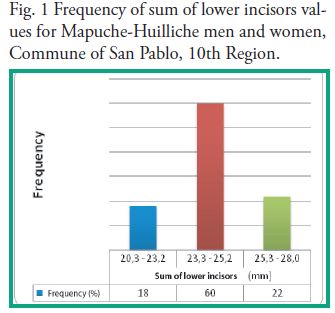
Fig.
1 Frequency of sum of lower incisors values for Mapuche-Huilliche men and
women, Commune of San Pablo, 10th Region.
The
sum of canines and premolars showed in maxilla a mean of 23.7 mm in men and
23.1 mm in women, and in mandible a mean of 22.9 mm in men and 22.1 mm in
women.
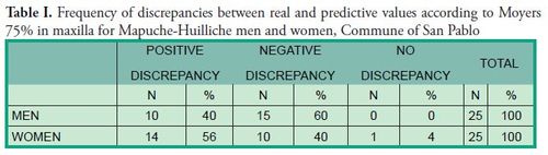
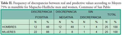
As
for the discrepancy between the real value of the sum of permanent canines and
premolars and the probability predicted with the Moyers tables at 75%, there
was an overestimation of 58% of the necessary space, that is to say, the sample
studied presented smaller dental sizes than the estimation made following the
Moyers analysis at 75%.
Upon
comparing both arches, the mandible was found to have a higher frequency of
overestimation (68%), whereas in maxilla the discrepancy percentage was more
even. When real and predictive values were compared in maxilla in both sexes, a
negative discrepancy of 60% was found in men and of 40% in women. The
distribution in mandible for men was similar regarding positive and negative
discrepancies, unlike in women, where positive discrepancies significantly
exceeded negative discrepancies with percentages of 88% and 8% respectively
(Tables I and II).
In maxilla, the greatest
differences between real and predictive values were observed in the ranges of
1.00 mm to 0.4 mm in men (72%) and of 0.5 mm to 0.9 mm in
women (64%). In mandible, the greatest differences between both values were
observed in the ranges of 1.00 mm to 0.9 mm in men (76%) and of 0.00 mm to
1.4 mm in women (76%).
Discussion
Many international
studies show different degrees of applicability of the Moyers prediction tables
developed
in a population of Caucasian children of Northern European descent (4) whose
bone and dental morphological characteristics differ greatly from those of the
population of this study. A study from Saudi Arabia
shows an overestimation of the size needed (6) and a study conducted in South
Africa (15) on a black population shows an underestimation of the space needed.
There are also significant differences between real values and Moyers
predictive values in Kenya (16). The variable results of several
international studies show that the extent of applicability of the method
varies in different ethnic groups (17). Therefore, it is suggested that these predictive tables should be
previously validated in a specific population to ensure their clinical
applicability (18–22). The criteria for
inclusion in this study do not include variables such as real crowding, a
history of tooth decay and loss of space in mixed and temporary dentition.
Perhaps they should be considered in further studies to minimize bias.
The high number of individuals of
the Mapuche ethnic group within the population that attends health centers in
the country entails an obligation for health professionals: they must know the
extent to which Western medical analysis and diagnostic methods are reliable
and applicable (11) .
In Chile, models of patients over
12 from Concepción and Antofagasta have been studied. The clinical and
statistical validity of the Moyers prediction tables at 75% was established in
both populations. The Moyers analysis is reliable in both sexes and has a
higher correlation in mandible (23). The results of
this study differ from other findings in Chile, as we have observed a higher
correlation in maxilla and in both sexes, and a significant overestimation in
mandible for women.
Conclusions
The Moyers prediction tables are applicable in maxilla for all the cases of this study. However, in mandible they are not applicable on women given the significant overestimation for canines and premolars according to the Moyers prediction tables at 75%.
References1. Hunter WS Application of analysis of crowding and spacing of the teeth. Dent. Clin. North Am. 1978; 22:563–77.
2. Huckaba GW. Arch size analysis and tooth size predition. Dent. Clin. North Am. 1964; 8: 431–40.
3. Staley R, O’Gorman T, Hoag J, Shelly T. Prediction of the widths of un-erupted canines and premolars. J. Am. Dental Assoc. 1984; 108(2):185–90.
4. Moyers RE. Handbook of orthodontics. 4th ed. Chicago: Year Book, 1998. 235–9
5. Zamora C, Medrano D. Análisis de Moyers. En: Compendio de cefalometría: análisis clínico y práctico. Venezuela: Ed. Amolca, 2004. 424-9
6. Al-Khadra BH Prediction of the size of un-erupted canines and premolars in a Saudi Arab population. Am. J. Orthod. Dentofac. Orthop. 1993;104(4):369–72.
7. Yuen K, Tang E, So L. Mixed dentition analysis for Hong Kong Chinese. Angle Orthod. 1998; 68(1):21–28.
8. Ferguson FS, Marco DJ, Sonnenburg EM, Shakun ML. The use of regression constants in estimating tooth size in the Negro population. Am. J. Orthod. 1978; 73(1):68–72.
9. van der Merwe S, Rossouw P, van Wyk Kotze T, Trutero H. An adaptation of the mixed dentition space analysis for a Western Cape Caucasian population. J.Dent. Assoc. South Africa. 1991; 46(9):475–9.
10. de Jong I, Missagia I . Dossier Etnias y Nación en América Latina: historia y comparación. Mem. Am. 2008; 16(1):11-7
11. Chile. Ministerio de Salud. Texto compilatorio, fondo de tesistas, Programa de Salud y Pueblos Indígenas, Servicio de salud Osorno, 2010.
12. Chile. Instituto Nacional de Estadísticas. [documento en línea] 2002 [fecha de acceso 19 de enero 2014]; URL disponible en:http://www.ine.cl/cd2002/sintesiscensal.pdf
13. Bengoa J. Historia del pueblo Mapuche (Siglo XIX y XX). Santiago de Chile: Lom Eds, 2000. 423p
14. Wilhelm E, Meyer W. Los Huilliches a través de sus apellidos: estudio etimológico de los patronímicos aborígenes sureños. Osorno: WM Rusca, 1952.
15. Schirmer UR, Wiltshire WA. Orthodontic probability tables for black patients of african descent: mixed dentition analysis. Am. J. Orthod. Dentofac. Orthop. 1997; 112:545-51.
16. Ngesa JL. Aplicability of tooth size predictions in the mixed dentition Analysis in a Kenyan sample. [Tesis]. Universidad de Western Cape, 2003. [Citado 16 de junio de 2014]. Disponible en: http://erepository.uonbi.ac.ke/handle/11295/16559
17. Buwembo W, Luboga S. Moyers’s method of mixed dentition analysis: a meta-analysis. Afri Health Sci. 2004; 41:63–66.
18. Buwembo W, Kutesa A, Muwazi L, Rwenyonyi CM. Prediction of width of un-erupted incisors, canines and premolars in a Ugandan population: a cross sectional study. BMC Oral Health 2012; 23;12:23.
19. Abu Alhaija ES, Qudeimat M A. Mixed dentition space analysis in a Jordanian population: comparison of two methods. Int. J. Paediatr. Dent. 2006;16(2):104-10.
20. Melgaço CA, Araújo MT, Ruellas AC. Applicability of three tooth size prediction methods for white Brazilians. Angle Orthod. 2006; 76(4):644-9.
21. Jaiswal AK, Paudel KR, Shrestha SL, Jaiswal S. Prediction of space available for unerupted permanent canine and premolars in a Nepalese population. J. Orthod. 2009; 36(4):253-9.
22. nik Tahere H, Majid S, Fateme M, Kharazi F, Javad M. Predicting the size of unerupted canines and premolars of the maxillary and mandibular quadrants in an Iranian population. J. Clin. Pediatr. Dent. 2007; 32(1):43-7.
23. Barrera V, Roa H, Oñate V, Pérez M A. Validación análisis de dentición mixta de Moyers en una muestra de la ciudad de Concepción y Antofagasta en niños chilenos. Rev. Fac. Odont. 2009; 8(11);17-25.

