


Services on Demand
Journal

Article
 text in
  English (pdf)
 Article in xml format
 Article references
Related links
Share

 Permalink
Odontoestomatología
On-line version ISSN 1688-9339
Odontoestomatología vol.16 no.24 Montevideo Nov. 2014
Morphological and morphometric study of the mental foramen using cone-beam CT in dentate adult patients
Cabanillas Padilla, Juan *
Quea Cahuana, Eduardo **
*Professor at Facultad de Odontología, Universidad de San Martín de Porres (USMP), Lima, Perú. Dental Surgeon
juancabanillaspadilla@gmail.com
**Professor at Facultad de Odontología, Universidad de San Martín de Porres (USMP), Lima, Perú. Dental Surgeon
ABSTRACT
Objective. To study the morphology and morphometry of the mental foramen using cone-beam CT in dentate adult patients. Methods. Transversal descriptive study in which 180 cone beam CTs were studied to analyze the distance between the upper and lower cortical areas of the mental foramen to the alveolar crest and the mandibular basal bone respectively, as well as the location, shape, size and presence of accessory holes. Results. It was found that the mean of the upper cortical area in relation to the alveolar crest was 15.00 mm and the mean of the lower cortical area to the mandibular basal bone was 13.75 mm. The most frequent location was the longitudinal axis of the second premolar (44.4% right side and 47.2% left side). The predominant shape was oval and the size was in the range of 2.00 mm to 2.99 mm. Accessory holes were present in 55.5% of cases. Conclusion. Knowing the exact location of the mental foramen and its variations helps to properly plan surgical procedures and to administer anesthesia effectively without damaging the neurovascular bundle.
Keywords: Mental foramen, Cone-beam computerized tomography.
Received on: 09 Mar 2014 – Accepted on: 27 Jun 2014
Introduction
The mandible stems from the first branchial arch, has mixed types of ossification and runs downwards and forward, as it develops from the condyle (1, 2).
Mandibular growth modifies the direction of the mental foramen. At birth, the neurovascular bundle runs forward through the hole, and in adults it runs backwards (3).
The mental foramen is an opening on the lateral part of the mandible. It is here that the inferior alveolar nerve branches into the mental nerve and the incisive nerve. These are terminal branches which supply sensory innervation to soft tissues in the vestibular area, the lower lip and the chin, up to the mandibular midline. The location of the mental foramen varies according to age. In children, before tooth eruption, it is located closer to the alveolar crest, whereas in adults it is, on average, between 13 mm and 15 mm above the inferior mandibular border. In individuals with bone resorption it is located closer to the alveolar crest, and it can even be found over it. Thus, this must considered when preparing full prosthetics, as the set may press on the mental nerve loop (1, 4, 5, 7, 18).
Cone-beam CT enables us to accurately determine the location, shape, and size of the mental foramen, as well as the presence of accessory holes. This allows for an accurate morphometric analysis, which enables us to develop a suitable treatment plan and to administer anesthesia effectively, thus we can conduct invasive procedures without damaging the mental foramen (6, 24). This technology also provides life-size images where precise lines and measurements can be made, as it works with isotropic voxels. Additionally, it provides images in all three planes (8-10).
Materials and methods
180 CTs of dentate adult patients of both sexes ranging between 20 and 50 years of age were analyzed (74 men and 106 women). They were taken at Centro de Diagnóstico por Imágenes, Lima, Perú. The tomographies were taken using Vatech equipment, Picasso Master model, with Easy Dent software, a field of view (FOV) of 20 x 19 mm, 70 Kv, 8 Ma, exposure time of 25 seconds and a focus point of 0.5 mm. The variables were studied on the transaxial plane through 1-mm cuts and 0.5-mm intervals.
The morphological and morphometric analysis of the mental foramen was conducted on both sides. The tomographies evaluated included all the teeth and a preserved alveolar crest. Tomographies with the following elements were excluded: periapical lesions, supernumerary teeth, orthodontics patients and bilateral absence of mental foramen.
The distances from the upper and lower cortical areas of the mental foramen to the alveolar crest and the mandibular basal bone respectively were analyzed, as well as the size of the hole on the transaxial cut. There we considered the distance between the upper and lower cortical areas of the structure, and the measurements were classified into ranges (Figures 1 and 2).
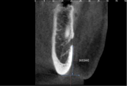
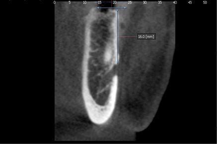
Fig. 1: Location of mental foramen on the transaxial plane
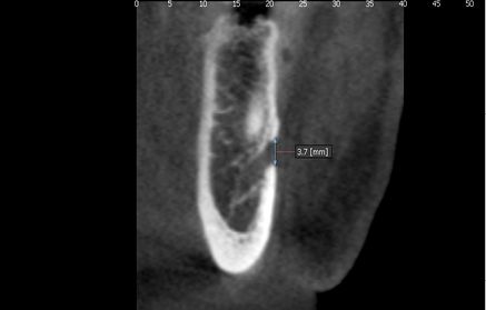
Fig. 2: Size of mental foramen
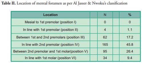
3D reconstruction was used to determine the location, shape and presence of accessory holes. To determine the location, the longitudinal axes of teeth were taken as reference as per Al Jasser & Nwoku's classification (21). Position 1: Mesial to first premolar; position 2: In line with first premolar; position 3: Between first and second premolars; position 4: In line with second premolar; position 5: Between second premolar and first molar, position 6: In line with first molar (Figure 3).
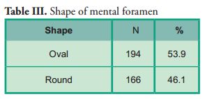
Oval and round shapes were taken as evaluation criteria, as well as the presence of accessory holes (Figure 4).
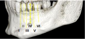
Figure 3: Location of mental foramen as per Al Jasser & Nwoku's classification
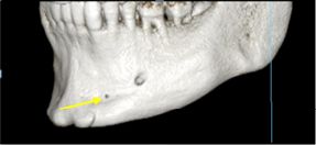
Figure 4: Presence of accessory mental foramina
Data processing and analysis was conducted using SPSS statistical software, version 15. Quantitative variables were presented in minimum values, maximum values, mean values and standard deviation. The Mann-Whitney U test was used to compare the differences between the right and left sides.
Qualitative values were presented via frequency distribution tables, and Pearson's chi square test and Fisher's exact test were used to compare the difference between the right and left sides. All tests had a significance level of 5%.
Results
The tomographies were evaluated by three calibrated examiners (Kappa 1.00).
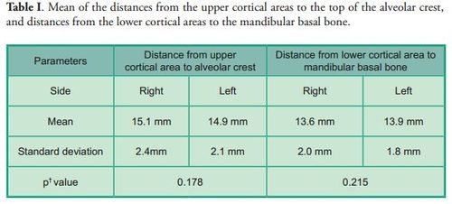
The distance from the upper and lower cortical areas of the mental foramen to the alveolar crest and the mandibular basal bone respectively was measured. No statistically significant differences were found between the right and left sides in either measurement (p*0.178), (p*0.215) (Table I).
The most frequent location of the mental foramen was the longitudinal axis of the second premolar on both sides (position IV), followed by positions V, III, VI and II. No mental foramen was found in position I, neither significant differences compared to the contralateral side (p*0.764) (Table II).
Oval was the most frequent shape, and no significant differences were observed compared with the contralateral side (p*0.057) (Table III).
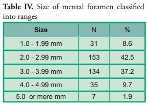
The size of the mental foramen was classified into five measuring ranges. Most (n=153; 42.5%) were included in the 2 mm to 2.99 mm range on both sides. No significant differences were observed compared with the contralateral side (p*0.623) (Table IV).
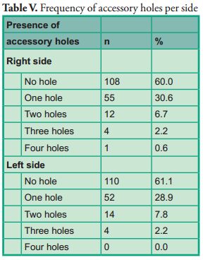
Accessory holes were found in 100 cases (55.5%) out of the 180 tomographies taken (Table V).
Discussion
This research found a distance from the upper cortical area of the mental foramen to the alveolar crest which is similar to the distance found in other studies (4, 11). Furthermore, other studies (6, 12) where measurements were taken on dry mandibles reported shorter distances, results which differ from the findings of this study.
The distance from the lower cortical area of the mental foramen to the mandibular basal bone was (13.6 mm ± 2.0 mm) and (13.9 mm ± 1.8 mm) on the right and left sides respectively, which, on average, is consistent with the results of other studies (4, 13, 14).
There is considerable debate in the literature regarding the location of the mental foramen in different ethnic groups. This study showed that this structure was found in all the samples between the root of the first premolar and the root of the first molar.
These results are similar to those of other studies (4, 6, 11, 13–17, 24), where the mental foramen was found on the longitudinal axis of the second premolar (Position IV). Other studies (7, 12, 18, 19) showed that the most frequent location of the mental foramen was position III, which differs from this study, as positions V and III ranked second and third respectively regarding frequency.
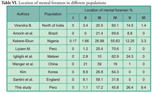
Several authors have studied the location of the mental foramen using the longitudinal axes of teeth as reference. However, there were variations in the results, which could be attributed to ethnic differences (Table VI).
Regarding the shape of the mental foramen, there are several studies whose results do not agree on the classification of shapes itself. However, other studies take the oval and round shapes as evaluation criteria. These studies (4, 6, 11, 12, 15, 22) show an oval shape in most cases, which is consistent with the present study. However, other authors (14, 16, 20, 24) disagree with our results as they report that the round shape is the most frequent one.
The size of the mental foramen was classified into 5 ranges. In most cases, they were located in the 2 mm to 2.99 mm range on both sides, with a frequency of 75 cases (41.7%) on the right side and 78 cases (43.3%) on the left side.
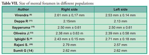
Several authors have evaluated the size of the mental foramen on dry mandibles. Their results are similar to the results of this study: within the 2 mm to 2.99 mm range (Table VII).
Accessory mental foramina were present in 55.5% of all cases, which differs from the results of most studies conducted on dry mandibles and evaluated using cone-beam CT (6, 14, 15, 20, 23). Singh R. & Srivastav observed the presence of accessory holes in 13% of the total population evaluated, while other studies (6, 14, 15, 23) showed a prevalence of 6.67%, 6.6%, 3.92% and 6.5% respectively.
All the variables studied are consistent with the results of Igbigbi & Lesbona, who evaluated the morphology and morphometry of the mental foramen in an African population.
Conclusion
It is concluded that the mental foramen in a dentate adult population is located, on average, 13.75 mm above the mandibular basal bone. The most frequent location was below the longitudinal axis of the second premolar, in most cases it was oval shaped and its size was within the 2 mm to 2.99 mm range. Accessory mental foramina were observed in over half the cases studied (55.5 %).
Cabanillas Padilla, Juan *
Quea Cahuana, Eduardo **
*Professor at Facultad de Odontología, Universidad de San Martín de Porres (USMP), Lima, Perú. Dental Surgeon
juancabanillaspadilla@gmail.com
**Professor at Facultad de Odontología, Universidad de San Martín de Porres (USMP), Lima, Perú. Dental Surgeon
ABSTRACT
Objective. To study the morphology and morphometry of the mental foramen using cone-beam CT in dentate adult patients. Methods. Transversal descriptive study in which 180 cone beam CTs were studied to analyze the distance between the upper and lower cortical areas of the mental foramen to the alveolar crest and the mandibular basal bone respectively, as well as the location, shape, size and presence of accessory holes. Results. It was found that the mean of the upper cortical area in relation to the alveolar crest was 15.00 mm and the mean of the lower cortical area to the mandibular basal bone was 13.75 mm. The most frequent location was the longitudinal axis of the second premolar (44.4% right side and 47.2% left side). The predominant shape was oval and the size was in the range of 2.00 mm to 2.99 mm. Accessory holes were present in 55.5% of cases. Conclusion. Knowing the exact location of the mental foramen and its variations helps to properly plan surgical procedures and to administer anesthesia effectively without damaging the neurovascular bundle.
Keywords: Mental foramen, Cone-beam computerized tomography.
Received on: 09 Mar 2014 – Accepted on: 27 Jun 2014
Introduction
The mandible stems from the first branchial arch, has mixed types of ossification and runs downwards and forward, as it develops from the condyle (1, 2).
Mandibular growth modifies the direction of the mental foramen. At birth, the neurovascular bundle runs forward through the hole, and in adults it runs backwards (3).
The mental foramen is an opening on the lateral part of the mandible. It is here that the inferior alveolar nerve branches into the mental nerve and the incisive nerve. These are terminal branches which supply sensory innervation to soft tissues in the vestibular area, the lower lip and the chin, up to the mandibular midline. The location of the mental foramen varies according to age. In children, before tooth eruption, it is located closer to the alveolar crest, whereas in adults it is, on average, between 13 mm and 15 mm above the inferior mandibular border. In individuals with bone resorption it is located closer to the alveolar crest, and it can even be found over it. Thus, this must considered when preparing full prosthetics, as the set may press on the mental nerve loop (1, 4, 5, 7, 18).
Cone-beam CT enables us to accurately determine the location, shape, and size of the mental foramen, as well as the presence of accessory holes. This allows for an accurate morphometric analysis, which enables us to develop a suitable treatment plan and to administer anesthesia effectively, thus we can conduct invasive procedures without damaging the mental foramen (6, 24). This technology also provides life-size images where precise lines and measurements can be made, as it works with isotropic voxels. Additionally, it provides images in all three planes (8-10).
Materials and methods
180 CTs of dentate adult patients of both sexes ranging between 20 and 50 years of age were analyzed (74 men and 106 women). They were taken at Centro de Diagnóstico por Imágenes, Lima, Perú. The tomographies were taken using Vatech equipment, Picasso Master model, with Easy Dent software, a field of view (FOV) of 20 x 19 mm, 70 Kv, 8 Ma, exposure time of 25 seconds and a focus point of 0.5 mm. The variables were studied on the transaxial plane through 1-mm cuts and 0.5-mm intervals.
The morphological and morphometric analysis of the mental foramen was conducted on both sides. The tomographies evaluated included all the teeth and a preserved alveolar crest. Tomographies with the following elements were excluded: periapical lesions, supernumerary teeth, orthodontics patients and bilateral absence of mental foramen.
The distances from the upper and lower cortical areas of the mental foramen to the alveolar crest and the mandibular basal bone respectively were analyzed, as well as the size of the hole on the transaxial cut. There we considered the distance between the upper and lower cortical areas of the structure, and the measurements were classified into ranges (Figures 1 and 2).
Fig. 1: Location of mental foramen on the transaxial plane
Fig. 2: Size of mental foramen
3D reconstruction was used to determine the location, shape and presence of accessory holes. To determine the location, the longitudinal axes of teeth were taken as reference as per Al Jasser & Nwoku's classification (21). Position 1: Mesial to first premolar; position 2: In line with first premolar; position 3: Between first and second premolars; position 4: In line with second premolar; position 5: Between second premolar and first molar, position 6: In line with first molar (Figure 3).
Oval and round shapes were taken as evaluation criteria, as well as the presence of accessory holes (Figure 4).
Figure 3: Location of mental foramen as per Al Jasser & Nwoku's classification
Figure 4: Presence of accessory mental foramina
Data processing and analysis was conducted using SPSS statistical software, version 15. Quantitative variables were presented in minimum values, maximum values, mean values and standard deviation. The Mann-Whitney U test was used to compare the differences between the right and left sides.
Qualitative values were presented via frequency distribution tables, and Pearson's chi square test and Fisher's exact test were used to compare the difference between the right and left sides. All tests had a significance level of 5%.
Results
The tomographies were evaluated by three calibrated examiners (Kappa 1.00).
The distance from the upper and lower cortical areas of the mental foramen to the alveolar crest and the mandibular basal bone respectively was measured. No statistically significant differences were found between the right and left sides in either measurement (p*0.178), (p*0.215) (Table I).
The most frequent location of the mental foramen was the longitudinal axis of the second premolar on both sides (position IV), followed by positions V, III, VI and II. No mental foramen was found in position I, neither significant differences compared to the contralateral side (p*0.764) (Table II).
Oval was the most frequent shape, and no significant differences were observed compared with the contralateral side (p*0.057) (Table III).
The size of the mental foramen was classified into five measuring ranges. Most (n=153; 42.5%) were included in the 2 mm to 2.99 mm range on both sides. No significant differences were observed compared with the contralateral side (p*0.623) (Table IV).
Accessory holes were found in 100 cases (55.5%) out of the 180 tomographies taken (Table V).
Discussion
This research found a distance from the upper cortical area of the mental foramen to the alveolar crest which is similar to the distance found in other studies (4, 11). Furthermore, other studies (6, 12) where measurements were taken on dry mandibles reported shorter distances, results which differ from the findings of this study.
The distance from the lower cortical area of the mental foramen to the mandibular basal bone was (13.6 mm ± 2.0 mm) and (13.9 mm ± 1.8 mm) on the right and left sides respectively, which, on average, is consistent with the results of other studies (4, 13, 14).
There is considerable debate in the literature regarding the location of the mental foramen in different ethnic groups. This study showed that this structure was found in all the samples between the root of the first premolar and the root of the first molar.
These results are similar to those of other studies (4, 6, 11, 13–17, 24), where the mental foramen was found on the longitudinal axis of the second premolar (Position IV). Other studies (7, 12, 18, 19) showed that the most frequent location of the mental foramen was position III, which differs from this study, as positions V and III ranked second and third respectively regarding frequency.
Several authors have studied the location of the mental foramen using the longitudinal axes of teeth as reference. However, there were variations in the results, which could be attributed to ethnic differences (Table VI).
Regarding the shape of the mental foramen, there are several studies whose results do not agree on the classification of shapes itself. However, other studies take the oval and round shapes as evaluation criteria. These studies (4, 6, 11, 12, 15, 22) show an oval shape in most cases, which is consistent with the present study. However, other authors (14, 16, 20, 24) disagree with our results as they report that the round shape is the most frequent one.
The size of the mental foramen was classified into 5 ranges. In most cases, they were located in the 2 mm to 2.99 mm range on both sides, with a frequency of 75 cases (41.7%) on the right side and 78 cases (43.3%) on the left side.
Several authors have evaluated the size of the mental foramen on dry mandibles. Their results are similar to the results of this study: within the 2 mm to 2.99 mm range (Table VII).
Accessory mental foramina were present in 55.5% of all cases, which differs from the results of most studies conducted on dry mandibles and evaluated using cone-beam CT (6, 14, 15, 20, 23). Singh R. & Srivastav observed the presence of accessory holes in 13% of the total population evaluated, while other studies (6, 14, 15, 23) showed a prevalence of 6.67%, 6.6%, 3.92% and 6.5% respectively.
All the variables studied are consistent with the results of Igbigbi & Lesbona, who evaluated the morphology and morphometry of the mental foramen in an African population.
Conclusion
It is concluded that the mental foramen in a dentate adult population is located, on average, 13.75 mm above the mandibular basal bone. The most frequent location was below the longitudinal axis of the second premolar, in most cases it was oval shaped and its size was within the 2 mm to 2.99 mm range. Accessory mental foramina were observed in over half the cases studied (55.5 %).
Conclusión
Se concluye que el agujero mentoniano en una población adulta dentada se ubica en promedio en 13.75 mm por encima de la basal mandibular; la ubicación más frecuente se evidenció debajo del eje longitudinal del segundo premolar, la forma predominante fue oval, el tamaño se ubicó en el rango de 2 mm a 2.99 mm y se evidenció que los agujeros mentonianos accesorios estuvieron presentes en más de la mitad de los casos estudiados. (55.5 %)
Referencias
1. Velayos JL. Anatomía de la Cabeza para Odontólogos. 4a ed. Buenos aires: Editorial Panamericana, 2007.
2. Solano Reina JE, Mendoza Mendoza A. Crecimiento Craneofacial y Desarrollo de las Arcadas Dentarias. En: Odontopediatria.Barcelona: Masson. 2004. p37–53.
3. Ries Centeno GA. Cirugía bucal: Patología, Clínica y Terapéutica. 9a ed. Buenos Aires: Editorial el Ateneo. 1987.
4. Igbigbi PS, Lesbona S. The Position and Dimensions of the Mental Foramen in Adult Malawian Mandibles. West African J Med. 2005; 24 (3): 184-189.
5. Hassan T, Fauzi M, Hassan D. Bilateral Absence of Mental Foramen: A Rare Variation. Int J Anatomic Variations [en línea] 2010; 3: 187-189. Citado el 8 de Enero 2014. Disponible en: http://www.ijav.org/2010/ijav_10_167-169.pdf
6. Budhiraja V, Rastogi R, Lalwani R, Goel P, Chandra S. Study of Position, Shape, and Size of Mental Foramen Utilizing Various Parameters in Dry Adult Human Mandibles From North India. Int Scholarly Res Notices [en línea] 2012; 1-5. Citado el 27 de Noviembre 2013. Disponible en: http://dx.doi.org/10.5402/2013/961429
7. Haghanifar S, Rokouei M. Radiographic Evaluation of the Mental Foramen in a Selected Iranian Population. Indian J Dent Res. [en línea] 2009; 20: 150-2. Citado el 17 de Diciembre 2013. Disponible en:
http://www.ijdr.in/article.asp?issn=0970-9290;year=2009;volume=20;issue=2;spage=150;epage=152;aulast=Haghanifar
8. Montoya K. Tomografía Cone Beam como Método de Diagnóstico Preciso y Confiable en Odontología [tesis] Universidad Veracruzana: 2011. Citado el 8 de Enero 2014. Disponible en: http://cdigital.uv.mx/bitstream/123456789/30959/1/MontoyaHernandez.pdf
9. Oviedo P. Tomografía Cone Beam Aplicado a la Endodoncia [tesis] Lima- Perú. Universidad Peruana Cayetano Heredia: 2010.
10. Ige M. Tomografía Computarizada Volumétrica: Cone Beam” [tesis] Lima- Perú. Universidad Peruana Cayetano Heredia: 2010.
11. Agarwal D, Gupta S. Morphometric Analysis of Mental Foramen in Human Mandibles of South Gujarat. People’s Journal of Scientific Research 2011; 15.4(1).
12. Oliveira J, Araujo A, Da Silva C, Sousa R, Lima F. Morphological and Morphometric Study of the Mental Foramen on the M-CP.18 Jianchenjiang Point. Int J Morpho [en línea] 2009; 27 (1): 231-238. Citado el 12 de Diciembre 2013. Disponible en http://www.scielo.cl/scielo.php?script=sci_pdf&pid=S0717-95022009000100039&lng=es&nrm=iso&tlng=en
13. Amorin M, Prado F, Borini C, Bittar T, Volpato M, Groppo F, et al. The Mental Foramen Position in Dentate and Edentulous Brazilian’s Mandible. Int J Morpho [en línea] 2008; 26 (4): 981-987. Citado el 21 de Enero 2014. Disponible en:
http://www.scielo.cl/scielo.php?script=sci_pdf&pid=S0717-95022008000400033&lng=es&nrm=iso&tlng=en
14. Gupta S, Soni JS. Study of Anatomical Variations and Incidence of Mental Foramen and Accessory Mental Foramen in Dry Human Mandibles. Natl J Med Res 2012; 2 (1): 28-30.
15. Ilayperuma I, Nanayakkara G, Palahepitiya N. Morphometric Analysis of the Mental Foramen in Adult Sri Lankan Mandibles. Int J Morphol [en línea] 2009; 27 (4): 1019-1024.Citado el 10 de Enero 2014. Disponible en:
http://www.scielo.cl/scielo.php?script=sci_pdf&pid=S0717-95022009000400010&lng=es&nrm=iso&tlng=en
16. Córdova L. Características Radiográficas del Foramen Mentoniano en Pacientes del Instituto de Salud Oral de la FAP del 2000 al 2008 [tesis] Lima- Perú. Universidad Nacional Federico Villarreal: 2009.
17. Kim IS, Kim SG, Kim YK, Kim JD. Position of the mental foramen in a Korean population: a clinical and radiographic study. Implant dentistry [en línea] 2006; 15 (4): 404-411. Citado el 9 de Enero 2014. Disponible en:
http://www.researchgate.net/publication/6627058_Position_of_the_mental_foramen_in_a_Korean_population_a_clinical_and_radiographic_study
18. Rupesh S, Winnier J, Sherin A, Tatu J, Prasad A, venugopal R. Radiographic Study of the Location of Mental Foramen in a Randomly Selected Asian Indian Population on Digital Panoramic Radiographs. J Med Sci [en línea] 2011; 11 (2): 90-95. Citado el 20 de Diciembre 2013. Disponible en: http://scialert.net/fulltext/?doi=jms.2011.90.95
19. Gungor K, Ozturk M, Semiz M, Brooks SL. Location of Mental Foramen in Turkish Population, Coll Antropol 2006; 30 (4): 801–805.
20. Singh R, Srivastav A. Study of position, shape, size and incidence of mental foramen and accessory mental foramen in Indian adult human skulls. Int J Morphol [en línea] 2010; 28 (4): 1141-1146. Citado el 21 de Enero 2014. Disponible en:
http://www.scielo.cl/scielo.php?script=sci_pdf&pid=S0717-95022010000400025&lng=es&nrm=iso&tlng=en
21. Al Jasser NM, Nwoku AL. Radiograpich Study of the mental foramen in a selected Saudi Population. Dentomaxillofacial Radiol [en línea] 1998; 27: 34-3. Citado el 15 Abril 2014. Disponible en: http://www.birpublications.org/toc/dmfr/27/6
22. Hasan T. Mental foramen morphology: a must knowin clinical dentistry. J Pak Dent Assoc. [en línea] 2012; 21 (03): 167-172. Citado el 21 de Abril 2014. Disponible en:
http://www.researchgate.net/publication/233790049_Morphology_of_the_mental_foramena_must_know_in_clinical_dentistry
23. Kalender A, Orhan K, Aksoy U. Evaluation of the Mental Foramen and Accessory Mental Foramen in Turkish Patients Using Cone-Beam Computed Tomography Imagen Reconstructed From a Volumetric Rendering Program. Clin Anat [en línea] 2012; 25 (5): 584–592 Citado el 23 de Abril 2014. Disponible en:
http://onlinelibrary.wiley.com/doi/10.1002/ca.21277/abstract?deniedAccessCustomisedMessage=&userIsAuthenticated=false
24. Sekerci A, Sahman H, Sisman Y, Aksu Y. Morphometric analysis of the mental foramen in a Turkish population based on multi-slice computed tomography. J Oral and Maxillofacial Radio [en línea] 2013; 1: 1-7. Citado el 28 de Abril 2014. Disponible en:
http://www.joomr.org/temp/JOralMaxillofacRadiol112-2975328_081553.

